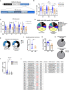
Neuro-PASC is characterized by enhanced CD4+ and diminished CD8+ T cell responses to SARS-CoV-2 Nucleocapsid protein
- PMID: 37313412
- PMCID: PMC10258318
- DOI: 10.3389/fimmu.2023.1155770
Neuro-PASC is characterized by enhanced CD4+ and diminished CD8+ T cell responses to SARS-CoV-2 Nucleocapsid protein
Erratum in
-
Corrigendum: Neuro-PASC is characterized by enhanced CD4+ and diminished CD8+ T cell responses to SARSCoV-2 Nucleocapsid protein.Front Immunol. 2023 Aug 24;14:1275925. doi: 10.3389/fimmu.2023.1275925. eCollection 2023. Front Immunol. 2023. PMID: 37691950 Free PMC article.
Abstract
Introduction: Many people with long COVID symptoms suffer from debilitating neurologic post-acute sequelae of SARS-CoV-2 infection (Neuro-PASC). Although symptoms of Neuro-PASC are widely documented, it is still unclear whether PASC symptoms impact virus-specific immune responses. Therefore, we examined T cell and antibody responses to SARS-CoV-2 Nucleocapsid protein to identify activation signatures distinguishing Neuro-PASC patients from healthy COVID convalescents.
Results: We report that Neuro-PASC patients exhibit distinct immunological signatures composed of elevated CD4+ T cell responses and diminished CD8+ memory T cell activation toward the C-terminal region of SARS-CoV-2 Nucleocapsid protein when examined both functionally and using TCR sequencing. CD8+ T cell production of IL-6 correlated with increased plasma IL-6 levels as well as heightened severity of neurologic symptoms, including pain. Elevated plasma immunoregulatory and reduced pro-inflammatory and antiviral response signatures were evident in Neuro-PASC patients compared with COVID convalescent controls without lasting symptoms, correlating with worse neurocognitive dysfunction.
Discussion: We conclude that these data provide new insight into the impact of virus-specific cellular immunity on the pathogenesis of long COVID and pave the way for the rational design of predictive biomarkers and therapeutic interventions.
Keywords: COVID-19 immunity; IL-6; T cell memory; immunoregulation; long COVID; neuro-PASC; proteomics.
Copyright © 2023 Visvabharathy, Hanson, Orban, Lim, Palacio, Jimenez, Clark, Graham, Liotta, Tachas, Penaloza-MacMaster and Koralnik.
Conflict of interest statement
Author GT was employed by Antisense Therapeutics, Ltd. The remaining authors declare that the research was conducted in the absence of any commercial or financial relationships that could be construed as a potential conflict of interest.
Figures


Similar articles
-
T cell responses to SARS-CoV-2 in people with and without neurologic symptoms of long COVID.medRxiv [Preprint]. 2022 Oct 21:2021.08.08.21261763. doi: 10.1101/2021.08.08.21261763. medRxiv. 2022. PMID: 34401886 Free PMC article. Preprint.
-
Plasma proteomics show altered inflammatory and mitochondrial proteins in patients with neurologic symptoms of post-acute sequelae of SARS-CoV-2 infection.Brain Behav Immun. 2023 Nov;114:462-474. doi: 10.1016/j.bbi.2023.08.022. Epub 2023 Sep 11. Brain Behav Immun. 2023. PMID: 37704012 Free PMC article.
-
Increased SARS-CoV-2 reactive low avidity T cells producing inflammatory cytokines in pediatric post-acute COVID-19 sequelae (PASC).Pediatr Allergy Immunol. 2023 Dec;34(12):e14060. doi: 10.1111/pai.14060. Pediatr Allergy Immunol. 2023. PMID: 38146118
-
The Neurological Manifestations of Post-Acute Sequelae of SARS-CoV-2 infection.Curr Neurol Neurosci Rep. 2021 Jun 28;21(9):44. doi: 10.1007/s11910-021-01130-1. Curr Neurol Neurosci Rep. 2021. PMID: 34181102 Free PMC article. Review.
-
Long COVID or Post-Acute Sequelae of SARS-CoV-2 Infection (PASC) and the Urgent Need to Identify Diagnostic Biomarkers and Risk Factors.Med Sci Monit. 2024 Sep 18;30:e946512. doi: 10.12659/MSM.946512. Med Sci Monit. 2024. PMID: 39289865 Free PMC article. Review.
Cited by
-
SARS-CoV-2-specific CD8+ T cells from people with long COVID establish and maintain effector phenotype and key TCR signatures over 2 years.Proc Natl Acad Sci U S A. 2024 Sep 24;121(39):e2411428121. doi: 10.1073/pnas.2411428121. Epub 2024 Sep 16. Proc Natl Acad Sci U S A. 2024. PMID: 39284068 Free PMC article.
-
A Step Forward in Long COVID Research: Validating the Post-COVID Cognitive Impairment Scale.Eur J Investig Health Psychol Educ. 2024 Dec 1;14(12):3001-3018. doi: 10.3390/ejihpe14120197. Eur J Investig Health Psychol Educ. 2024. PMID: 39727505 Free PMC article.
-
The Omics Landscape of Long COVID-A Comprehensive Systematic Review to Advance Biomarker, Target and Drug Discovery.Allergy. 2025 Apr;80(4):932-948. doi: 10.1111/all.16526. Epub 2025 Mar 14. Allergy. 2025. PMID: 40084919 Free PMC article.
-
Role of toll-like receptors in post-COVID-19 associated neurodegenerative disorders?Front Med (Lausanne). 2025 Mar 26;12:1458281. doi: 10.3389/fmed.2025.1458281. eCollection 2025. Front Med (Lausanne). 2025. PMID: 40206484 Free PMC article. Review.
-
Cerebromicrovascular mechanisms contributing to long COVID: implications for neurocognitive health.Geroscience. 2025 Feb;47(1):745-779. doi: 10.1007/s11357-024-01487-4. Epub 2025 Jan 7. Geroscience. 2025. PMID: 39777702 Free PMC article. Review.
References
-
- W.H. Organization . WHO coronavirus (COVID-19) dashboard, world health organization. World Health Organization Website; (2023). Available at: https://covid19.who.int/.
Publication types
MeSH terms
Substances
Grants and funding
LinkOut - more resources
Full Text Sources
Medical
Molecular Biology Databases
Research Materials
Miscellaneous

