. 2023 Jun;13(6):e1296.
doi: 10.1002/ctm2.1296.

BMP6 knockdown enhances cardiac fibrosis in a mouse myocardial infarction model by upregulating AP-1/CEMIP expression

Affiliations

BMP6 knockdown enhances cardiac fibrosis in a mouse myocardial infarction model by upregulating AP-1/CEMIP expression

Guiping Lu et al. Clin Transl Med. 2023 Jun.

Abstract

Background: The cardiac repair process following a myocardial infarction is a key factor in patient prognosis. In this repair process, cardiac fibrosis takes a critically important role. Among those featured genes for fibrosis, transforming growth factor beta (TGF-β) is known to be involved in the fibrosis in various organs. And bone morphogenetic protein (BMP)6 belongs to the TGF-β superfamily. Although BMPs are known to play exclusive roles in cardiac repair processes, the character of BMP6 in cardiac remodelling remains unclear.

Purpose: This study aimed to investigate how BMP6 functioned in cardiac fibrosis following myocardial infarction (MI).

Results: In this paper, we demonstrated that BMP6 expression was upregulated after myocardial infarction in wild-type (WT) mice. Furthermore, BMP6-/- mice showed a more significant decline in cardiac function and lower survival curves after MI. An enlarged infarct area, increased fibrosis and more pronounced inflammatory infiltration were observed in BMP6-/- mice compared to WT mice. The expression of collagen I, collagen III and α-SMA was increased in BMP6-/- mice. In vitro, through gain-of-function and loss-of-function experiments, it was demonstrated that BMP6 decreases collagen secretion in fibroblasts. Mechanistically, knocking down BMP6 promoted AP-1 phosphorylation, which in turn promotes CEMIP expression, led to an acceleration in the progression of cardiac fibrosis. Finally, it was found that rhBMP6 would alleviate ventricular remodelling abnormalities after myocardial infarction.

Conclusion: Therefore, BMP6 may be a novel molecular target for improving myocardial fibrosis and cardiac function after myocardial infarction.

Keywords: bone morphogenetic protein 6; cardiac fibrosis; myocardial infarction.


Conflict of interest statement

The authors declare they have no conflicts of interest.

Figures
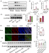
FIGURE 1
FIGURE 1
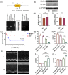
BMP6 expression is upregulated in a mouse myocardial infarction model. (A) The protein expression of BMP6 was detected from cardiac muscle by Western blot on day 14 post MI, which had original tissues from both border and remote of infarct region in wild‐type (WT) mice (n = 6). (B and C) Quantification of A‐plot results. (D) On the days 3 and 7 post MI, Western blot was used to detect the protein BMP6 in the infarct border zone (n = 6). (E) Quantification of D‐plot results. (F) The ELISA was used to detect serum BMP6 in sham‐operated group and MI group mice (n = 4). (G) Immunofluorescence detection of BMP6 expression after MI (heart section stained with an anti‐BMP6 is green, anti‐vimentin is red and DAPI is blue, scar bar: 50 μm (n = 6). (H) Quantification of G‐plot results. (I) The expression of BMP6 was detected from hypoxia‐induced cardiac fibroblasts using Western blot (n = 6). (J) Quantification of I‐plot results. All data are mean ± SD. One‐way ANOVA followed by Tukey's multiple comparisons test for (B), (C), (E) and (J). Student's t‐test for (F) and (H). Statistical significance was defined as *p < .05; **p < .01; ***p < .001.
FIGURE 2
FIGURE 2
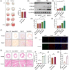
Knockout of BMP6 exacerbates cardiac failure after myocardial infarction. (A) Pattern map and genetic genotype identification results in BMP6−/− mice. (B) The expression BMP4 and BMP5 by Western blot in WT and BMP6−/− mice (n = 6). (C and D) Quantification of BMP4 and BMP5 protein expression. (E) Survival curves of WT mice and BMP6−/− mice on day 28 post MI. (F) WT mice and BMP6−/− mice were divided into sham‐operated and MI groups, with representative echocardiograms in different groups (n = 4). (G–J) WT mice and BMP6−/− mice were divided into sham‐operated and MI groups, and the left ventricular ejection fraction (LVEF), left ventricular fractional shortening (LVFS), left ventricular end‐systolic diameter (LVESD) and left ventricular end‐diastolic diameter (LVEDD) of mice in each group. All data are mean ± SD. One‐way ANOVA followed by Tukey's multiple comparisons test for (G–J). Student's t‐test for (C) and (D). Mantel–Cox test for (E). Statistical significance was defined as *p < .05; **p < .01; ***p < .001; ns = not significant. WT: wild‐type.
FIGURE 3
FIGURE 3
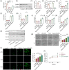
Knockout of BMP6 exacerbates myocardial fibrosis and inflammatory infiltration after myocardial infarction. (A) TTC staining 24 h post MI surgery in WT mice and BMP6−/− mice (left panel); percentage of risk area after coronary artery ligation in mice (right panel) (n = 3). (B) To detect the protein expression of collagen on cardiac fibrosis by Western blot in WT mice, BMP6−/− mice in sham and MI groups (n = 4). (C–E) Quantification of each protein expression. (F) Paraffin sections were subjected to Sirius red staining to detect cardiac collagen fibres from WT mice and BMP6−/− mice, which were divided into sham and MI groups, respectively (collagen fibres in red, scar bar: 50 μm) (n = 3). (G) Paraffin sections were subjected to HE staining (scar bar: 50 μm) (n = 3). (H) Immunofluorescence detection of α‐SMA protein in different groups (scar bar: 50 μm) (n = 3). (I) Quantification of fibrosis area of heart in different groups. (J) Quantification of infarct size by ImageJ in heart. (K) Quantification of H‐plot results. All data are mean ± SD. One‐way ANOVA followed by Tukey's multiple comparisons test for (C–E) and (I). Student's t‐test for (A), (J) and (K). Statistical significance was defined as *p < .05; **p < .01; ***p < .001; ns = not significant.
FIGURE 4
FIGURE 4
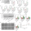
Knockdown of BMP6 exacerbates hypoxia‐induced injury in cardiac fibroblasts, which mainly promotes fibroblast migration, differentiation and proliferation. (A) In vitro, siRNA‐NC and siRNA‐BMP6 were transfected into cardiac fibroblasts, then we detected the mRNA expression of BMP6 (n = 3). (B) Verification of siRNA‐BMP6 knockdown at the protein level (n = 4). (C) Quantification of B‐plot results. (D–G) Fibroblasts were transfected with siRNA‐BMP6 following hypoxia state, and Q‐PCR was performed to detect the expression of BMP6, Col1a1, Col3a1 and FN1 mRNA in each group (n = 6). (H) Fibroblasts were transfected with siRNA‐BMP6 in hypoxia‐induced injury, and the expression of Col1a1, Col3a1 and α‐SMA protein was detected by Western blot (n = 4). (I–K) Quantification of H‐plot results. (L) Immunofluorescence staining the marker of α‐SMA in cardiac fibroblasts (n = 3) and quantification of L‐plot fluorescence staining results. (M) Cell scratching assay to detect fibroblast migration (n = 4) and quantification of M‐plot results. (N) CCK8 assay to detect fibroblast proliferation. All data are mean ± SD. One‐way ANOVA followed by Tukey's multiple comparisons test for (A), (D–G) and (I–N). Student's t‐test for (C). Statistical significance was defined as *p < .05; **p < .01; ***p < .001.
FIGURE 5
FIGURE 5
Overexpression of BMP6 reduces fibroblast over proliferation and migration in hypoxia‐induced cardiac fibroblasts. (A) Detection of BMP6 protein expression in fibroblasts after transfection with overexpressing adenovirus for 48 h (n = 6). (B) Quantification of A plot. (C–G) Detection of α‐SMA, Col3a1, FN1 and CCN2 mRNA level following overexpression of BMP6 in hypoxic state and anoxic state (n = 4). (H and I) After overexpression of BMP6, Q‐PCR was performed to detect the mRNA expression of IL‐6 and IL‐18 (n = 3). (J) The proteins of different collagens were detected by Western blot in hypoxic‐induced injuries from cardiac fibroblasts. (K–M) Quantification of J‐plot results. (N) After overexpression of BMP6, the results of cell migration in each group under hypoxic and anoxic conditions (left) and quantification of N‐plot in cell scratching experiments (right). (O) CCK8 assay to detect fibroblast proliferation. All data are mean ± SD. One‐way ANOVA followed by Tukey's multiple comparisons test for (C–I) and (K–O). Student's t‐test for (B). Statistical significance was defined as *p < .05; **p < .01; ***p < .001. Ad: adenovirus.
FIGURE 6
FIGURE 6
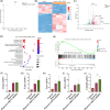
RNA‐seq indicates that cell migration‐inducing protein (CEMIP) is involved in the regulation of ventricular remodelling by BMP6. (A) PCA of hypoxic group with hypoxia+siRNA‐BMP6 composed of fibroblasts. (B) Heatmap of differential genes in the hypoxic and hypoxic+siRNA‐BMP6 groups. (C) A volcano plot of differentially expressed genes in the hypoxia group versus the hypoxia+siRNA‐BMP6 group is shown, with upregulated genes in the red, and downregulated genes in the green (n = 4). (D) GO‐C enrichment for differential genes. (E) GSEA analysis diagram of the relationship between BMP6 knockdown and genes related to cardiac fibrosis under the situation of hypoxia. (F–J) Analysis of mRNA of Gbp4/Tam1/Fpr2/Zbp1/CEMIP gene in different groups (n = 3). All data are mean ± SD. One‐way ANOVA followed by Tukey's multiple comparisons test for (F–J). Statistical significance was defined as *p < .05; **p < .01; ***p < .001.
FIGURE 7
FIGURE 7
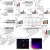
BMP6 regulates CEMIP through c‐Jun phosphorylation to improve myocardial fibrosis. (A) The protein level of CEMIP was detected by Western blot post MI (n = 6). (B) Quantification of A‐plot results in the different groups. (C) To verify the results of siRNA‐CEMIP by Western blot (n = 4). (D). Quantification of C‐plot results. (E) To detect collagen expression by Western blot in cardiac fibroblasts under different conditions (n = 3). (F–H) Quantification of E‐plot results. (I) Expression of phospho‐c‐Jun in WT mice versus BMP6 knockout mice in different groups (n = 3). (J) Immunohistochemical detection of phospho‐c‐Jun expression in different groups. (K) Expression results of c‐fos, c‐Jun after T5224 stimulation of fibroblasts and the quantification of K‐plot results. (L) Expression of CEMIP under different states in cardiac fibroblasts (n = 3). (M) Quantification of L‐plot results. (N) Immunofluorescence staining for co‐localization of AP‐1/CEMIP. (O) Quantification of N‐plot. All data are mean ± SD. Student's t‐test for (B). One‐way ANOVA followed by Tukey's multiple comparisons test for (D), (F–K) and (M). Statistical significance was defined as *p < .05; **p < .01; ***p < .001.
FIGURE 8
FIGURE 8
Recombinant human protein BMP6 (rhBMP6) enhances cardiac function after myocardial infarction in WT mice. (A) The WT mice were divided into two groups: WT mice alone and WT mice + rhBMP6 tail vein administration group. The echocardiograms of each group were obtained after creating models of MI or conducting sham procedures on mice. (B–E) LVEF, LVFS, LVESD, LVEDD in different groups of mice (n = 3). (F) To detect the collagen deposition by Western blot in MI mice (n = 3). (G) Quantification of F‐plot results. All data are mean ± SD. One‐way ANOVA followed by Tukey's multiple comparisons test for (B–E) and (G). Statistical significance was defined as *p < .05; **p < .01; ***p < .001.
FIGURE 9
FIGURE 9
Mechanism of BMP6 regulates myocardial fibrosis through AP‐1/CEMIP. • Knocking out BMP6 promotes AP‐1 phosphorylation in hypoxia‐induced cardiac fibroblasts. • The expression of CEMIP is upregulated post MI, which is affected by AP‐1/BMP6. • rhBMP6 affects the process of cardiac remodelling, which may be a potential therapeutic target in the cardiac remodelling post MI.

References

    1. Puymirat E, Simon T, Cayla G, et al. Acute myocardial infarction: changes in patient characteristics, management, and 6‐month outcomes over a period of 20 years in the FAST‐MI program (French Registry of acute ST‐elevation or non‐ST‐elevation myocardial infarction) 1995 to 2015. Circulation. 2017;136(20):1908‐1919. - PubMed
    1. Yeh RW, Sidney S, Chandra M, et al. Population trends in the incidence and outcomes of acute myocardial infarction. N Engl J Med. 2010;362(23):2155‐2165. - PubMed
    1. Prabhu SD, Frangogiannis NG. The biological basis for cardiac repair after myocardial infarction: from inflammation to fibrosis. Circ Res. 2016;119(1):91‐112. - PMC - PubMed
    1. Talman V, Ruskoaho H. Cardiac fibrosis in myocardial infarction‐from repair and remodeling to regeneration. Cell Tissue Res. 2016;365(3):563‐581. - PMC - PubMed
    1. Chen D, Zhao M, Mundy GR. Bone morphogenetic proteins. Growth Factors. 2004;22(4):233‐241. - PubMed

Publication types

LinkOut - more resources