. 2023 Sep;60(9):5395-5410.
doi: 10.1007/s12035-023-03435-3. Epub 2023 Jun 14.

Reduced Cerebellar BDNF Availability Affects Postnatal Differentiation and Maturation of Granule Cells in a Mouse Model of Cholesterol Dyshomeostasis

Affiliations

Reduced Cerebellar BDNF Availability Affects Postnatal Differentiation and Maturation of Granule Cells in a Mouse Model of Cholesterol Dyshomeostasis

Micaela Lucarelli et al. Mol Neurobiol. 2023 Sep.

Abstract

Niemann-Pick type C1 (NPC1) disease is a lysosomal lipid storage disorder due to mutations in the NPC1 gene resulting in the accumulation of cholesterol within the endosomal/lysosomal compartments. The prominent feature of the disorder is the progressive Purkinje cell degeneration leading to ataxia.In a mouse model of NPC1 disease, we have previously demonstrated that impaired Sonic hedgehog signaling causes defective proliferation of granule cells (GCs) and abnormal cerebellar morphogenesis. Studies conducted on cortical and hippocampal neurons indicate a functional interaction between Sonic hedgehog and brain-derived neurotrophic factor (BDNF) expression, leading us to hypothesize that BDNF signaling may be altered in Npc1 mutant mice, contributing to the onset of cerebellar alterations present in NPC1 disease before the appearance of signs of ataxia.We characterized the expression/localization patterns of the BDNF and its receptor, tropomyosin-related kinase B (TrkB), in the early postnatal and young adult cerebellum of the Npc1nmf164 mutant mouse strain.In Npc1nmf164 mice, our results show (i) a reduced expression of cerebellar BDNF and pTrkB in the first 2 weeks postpartum, phases in which most GCs complete the proliferative/migrative program and begin differentiation; (ii) an altered subcellular localization of the pTrkB receptor in GCs, both in vivo and in vitro; (iii) reduced chemotactic response to BDNF in GCs cultured in vitro, associated with impaired internalization of the activated TrkB receptor; (iv) an overall increase in dendritic branching in mature GCs, resulting in impaired differentiation of the cerebellar glomeruli, the major synaptic complex between GCs and mossy fibers.

Keywords: Cerebellar glomerulus; Cerebellum; Niemann-Pick type C1 disease; Npc1; TrkB receptor.


Conflict of interest statement

The authors declare no competing interests.

Figures

Fig. 1
Fig. 1
Npc1nmf164 mice display a reduced BDNF expression in the postnatal developing cerebellum. Relative quantification of BDNF mRNA (A) and representative immunoblots of BDNF protein expression levels (B) in wt (empty bars) and Npc1nmf164 (full bars) mice at increasing time points. The BDNF transcript and protein levels were normalized to the expression levels of the housekeeping gene S16 (unpaired t test * p < .05 versus wt. ** p < .01 versus wt) and β-actin (unpaired t test **p < .01), respectively. Histograms indicate the abundance expressed as mean ± SD (5 mice/group).
Fig. 2
Fig. 2
Npc1nmf164 mice display abnormal BDNF protein localization along cerebellar cortex development. Representative fields of parasagittal sections of wt and Npc1nmf164 mouse cerebella are shown in the figure. Sections of PN11, 15, and 30 of wt and Npc1nmf164 mice (3–4 mice/group; 3–4 sections/mouse) were immunostained with anti-BDNF antibody (brown) and nuclei counterstained with methyl green. Higher magnifications of the PN11 fields are shown in upper panels. EGL, external granular layer; ML, molecular layer; PCL, Purkinje cell layer; IGL, internal granular layer; asterisks point mossy fibers; white arrowheads point PCs. scale bars: 40 μm
Fig. 3
Fig. 3
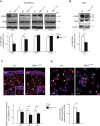
Abnormal pTrkB receptor expression and localization are observed in the early stage of cerebellar development and in vitro GCs of Npc1nmf164 mice. A, B Representative immunoblots of pTrkB protein expression in extracts from wt and Npc1nmf164 mice at different stages of cerebellar development (PN8,11, 15, and 30) and in DIV0 GCs. Western blot quantification of pTrkB in wt (white bars) and Npc1nmf164 (black bars) mice. Histograms indicate the abundance (mean ± SD) of each protein determined by the densitometry of protein bands obtained by taking total TrkB (TrkB) as an internal reference. Arrowhead indicates TrkB truncated form present in GCs. Significance is shown as a p value calculated using an unpaired t test. *p < .05 versus wt. **p < .01 versus wt (n = 5 mice/group). C, D Representative fields of confocal images are shown in the figure. Detection of pTrkB receptor (red) and nuclei (Hoechst 33258, blue) in PN8 cerebellar sections and in DIV3 GCs. The GCs in the EGL are magnified in the boxed regions of panel C. Scale bar: 20 μm. Comparison of the mean fluorescence intensity (y-axis) generated by staining sagittal sections of PN8 cerebellar cortex layers of wt and Npc1nmf164 mice with antibodies against pTrkB receptor. D GCs expressing pTrkB receptors in the soma (arrowheads) and in the leading process (asterisks) are indicated. Data shown represent the means ± SD and were evaluated by unpaired t test. **p < .01 versus wt (n = 3 mice/group). ML, molecular layer; PC, Purkinje cell; IGL, internal granular layer. Quantitative analysis of in vitro GCs with BDNF-dependent pTrkB polarization in wt and Npc1 mutant mice is shown as a p value calculated using an unpaired t test. *p < .05 versus wt
Fig. 4
Fig. 4
Reduced pTrkB localization in early endosomes of Npc1nmf164 cerebella. A BDNF-dependent polarization and colocalization of pTrkB and early endosome antigen 1 (EEA1) were visualized by immunostaining with anti-pTrkB (green) and anti-EEA1 (red), respectively, in BDNF stimulated DIV3 GCs of wt and Npc1nmf164 mice. Scale bar: 20 μm; 10 μm. B Representative immunoblots of pTrkB protein expression in cerebellar extracts from wt and Npc1nmf164 mice at PN8. Western blot quantification of pTrkB and EEA1 in wt (white bars) and Npc1nmf164 (black bars) mice. Histograms indicate the abundance (mean ± SD) of each protein determined by the densitometry of protein bands obtained taking EEA1, for endosomal fraction, and β-actin, for total homogenate, as internal reference. Significance is shown as a p value calculated using an unpaired t test. *p < .05 versus wt (n = 3 mice/group)
Fig. 5
Fig. 5
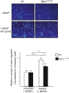
In the Npc1 mutant mice, in vitro cultured GCs display a reduced BDNF chemotaxis response. Purified wt and Npc1nmf164 GCs were cultured for 24 h in Boyden chambers without (−BDNF) or with (+BDNF) 40 ng/mL BDNF in the lower chambers. GCs that migrated through the porous membrane into the lower chamber were directly quantified by fluorescence microscopy. Representative fields of migrated GCs, with nuclei staining with 0.5 μg/mL Hoechst-33258. Scale bar 20 μm. The results from five separate experiments, expressed as number of migrated cells normalized to untreated cells, are shown in the histograms. Treated Npc1nmf164 GCs (black bars) display a significant reduced chemotaxis response to BDNF compared to the treated wt cells (white bars). *p< .05 versus wt (n = 5 mice/group)
Fig. 6
Fig. 6
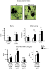
GCs of Npc1nmf164 mice exhibit abnormal dendritic morphology in the IGL layer. Representative fields of Golgi-impregnated GCs from the PN30 IGL of wt (left) and Npc1nmf164 (right) mice. (Scale bar: 10μm). Soma and dendrites of Golgi-stained neurons were traced, and morphological parameters were analyzed using Neurolucida Software. Unpaired Student’s t test analysis of soma (top left); number of dendrites, nodes, and ends (top right); total dendritic length, surface, and, volume (bottom left); and average diameter (bottom right) of cerebellar GCs were represented by histograms. Data are shown as mean ± SD; *p < .05; **p < .01; ***p < .001. (n cell for each genotype = 10). wt = white bars; Npc1nmf164 = black bars
Fig. 7
Fig. 7
Morphological defects of cerebellar MF terminals maturation in the IGL of Npc1nmf164 mice. Synaptophysin (Syp) immunohistochemistry of cerebellar sagittal sections from three independently bred pairs of PN11 and PN30 wt and Npc1 mutant mice, counterstained with methyl green (scale bar: 20 μm). The number of cells and morphological parameters (area and tortuosity) were analyzed with the Neurolucida Software. Moreover, the number of GCs and the distance between the GCs and among the GCs-glomeruli rosette were also analyzed in the same fields. The Npc1nmf164 mice exhibit fewer, smaller, and less complex Syp-positive area at the levels of glomeruli rosette in both stages analyzed, which was associated with a reduced number of GCs. Histograms represent statistical analysis with unpaired Student’s t test. Data are shown as mean ± SD *p < .05; **p < .01. wt = white bars; Npc1nmf164 = black bars. Arrowheads = MF terminals, ML, molecular layer; PC, Purkinje cell; IGL, internal granular layer

Similar articles

Cited by

References

    1. Pentchev PG, Brady RO, Blanchette-Mackie EJ, Vanier MT, Carstea ED, Parker CC, Goldin E, Roff CF. The Niemann-Pick C lesion and its relationship to the intracellular distribution and utilization of LDL cholesterol. Biochim Biophys Acta. 1994;1225:235–243. doi: 10.1016/0925-4439(94)90001-9. - DOI - PubMed
    1. Vanier MT. Niemann-Pick disease type C. Orphanet J Rare Dis. 2010;5:16. doi: 10.1186/1750-1172-5-16. - DOI - PMC - PubMed
    1. Palladino G, Loizzo S, Fortuna A, Canterini S, Palombi F, Erickson RP, Mangia F, Fiorenza MT. Visual evoked potentials of Niemann-Pick type C1 mice reveal an impairment of the visual pathway that is rescued by 2-hydroxypropyl-ß-cyclodextrin. Orphanet J Rare Dis. 2015;10:133. doi: 10.1186/s13023-015-0348-0. - DOI - PMC - PubMed
    1. Boyle BR, Melli SE, Altreche RS, Padron ZM, Fak Y, Kim S, Vasquez MD, Carone DM, Carone BR, Soto I. NPC1 deficiency impairs cerebellar postnatal development of microglia and climbing fiber refinement in a mouse model of Niemann-Pick disease type C. Development. 2020;147:dev189019. doi: 10.1242/dev.189019. - DOI - PMC - PubMed
    1. Oddi S, Caporali P, Dragotto J, et al. The endocannabinoid system is affected by cholesterol dyshomeostasis: Insights from a murine model of Niemann Pick type C disease. Neurobiol Dis. 2019;130:104531. doi: 10.1016/j.nbd.2019.104531. - DOI - PubMed

LinkOut - more resources