. 2023 Jun 16;9(24):eadf0661.
doi: 10.1126/sciadv.adf0661. Epub 2023 Jun 14.

CD62L expression marks SARS-CoV-2 memory B cell subset with preference for neutralizing epitopes

Affiliations

CD62L expression marks SARS-CoV-2 memory B cell subset with preference for neutralizing epitopes

Taishi Onodera et al. Sci Adv. .

Abstract

Severe acute respiratory syndrome coronavirus 2-neutralizing antibodies primarily target the spike receptor binding domain (RBD). However, B cell antigen receptors (BCRs) on RBD-binding memory B (Bmem) cells have variation in the neutralizing activities. Here, by combining single Bmem cell profiling with antibody functional assessment, we dissected the phenotype of Bmem cell harboring the potently neutralizing antibodies in coronavirus disease 2019 (COVID-19)-convalescent individuals. The neutralizing subset was marked by an elevated CD62L expression and characterized by distinct epitope preference and usage of convergent VH (variable region of immunoglobulin heavy chain) genes, accounting for the neutralizing activities. Concordantly, the correlation was observed between neutralizing antibody titers in blood and CD62L+ subset, despite the equivalent RBD binding of CD62L+ and CD62L- subset. Furthermore, the kinetics of CD62L+ subset differed between the patients who recovered from different COVID-19 severities. Our Bmem cell profiling reveals the unique phenotype of Bmem cell subset that harbors potently neutralizing BCRs, advancing our understanding of humoral protection.


Figures

Fig. 1.
Fig. 1.. Linking transcriptional B cell profiling with antibody function.
(A) Schematic diagram of experimental workflow for linking transcriptional B cell profiling with antibody function. COVID-19 convalescent peripheral blood mononuclear cells (PBMCs) were subjected to fluorescence-activated cell sorting (FACS) using DNA-barcoded antigen probes and single-cell transcriptome analysis (LIBRA-seq). The antibody neutralizing activities were determined using reconstructed mAbs and were linked to LIBRA-seq data. scRNA-seq, single-cell RNA sequencing; scV(D)J-seq, single-cell V(D)J sequencing. (B) FACS gating strategy for sorting before LIBRA-seq analysis. FSC, forward scatter; SSC, side scatter. (C) The barcode counts from antigen probes were plotted in a similar format to the FACS profile. Probe-positive cells were as gated, and bright and dim cells were defined as above and below 33 percentiles in the positive compartment (dashed line). Recombinant mAbs were reconstructed from bright and dim cells among S trimer+RBD+, non-RBD S trimer+, and RBD+ only (n = 191). (D) Antigen-binding activities of reconstructed mAbs were assessed by FACS using mAb-coated beads.
Fig. 2.
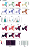
Fig. 2.. Multiple B cell clusters with distinct transcriptional profiling.
(A) UMAP plot displaying the cells defined by their single-cell transcriptome analysis. The cells are colored based on their cluster assignments by the Louvain clustering algorithm. The distribution of S trimer+RBD+, non-RBD S trimer+, and immunoglobulin subclasses was highlighted (top). The distribution of CD27+/CD21+/FcRL5+ cells and cells with different disease severities was highlighted in different colors (bottom). (B) The normalized counts for the indicated transcripts are shown in the violin plot from each cluster. (C) The distribution of the indicated transcripts for each cluster is highlighted in different colors. (D) Heatmap representation of VH gene fraction for each cluster. (E) Cells in public BCR clonotypes for each cluster are highlighted in colors. Different colors indicate different donors.
Fig. 3.
Fig. 3.. B cell mapping with potently neutralizing antibody identifies CD62L phenotype.
(A) RBD binders with bright intensity (n = 127) were tested for the neutralizing activities and highlighted in UMAP based on the NT activities (NT high and NT med). (B) Percentages of the indicated NT activities are shown in cluster 0/1 (n = 8), 2 (n = 76), 3 (n = 10), and 4 (n = 33). (C) Distribution of NT activities in different disease severities. (D) Distribution of CD62L+ and CD62L cells is highlighted in UMAP. (E) Expression levels of CD62L among the cells in different clusters are shown on the basis of the transcript counts. Statistical difference was evaluated between the cells in each cluster and all cells. (F) CD62L and CD19 expression levels are comparatively plotted on the basis of neutralizing activities. Statistical difference was evaluated between the cells in each group and all cells. (G) S trimer+RBD+IgG+ Bmem cells were fractionated into either CD62L+ or CD62L subset and subjected to single-cell culture. Pie charts represent the ratios of mAb clones with NT activity. Statistical analyses were performed using the chi-square test in (B) and (C), Kruskal-Wallis test followed by Dunn’s multiple comparison test in (E) and (F), Fisher’s exact test in (G), and Jackknife resampling was performed to assure the statistical significance in (G). *P < 0.05, **P < 0.01, and ****P < 0.0001. ns, not significant (P ≥ 0.05).
Fig. 4.
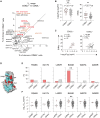
Fig. 4.. IgV genes and epitope preference of CD62L+ Bmem subset.
(A) Numbers of VH genes detected from the single-cell transcriptome analysis were plotted in IgG1+CD62L+ and CD62L Bmem subsets among RBD binders (n = 782). The VH genes were highlighted on the basis of the number of sequences deposited into the CoV-AbDab database (https://opig.stats.ox.ac.uk/webapps/covabdab/). (B) IC50 and numbers of somatic hypermutations (SHMs) per VH + VL genes from RBD-binding, NT+ IgG clones derived from IgG+CD62L+ (n = 22) and CD62L (n = 50) Bmem cell subsets were plotted. Each dot represents the data from an individual IgG clone. (C) Spearman correlations between IC50 and somatic hypermutations in each subset are plotted. R and P values are indicated. (D) RBD structure (Protein Data Bank: 6LZG) highlighted with human ACE2 binding region (light blue) and amino acid positions of single-mutant RBDs (red) was shown. (E) IgG clones derived from the indicated Bmem cells were subjected to the binding assay against six RBD mutants bearing a single mutation. The IgG clones that lost the binding to the indicated RBD mutants only were identified and the percentages are shown. (F) NT activities of IgG clones that lost the binding to RBD mutants were comparably plotted with other IgG clones. Statistical analyses were performed using Spearman’s rank-order correlation test in (B), Fisher’s exact test in (D), and Mann-Whitney test in (B) and (E). **P < 0.01.
Fig. 5.
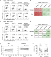
Fig. 5.. The functional relevance of CD62L+ Bmem cell subset.
(A to D) RBD-binding IgG+ Bmem cells were divided into four populations (resting, CD27lo, activated, and atypical). The gating strategies for the Bmem cell subsets are shown in fig. S4. The numbers of CD62L+ (A and B) or CD62L (C and D) subsets in individual populations were plotted against RBD-binding IgG tiers or plasma dilution factors for IC50. (E) The numbers of RBD-binding IgG+ Bmem cells were enumerated in the T1 and T2 periods in the mild and moderate patients. (F) CD62L+ ratios among the RBD-binding IgG+ Bmem cells were plotted in the T1 and T2 from mild and moderate patients. Each dot represents the data from an individual donor. Statistical analyses were performed using the Spearman’s rank-order correlation test in (A) to (D), Wilcoxon test in (E) and (F). *P < 0.05, **P < 0.01, ***P < 0.001, and ****P < 0.0001.

References

    1. T. Kurosaki, K. Kometani, W. Ise, Memory B cells. Nat. Rev. Immunol. 15, 149–159 (2015). - PubMed
    1. R. R. Goel, M. M. Painter, S. A. Apostolidis, D. Mathew, W. Meng, A. M. Rosenfeld, K. A. Lundgreen, A. Reynaldi, D. S. Khoury, A. Pattekar, S. Gouma, L. Kuri-Cervantes, P. Hicks, S. Dysinger, A. Hicks, H. Sharma, S. Herring, S. Korte, A. E. Baxter, D. A. Oldridge, J. R. Giles, M. E. Weirick, C. M. McAllister, M. Awofolaju, N. Tanenbaum, E. M. Drapeau, J. Dougherty, S. Long, K. D'Andrea, J. T. Hamilton, M. McLaughlin, J. C. Williams, S. Adamski, O. Kuthuru; UPenn COVID Processing Unit, I. Frank, M. R. Betts, L. A. Vella, A. Grifoni, D. Weiskopf, A. Sette, S. E. Hensley, M. P. Davenport, P. Bates, E. T. L. Prak, A. R. Greenplate, E. J. Wherry, mRNA vaccines induce durable immune memory to SARS-CoV-2 and variants of concern. Science 374, abm0829 (2021). - PMC - PubMed
    1. D. S. Khoury, D. Cromer, A. Reynaldi, T. E. Schlub, A. K. Wheatley, J. A. Juno, K. Subbarao, S. J. Kent, J. A. Triccas, M. P. Davenport, Neutralizing antibody levels are highly predictive of immune protection from symptomatic SARS-CoV-2 infection. Nat. Med. 27, 1205–1211 (2021). - PubMed
    1. A. Sette, S. Crotty, Adaptive immunity to SARS-CoV-2 and COVID-19. Cell 184, 861–880 (2021). - PMC - PubMed
    1. E. Andreano, E. Nicastri, I. Paciello, P. Pileri, N. Manganaro, G. Piccini, A. Manenti, E. Pantano, A. Kabanova, M. Troisi, F. Vacca, D. Cardamone, C. De Santi, J. L. Torres, G. Ozorowski, L. Benincasa, H. Jang, C. Di Genova, L. Depau, J. Brunetti, C. Agrati, M. R. Capobianchi, C. Castilletti, A. Emiliozzi, M. Fabbiani, F. Montagnani, L. Bracci, G. Sautto, T. M. Ross, E. Montomoli, N. Temperton, A. B. Ward, C. Sala, G. Ippolito, R. Rappuoli, Extremely potent human monoclonal antibodies from COVID-19 convalescent patients. Cell 184, 1821–1835.e16 (2021). - PMC - PubMed