. 2023 Jun 13;18(6):1308-1324.
doi: 10.1016/j.stemcr.2023.05.007.

Parallel use of human stem cell lung and heart models provide insights for SARS-CoV-2 treatment

Affiliations

Parallel use of human stem cell lung and heart models provide insights for SARS-CoV-2 treatment

Rajeev Rudraraju et al. Stem Cell Reports. .

Abstract

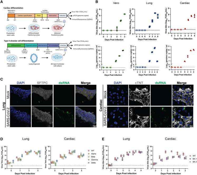
Severe acute respiratory syndrome coronavirus 2 (SARS-CoV-2) primarily infects the respiratory tract, but pulmonary and cardiac complications occur in severe coronavirus disease 2019 (COVID-19). To elucidate molecular mechanisms in the lung and heart, we conducted paired experiments in human stem cell-derived lung alveolar type II (AT2) epithelial cell and cardiac cultures infected with SARS-CoV-2. With CRISPR-Cas9-mediated knockout of ACE2, we demonstrated that angiotensin-converting enzyme 2 (ACE2) was essential for SARS-CoV-2 infection of both cell types but that further processing in lung cells required TMPRSS2, while cardiac cells required the endosomal pathway. Host responses were significantly different; transcriptome profiling and phosphoproteomics responses depended strongly on the cell type. We identified several antiviral compounds with distinct antiviral and toxicity profiles in lung AT2 and cardiac cells, highlighting the importance of using several relevant cell types for evaluation of antiviral drugs. Our data provide new insights into rational drug combinations for effective treatment of a virus that affects multiple organ systems.

Keywords: COVID-19; SARS-CoV-2; antiviral drugs; human stem cell-derived lung and heart models.


Conflict of interest statement

Conflict of interests E.P. is a co-founder and scientific advisor and holds equity in, and N.C. has a paid position with, Dynomics, a biotechnology company focused on the development of heart failure therapeutics. J.M.P. is a co-founder and shareholder of Mogrify, Ltd., a cell therapy company.

Figures

None
Graphical abstract
Figure 1
Figure 1
SARS-CoV-2 infection of cardiac and lung AT2 cells (A) Schematic of cell differentiation and infection protocols. (B) Viral titers and genome copies in SARS-CoV-2 (VIC01)-infected Vero, lung AT2 (H9), and cardiac (NKX2-5) culture supernatants. Data are presented as mean ± SD. Results are representative of two independent experiments each with 2 technical replicates. (C) Representative fluorescent confocal microscopy images of dsRNA (green) expression in infected lung AT2 (SFTPC-positive) and cardiac (cTNT-positive) cells at 3 dpi. (D and E) Titers in supernatants from cardiac (NKX2-5) and lung AT2 cells infected with VIC01 (WT) compared with cells infected with Alpha, Beta, Gamma, and Delta variants (D) or Omicron (BA.1 and BA.2) variants (E). Data are presented as mean ± SD. Results are representative of two independent experiments each with 3 technical replicates. See also Figure S1.
Figure 2
Figure 2
SARS-CoV-2 infection is ACE2 dependent but further steps in entry and antiviral sensitivity are different between lung AT2 and cardiac cells (A) Virus titer in supernatants from SARS-CoV-2 (VIC01)-infected H9-derived WT and ACE2 KO cardiac and lung AT2 cells. (B) Virus titer at 3 dpi in supernatants from lung AT2 (H9) and cardiac cells (NKX2-5) treated with two α-ACE2 antibodies or a human IgG1 isotype control before infection with SARS-CoV-2. (C) Virus titer at 3 dpi in supernatant from cardiac cells (NKX2-5, red triangles) and lung AT2 cells (blue squares) infected with SARS-CoV-2 (VIC01) in the presence of camostat, CA-074 Me, or DMSO (vehicle control). (D and E) Virus titers and genome copies at 3 dpi in supernatant from Vero, lung AT2, and cardiac cells (NKX2-5) infected with SARS-CoV-2 VIC01 in the presence of various concentrations of Remdesivir (D) or NHC (E). Data are presented as mean ± SD. Results are representative of two independent experiments each with 3 technical replicates. See also Figure S2 and S3.
Figure 3
Figure 3
Cardiac cells have a stronger interferon signature than lung AT2 cells following SARS-CoV-2 infection (A) Principal-component analysis (PCA) plot of uninfected (mock) and SARS-CoV-2-infected (virus) cardiac (H9) and lung AT2 cells at 1 and 3 dpi. (B) Venn diagram depicting the number of overlapping and unique differentially expressed genes compared with mock. (C) Extended plot of top enriched WP terms from total cardiac, total lung, intersect, lung unique, and cardiac unique differentially expressed genes. Circle size is proportional to the number of genes that matched the pathway, and color represents the logp value as calculated by Metascape. (D) Heatmap in log2 fold change (FC) of representative interferon genes (compared with representative mock samples at 1 dpi) separated by type as defined by WikiPathways. Columns on the right show the concordance between direction of FC of RNA-seq data and the LegendPlex/Taqman assays. For 1 and 3 dpi, 3 lung samples and 2 cardiac samples were analyzed per group (mock, virus). (E) Cytokine concentrations in supernatants of mock- and SARS-CoV-2-infected cardiac and lung AT2 cells at 3 dpi. Data are presented as mean ± SD. Results are representative of two independent experiments each with 3 technical replicates. See also Figure S4.
Figure 4
Figure 4
SARS-CoV-2 infection alters different signaling networks in heart and lung organoids (A) Schematic of phosphoproteomics workflow. (B) Number of phosphorylated peptides, sites, and proteins found in 3 samples. (C) PCA plot of median-normalized phosphoproteome replicates from uninfected (mock) and SARS-CoV-2-infected (CoV) cardiac (H9) and lung AT2 cells at 18 and 24 h post-infection. (D and E) Number of significantly (adjusted p [padj] < 0.05, FC > 1.5) regulated phosphopeptides per timepoint (D) and their overlap (E) between cell types. (F and G) Top 5 predicted drivers following SARS-CoV-2 infection in cardiac cells (F) and in lung cells (G) at 72 h post-infection. padj are calculated based on the computation of Z score probability distributions of a molecule to be a driver and adjusted using the Benjamini and Hochberg algorithm. (H) GO cellular components enriched in at least one condition (p < 0.05) that are related to endosomes. (I) Kinases with enriched substrates, which were selected for targeting. (J) Abundance of the PKC substrate MARCKS S170. (K) Abundance of the SRPK1 substrate SARS-CoV-2 nucleocapsid protein S206. See also Figure S4.
Figure 5
Figure 5
Efficacy of kinase inhibitors against SARS-CoV-2 replication varies between lung AT2 and cardiac cells Cell viability (percentage relative to vehicle control, black lines) and inhibition of SARS-CoV-2 growth (percentage relative to vehicle control) in the presence of CDK (A), CHK (B), PKC (C), and SRPK1 (D) inhibitors in lung AT2 cells (H9) and cardiac cells (NKX2-5) at 2 dpi. Dotted black lines indicate 50% virus inhibition or cell viability. Results are representative of two independent experiments each with 3 technical replicates. See also Figure S5.

Update of

References

    1. Anderson D.J., Kaplan D.I., Bell K.M., Koutsis K., Haynes J.M., Mills R.J., Phelan D.G., Qian E.L., Leitoguinho A.R., Arasaratnam D., et al. NKX2-5 regulates human cardiomyogenesis via a HEY2 dependent transcriptional network. Nat. Commun. 2018;9:1373. doi: 10.1038/s41467-018-03714-x. - DOI - PMC - PubMed
    1. Bailey A.L., Dmytrenko O., Greenberg L., Bredemeyer A.L., Ma P., Liu J., Penna V., Winkler E.S., Sviben S., Brooks E., et al. SARS-CoV-2 infects human engineered heart tissues and models COVID-19 myocarditis. JACC Basic Transl Sci. 2021;6:331–345. doi: 10.1016/j.jacbts.2021.01.002. - DOI - PMC - PubMed
    1. Bojkova D., Wagner J.U.G., Shumliakivska M., Aslan G.S., Saleem U., Hansen A., Luxán G., Günther S., Pham M.D., Krishnan J., et al. SARS-CoV-2 infects and induces cytotoxic effects in human cardiomyocytes. Cardiovasc. Res. 2020;116:2207–2215. doi: 10.1093/cvr/cvaa267. - DOI - PMC - PubMed
    1. Bouhaddou M., Memon D., Meyer B., White K.M., Rezelj V.V., Correa Marrero M., Polacco B.J., Melnyk J.E., Ulferts S., Kaake R.M., et al. The global phosphorylation landscape of SARS-CoV-2 infection. Cell. 2020;182:685–712.e19. doi: 10.1016/j.cell.2020.06.034. - DOI - PMC - PubMed
    1. Chen J., Gao K., Wang R., Wei G.W. An iTSC-derived placental model of SARS-CoV-2 infection reveals ACE2-dependent susceptibility in syncytiotrophoblasts. bioRxiv. 2021 doi: 10.1101/2021.10.27.465224. - DOI - PubMed

Publication types

Substances