. 2023 Sep:186:109741.
doi: 10.1016/j.radonc.2023.109741. Epub 2023 Jun 12.

Ultra-high dose-rate proton FLASH improves tumor control

Affiliations

Ultra-high dose-rate proton FLASH improves tumor control

Samriddhi Shukla et al. Radiother Oncol. 2023 Sep.

Abstract

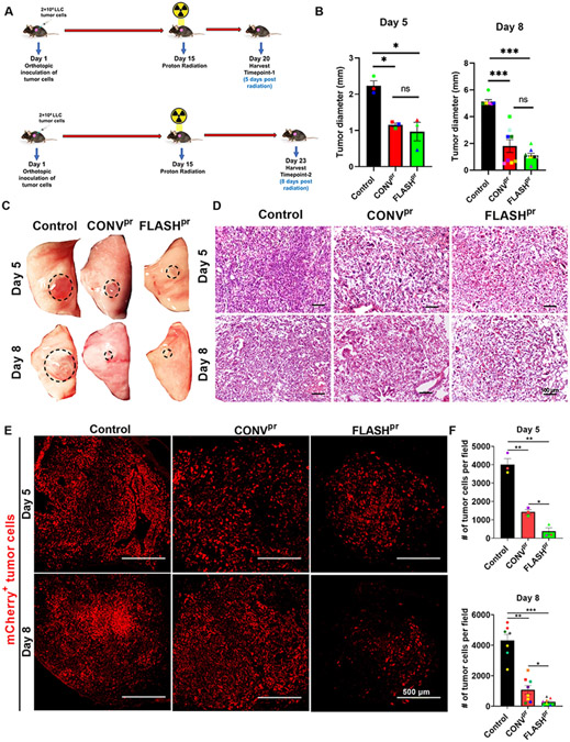
Background and purpose: Proton radiotherapy (PRT) offers potential benefits over other radiation modalities, including photon and electron radiotherapy. Increasing the rate at which proton radiation is delivered may provide a therapeutic advantage. Here, we compared the efficacy of conventional proton therapy (CONVpr) to ultrahigh dose-rate proton therapy, FLASHpr, in a mouse model of non-small cell lung cancers (NSCLC).

Materials and methods: Mice bearing orthotopic lung tumors received thoracic radiation therapy using CONVpr (<0.05 Gy/s) and FLASHpr (>60 Gy/s) dose rates.

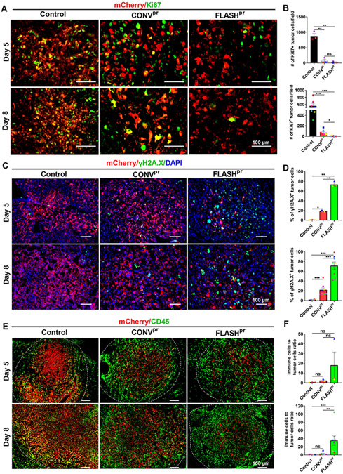
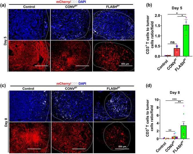
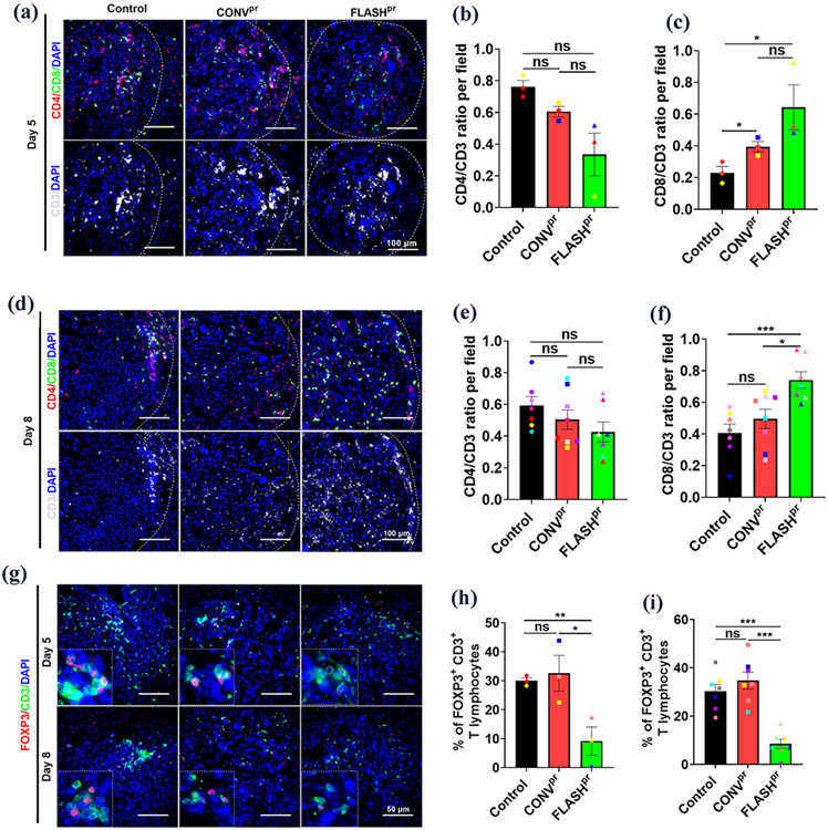
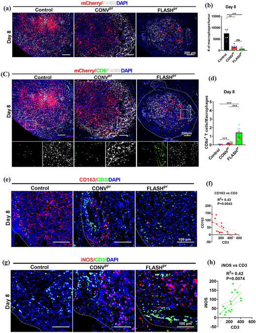
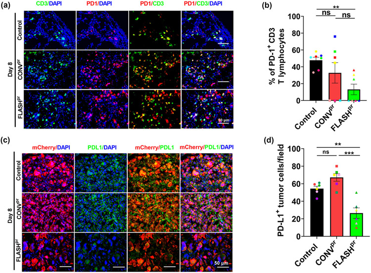
Results: Compared to CONVpr, FLASHpr was more effective in reducing tumor burden and decreasing tumor cell proliferation. Furthermore, FLASHpr was more efficient in increasing the infiltration of cytotoxic CD8+ T-lymphocytes inside the tumor while simultaneously reducing the percentage of immunosuppressive regulatory T-cells (Tregs) among T-lymphocytes. Also, compared to CONVpr, FLASHpr was more effective in decreasing pro-tumorigenic M2-like macrophages in lung tumors, while increasing infiltration of anti-tumor M1-like macrophages. Finally, FLASHpr treatment reduced expression of checkpoint inhibitors in lung tumors, indicating reduced immune tolerance.

Conclusions: Our results suggest that FLASH dose-rate proton delivery modulates the immune system to improve tumor control and might thus be a promising new alternative to conventional dose rates for NSCLC treatment.

Keywords: FLASH; Lung cancer; Mouse model; Proton radiotherapy; Ultrahigh dose-rate proton.


Conflict of interest statement

Declaration of Competing Interest The authors declare that they have no known competing financial interests or personal relationships that could have appeared to influence the work reported in this paper.

Figures

Figure 1.
Figure 1.. FLASHpr has better efficacy in reducing lung tumor burden compared to CONVpr.
(A) Experimental schematic for LLC orthotopic model of lung cancer, and timeline of irradiation and tissue harvest. (B) Mice treated with FLASHpr and CONVpr radiation have decreased lung tumor diameters compared to control tumor bearing mice. Tumors were measured using calipers at day 5 (3 mice per group) and day 8 (8 mice per group). (C) Representative images of tumor-bearing left lung lobe from each group at day 5 and day 8 post radiation. (D) Representative images after H&E staining of lung tumors at day 5 and day 8. (Scale bar 100 μm). (E-F) Immunofluorescence staining for mCherry-labelled tumor cells shows significant reduction of tumor burden at day 5 and day 8 in FLASHpr group compared to control and CONVpr. (Scale bar 500 μm). *, P<0.05; **, P<0.01; ***, P<0.001.
Figure 2.
Figure 2.. FLASH decreases the number of proliferating tumor cells compared to conventional dose-rate proton treatment.
(A-B) Number of Ki67 positive tumor cells is decreased in lung tumors treated with FLASHpr compared to CONVpr. Number of mCherry and Ki67 double positive cells per field was counted for each tumor and presented as average ± SEM (Scale bar 100 μm). Each dot represents one biological replicate. (C-D) FLASHpr increases the percentage of mCherry/γH2A.X-double positive tumor cells compared to CONVpr. Nuclei are counterstained with DAPI. Percentage of double positive cells were calculated for each tumor and presented as mean percentage ± SEM (Scale bar 100 μm). Each dot represents one biological replicate. (E-F) FLASHpr increases the CD45+ immune cells to mCherry+ tumor cell ratio in lung tumors. Number of mCherry+ tumor cells and CD45+ immune cells were counted for each tumor and presented as the mean ratio of immune cells to tumor cells ± SEM (Scale bar 100 μm). Each dot represents one biological replicate. N = 3-8 mice per group; *, P<0.05; **, P<0.01; ***, P<0.001.
Figure 3.
Figure 3.. FLASH proton irradiation increase infiltration of CD3+ T-cells into tumors.
(A-B) Immunostaining with anti-CD3 antibody shows increased ratio between the number of T-lymphocytes and tumor cells in FLASHpr-treated mice compared to CONVpr at day 5 post radiation. The lower panel shows corresponding mCherry-positive tumor. Each dot represents the number of CD3+ T-cells/field for one biological replicate and data is presented as group mean ± SEM. N=3 mice per group (Scale bar 500 μm). (C-D) FLASHpr increases the ratio of T-cells to tumor cells at day 8 after irradiation. Each dot represents the ratio of CD3+ T-cells to tumor cells for one biological replicate and data is presented as group mean ± SEM. (Scale bar 500 μm). N=8 mice per group. *, P<0.05; **, P<0.01; ***, P<0.001.
Figure 4.
Figure 4.. FLASHpr increases infiltration of CD8+ cytotoxic T-cell into the tumor and reduces the number of Tregs.
(A-C) Co-localization studies show the distribution of CD3+ T-cells (white), CD4+ helper T-cells (red) and CD8+ cytotoxic T-cells (green) inside lung tumors of different treatment groups at day 5 post radiation. Average numbers of CD4+, CD8+ and CD3+ T-lymphocytes in the lung tumors of different treatment groups were calculated for each biological replicate and presented as mean ratio ± SEM (Scale bar 100 μm). N=3 per group. (D-F) Co-localization studies show the distribution of CD4+ helper T-cells (red) and CD8+ cytotoxic T-cells (green) inside lung tumors of different treatment groups at day 8 post radiation. Average numbers of CD4+, CD8+ and CD3+ T-lymphocytes in the lung tumors of different treatment groups were calculated for each biological replicate and presented as mean ratio ± SEM (Scale bar 100 μm). N=8 per group. (G-I) Co-localization studies show the distribution of FOXP3+ Tregs (red) among CD3+ T-lymphocytes (green) inside lung tumors of different proton-irradiated groups at day 5 and day 8 (Scale bar 50 μm). Graph shows the percentage of FOXP3+ Tregs among total tumor associated CD3+ T-lymphocytes. Percentage of FOXP3+ Tregs for each biological replicate was counted and presented as mean ± SEM. N=3-8 per group. *, P<0.05; **, P<0.01; ***, P<0.001.
Figure 5.
Figure 5.. FLASHpr reduces the number of macrophages and increases the infiltration of T cells in tumors.
(A-B) Representative images show distribution of F4/80+ macrophages (white) in proton-treated and untreated lung tumors at day 8. LLC tumor cells are shown in red and nuclei are counterstained with DAPI (Scale bar 200 μm). Number of mCherry+ tumor cells and F4/80+ macrophages were counted for each biological replicate and presented as the ratio of macrophages to tumor cells per field ± SEM. (C-D) Co-localization studies show the distribution of CD8+ cytotoxic T-cells (green) and F4/80+ tumor-associated macrophages (white) among the mCherry-labelled tumor cells (red) in lung tumors of different treatment groups at day 8 (Scale bar 200 μm). Inserts are 20X images showing CD8+ cytotoxic T cells (green) and F4/80+ tumor-associated macrophages (white). Graphs show the ratio of CD8+ T-lymphocytes to F4/80+ macrophages per tumor. Data is representative of complete tumor fields and presented as mean ratio ± SEM. N= 8 per group. *, P<0.05; **, P<0.01; ***, P<0.001. (E-F) Co-localization studies demonstrate the decrease in the number of CD163+ cells (red) and the increase in the number of CD3+ T-lymphocytes (green) in the irradiated lung tumors compared to untreated tumors. Representative images of CD3+ T-cells and CD163+ cells at day 8 (Scale bar 100 μm). Graph shows correlation between the numbers of CD3+ T-cells and CD163+ M2-like macrophages in lung tumors. (G-H) Co-localization studies demonstrate the increase in the number of iNOS+ cells (red) and the increase in the number of CD3+ T-lymphocytes (green) in the radiation-treated lung tumors compared to untreated tumors. Representative images of CD3+ T-cells and iNOS+ cells distribution at day 8 (Scale bar 100 μm). Graph shows correlation between the number of CD3+ T-cells and iNOS+ M1-like macrophages in the lung tumors.
Figure 6.
Figure 6.. FLASHpr inhibits expression of checkpoint inhibitors PD-1 and PD-L1.
(A-B) Co-localization studies show the expression of PD-1 (red) among CD3+ T-lymphocytes (green) in lung tumors of different treatment groups at day 8 (Scale bar 50 μm). Nuclei were counterstained with DAPI. At least five tumors per group were used to calculate percentage of PD-1+ T-lymphocytes and presented as mean ratio ± SEM. N=5-7 per group. *, P<0.05; **, P<0.01; ***, P<0.001. (C-D) Co-localization studies show the expression of PD-L1 (green) among lung tumor cells (red) in different treatment groups at day 8 (Scale bar 50 μm). Nuclei were counterstained with DAPI. At least five tumors per group were used to calculate percentage of PD-1+ T-lymphocytes and presented as mean ratio ± SEM. *, P<0.05; **, P<0.01; ***, P<0.001.

References

    1. Graves PR, Siddiqui F, Anscher MS, Movsas B. Radiation pulmonary toxicity: from mechanisms to management. Semin Radiat Oncol. 2010;20(3):201–207. - PubMed
    1. Ramella S, D'Angelillo RM. Proton beam or photon beam radiotherapy in the treatment of non-small-cell lung cancer. Lancet Oncol. 2020;21(7):873–875. - PubMed
    1. Palma DA, Senan S, Tsujino K, et al. Predicting radiation pneumonitis after chemoradiation therapy for lung cancer: an international individual patient data meta-analysis. Int J Radiat Oncol Biol Phys. 2013;85(2):444–450. - PMC - PubMed
    1. Lu C, Lei Z, Wu H, Lu H. Evaluating risk factors of radiation pneumonitis after stereotactic body radiation therapy in lung tumor: Meta-analysis of 9 observational studies. PLoS One. 2018;13(12):e0208637. - PMC - PubMed
    1. Stauder MC, Macdonald OK, Olivier KR, et al. Early pulmonary toxicity following lung stereotactic body radiation therapy delivered in consecutive daily fractions. Radiother Oncol. 2011;99(2):166–171. - PubMed

Publication types