. 2023 Aug:112:220-234.
doi: 10.1016/j.bbi.2023.06.011. Epub 2023 Jun 12.

Fibroblast-derived PI16 sustains inflammatory pain via regulation of CD206+ myeloid cells

Affiliations

Fibroblast-derived PI16 sustains inflammatory pain via regulation of CD206+ myeloid cells

Rachelle Garrity et al. Brain Behav Immun. 2023 Aug.

Abstract

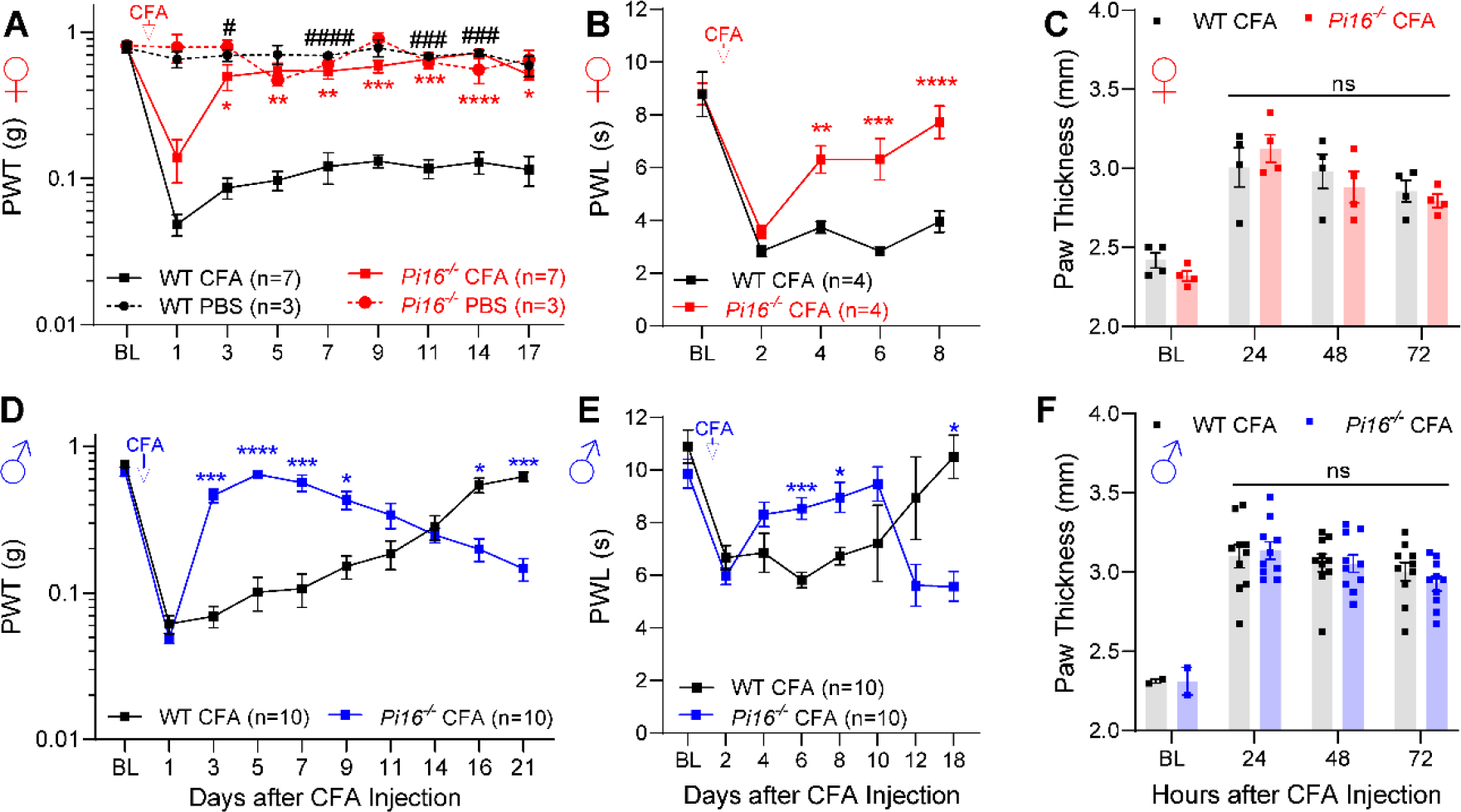
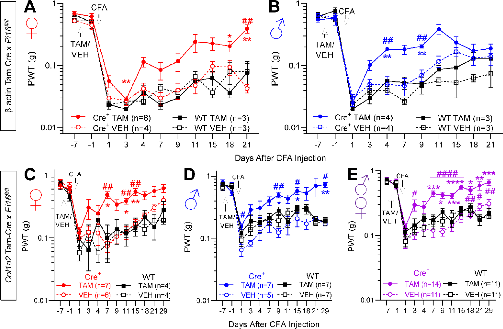
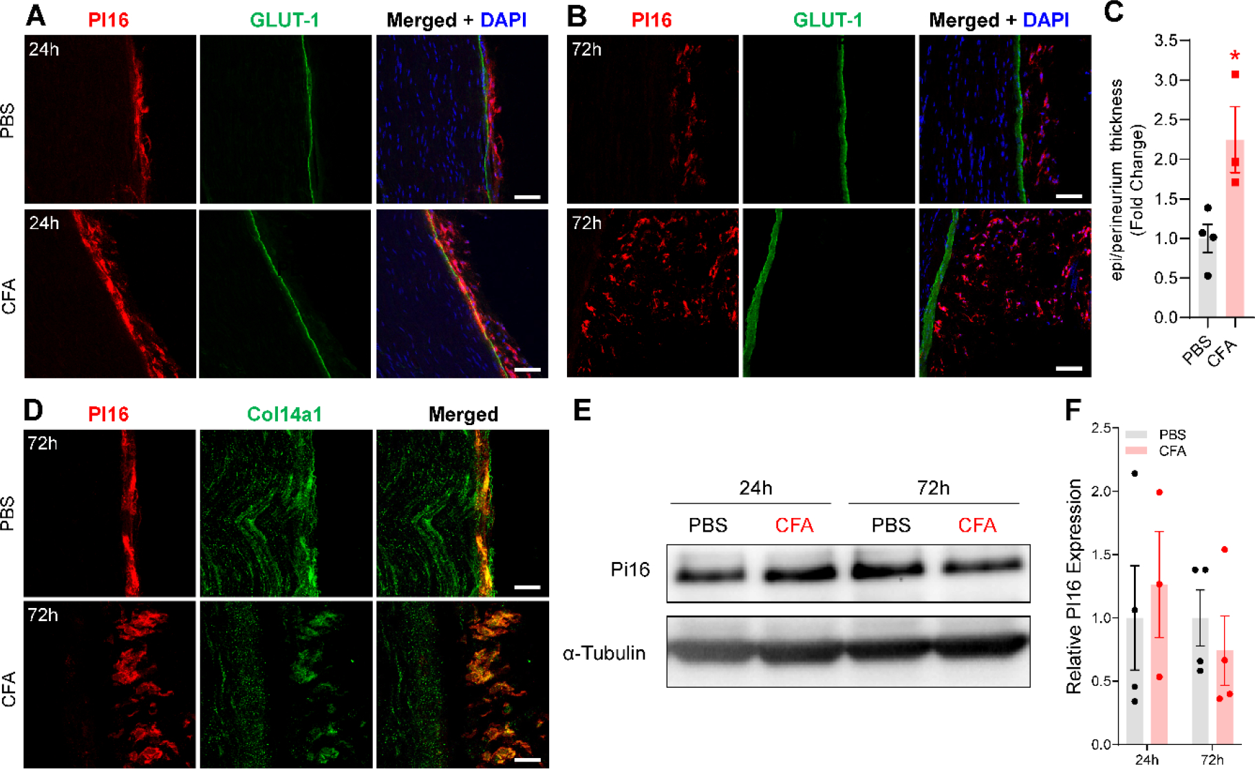
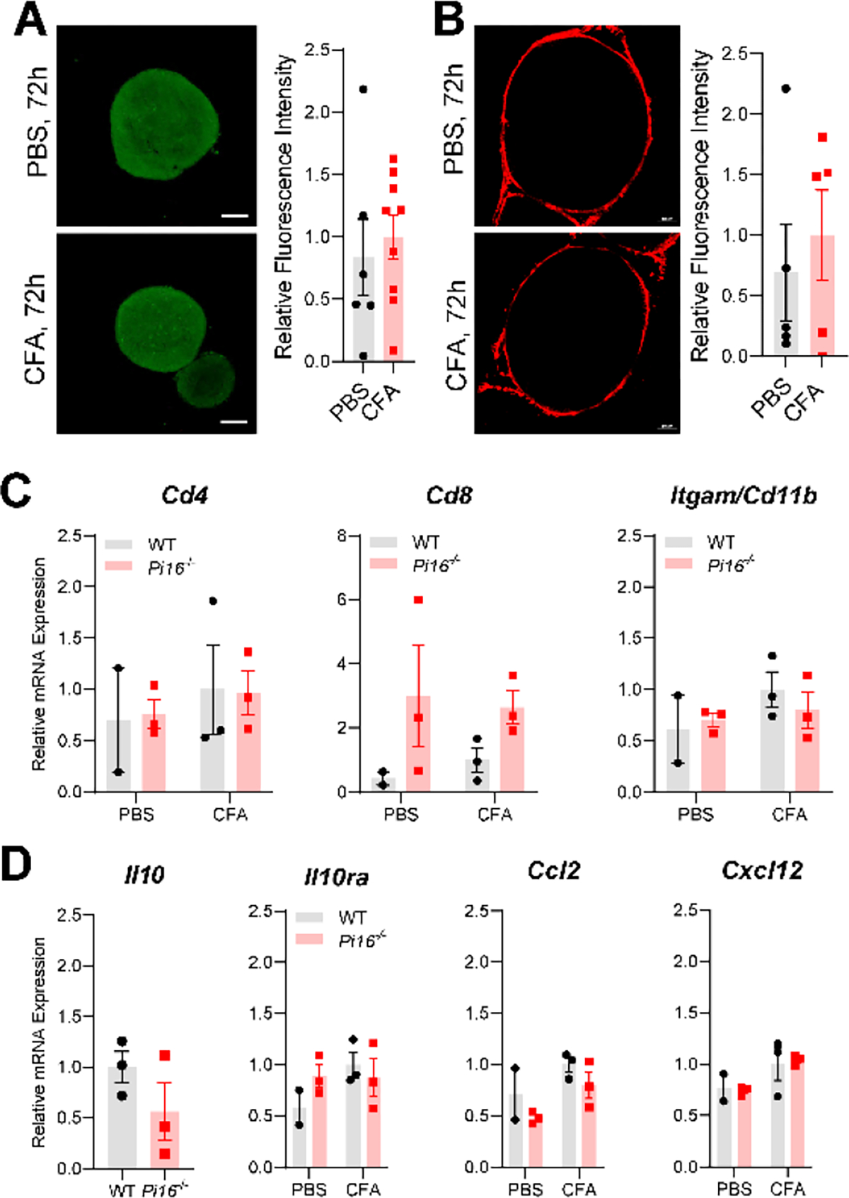
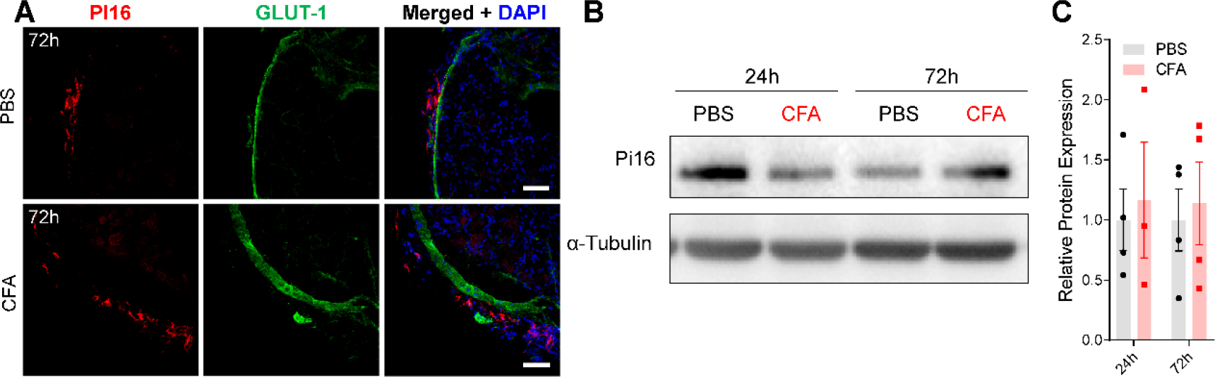
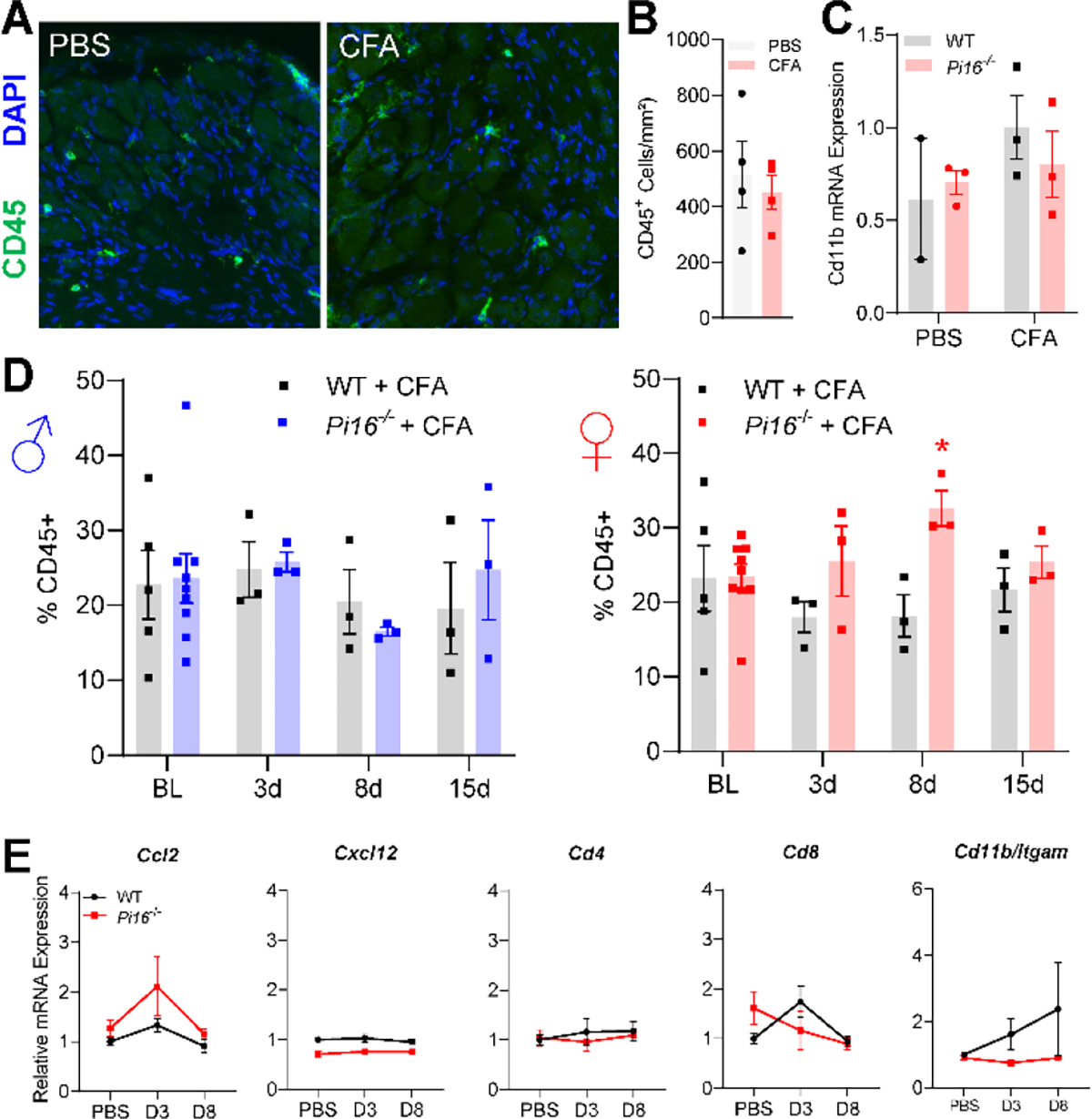
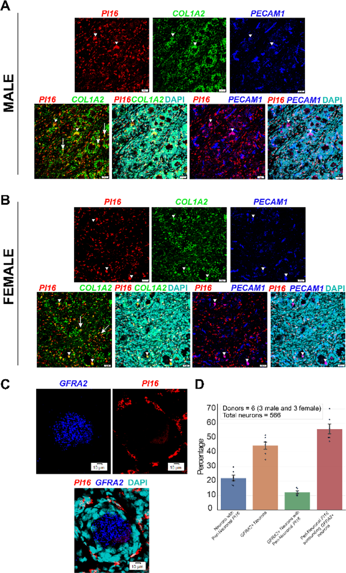
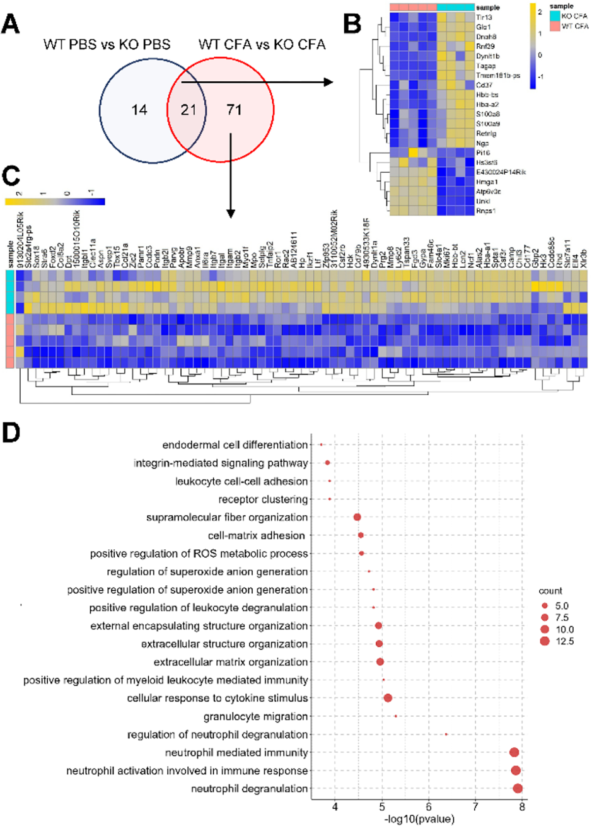
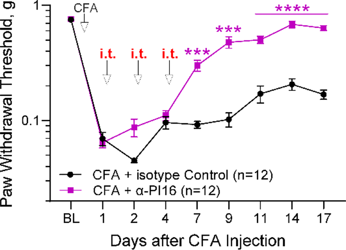
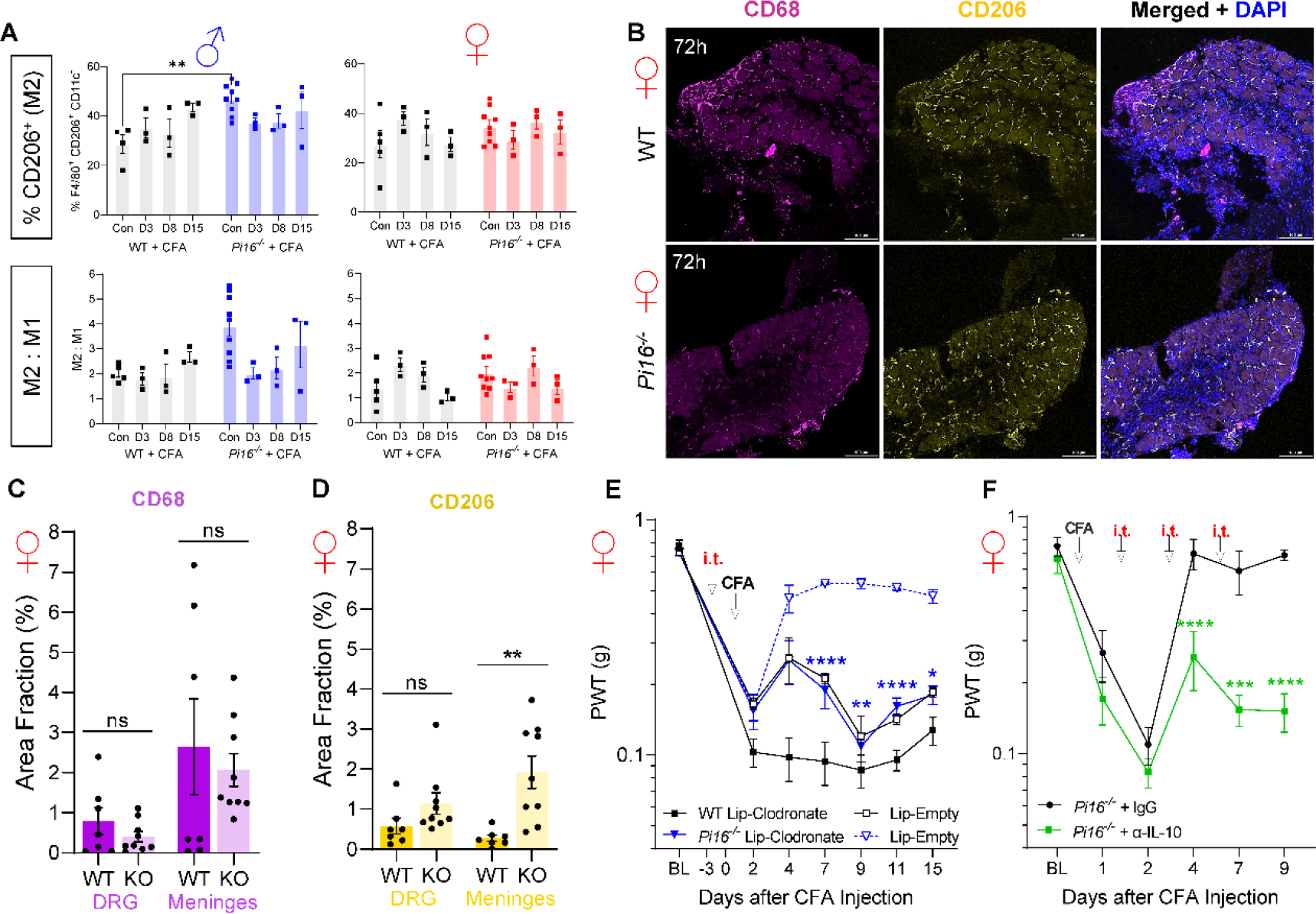
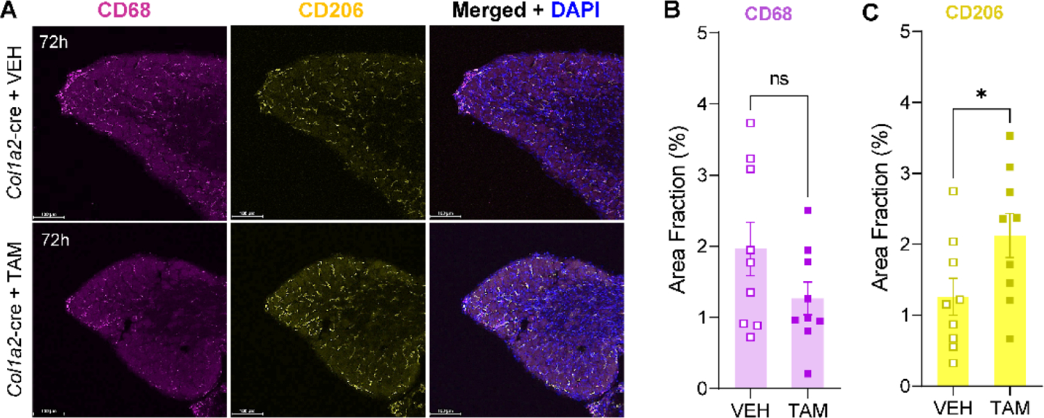
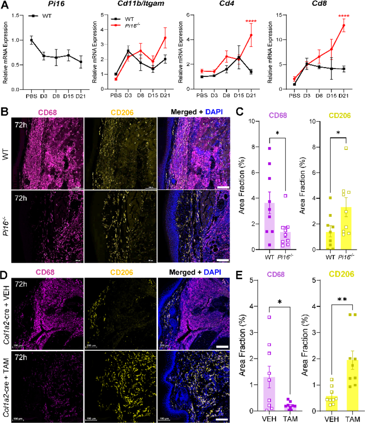
Originally identified in fibroblasts, Protease Inhibitor (PI)16 was recently shown to be crucial for the development of neuropathic pain via effects on blood-nerve barrier permeability and leukocyte infiltration, though its impact on inflammatory pain has not been established. Using the complete Freund's Adjuvant inflammatory pain model, we show that Pi16-/- mice are protected against sustained inflammatory pain. Accordingly, intrathecal delivery of a PI16 neutralizing antibody in wild-type mice prevented sustained CFA pain. In contrast to neuropathic pain models, we did not observe any changes in blood-nerve barrier permeability due to PI16 deletion. Instead, Pi16-/- mice display reduced macrophage density in the CFA-injected hindpaw. Furthermore, there was a significant bias toward CD206hi (anti-inflammatory) macrophages in the hindpaw and associated dorsal root ganglia. Following CFA, intrathecal depletion of CD206+ macrophages using mannosylated clodronate liposomes promoted sustained pain in Pi16-/- mice. Similarly, an IL-10 neutralizing antibody also promoted sustained CFA pain in the Pi16-/ when administered intrathecally. Collectively, our results point to fibroblast-derived PI16 mediating substantial differences in macrophage phenotype in the pain neuroaxis under conditions of inflammation. The co-expression of PI16 alongside fibroblast markers in human DRG raise the likelihood that a similar mechanism operates in human inflammatory pain states. Collectively, our findings may have implications for targeting fibroblast-immune cell crosstalk for the treatment of chronic pain.

Keywords: Blood-nerve barrier; CD206; Complete Freund’s Adjuvant; Fibroblast; IL-10; Inflammation; Macrophage; Meninges; PI16; Pain.


Conflict of interest statement

Declaration of Competing Interest The authors declare that they have no known competing financial interests or personal relationships that could have appeared to influence the work reported in this paper.

Figures

Fig. 1.
Fig. 1.
Role of Pi16 deletion in inflammatory pain. (A, D) Inflammatory pain was induced by intraplantar CFA injection. Mechanical allodynia was assessed at baseline and over time using von Frey filaments in female (A) and male (D) mice. Mixed-effects analysis, Šidák’s multiple comparisons test: WT CFA vs Pi16−/− CFA: *P<0.05, **P<0.01, ***P<0.005, ****P<0.0001, WT PBS vs WT CFA: #P<0.05, ###P<0.01, ####P<0.0001. (B, E) Thermal sensitivity was assessed at baseline and over time in female (B) and male (E) mice using Hargreaves’ test. Mixed-effects analysis, Šidák’s multiple comparisons test: *P<0.05, **P<0.01, ***P<0.005, ****P<0.0001. (C, F) Hind paw thickness at baseline and over time following intraplantar CFA injection in female (C) and male (F) mice was measured using a Digimatic micrometer. Two-way ANOVA, Šidák’s multiple comparisons test.
Fig. 2.
Fig. 2.
Hindpaw von Frey following CFA in systemic inducible Pi16−/− (A-B) and fibroblast-specific inducible Pi16−/− (C-D). Cre-positive (Cre+) or wild-type littermate controls (WT) were treated with vehicle (VEH) or tamoxifen (TAM) between baseline measurements 1 and 2 (days −7 and −1 relative to intraplantar CFA). (A, B) Systemic inducible deletion of PI16 protects females from acute and prolonged CFA-induced hypersensitivity, whereas males only show attenuation of pain up to 11–15 days post-CFA. (C, D) Inducible deletion of PI16 from Col1a2 fibroblasts protects female (C) and male (D) mice from prolonged CFA-induced mechanical hypersensitivity. Merged results from (C) and (D) are depicted in panel (E). Two-way repeated-measures ANOVA, Šidák’s multiple comparisons tests: Cre+ + TAM vs WT + TAM: *P<0.05, **P<0.01, ***P<0.005, ****P<0.0001. Cre+ + TAM vs Cre+ + VEH: #P<0.05, ##P<0.01, ####P<0.0001.
Fig. 3.
Fig. 3.
Effect of intraplantar CFA on PI16 protein expression. (A) Representative confocal image of sciatic nerve from PBS and CFA-treated female mice on day 1 of treatment showing immunostaining of PI16 (red) with outermost perineurial marker GLUT1 (green; B) from PBS and CFA-injected mice 3 days post-injection showing immunostaining of PI16 (red) with outermost perineurial marker GLUT1 (green). (C) Quantification of epineurium/perineurium thickness measured from perineurial marker GLUT1. Statistical analysis was performed using t-test: *P<0.05. (D) Representative image of PI16 (red) and Col14a1 (green) staining in the sciatic nerve on day 3 after CFA injection. Merged images show the co-localization of PI16 and Col14a1 (yellow), merged images in CFA show expansion of Col14a1 positive fibroblast in the epi/perineurium of the sciatic nerve co-expressing PI16. Immunofluorescence data are representative of n=4, each group (Scale bar 50 μm). (E) Representative Western blot and quantification of sciatic nerve PI16 24 or 72h after CFA injection in WT mice, quantified in (F).
Fig. 4.
Fig. 4.
CFA does not affect sciatic nerve blood-nerve barrier permeability or inflammation. Representative image of sodium fluorescein (A) and Evans Blue Albumin (B) permeability in the sciatic nerves of female mice on day 3 following CFA or PBS controls (n=5, Scale bar 50 μm). (C) Quantification of leukocyte markers in the sciatic nerve on day 3 following CFA or PBS injection. (D) Quantification of cytokines/chemokines in the sciatic nerve on day 3 following CFA or PBS injection.
Fig. 5.
Fig. 5.
(A) Representative confocal image of DRG from PBS and CFA-injected female mice 3 days post-injection showing immunostaining of PI16 (red) with outermost perineurial marker GLUT1 (green). Immunofluorescence data are representative of n=4, each group (Scale bar: 50 μm). Representative Western blot (B) and quantification (C) of DRG PI16, 24 or 72h after CFA injection in WT female mice.
Fig. 6.
Fig. 6.
(A) Representative confocal images of DRG from PBS and CFA mice 3 days post-injection showing immunostaining of CD45 (green). DAPI (blue). Scale bar: 50 μm. CD45 density quantified in (B). (C) qPCR of Cd11b in WT and Pi16−/− DRG 3d after PBS or CFA injection shows no significant differences in transcript level. (D) Flow cytometry of lumbar DRG shows elevated CD45+ cell numbers selectively in female Pi16−/− at 8d post-CFA injection. (E) qPCR shows no significant differences between WT and Pi16−/− inflammatory marker expression in DRG after intraplantar CFA injection.
Fig. 7.
Fig. 7.
PI16 mRNA expression colocalizes with fibroblast marker COL1A2 in human DRG and surrounds GFRA2+ neurons. PI16 (red) expression overlaps with a subset of COL1A2+ fibroblasts (green) but not with endothelial cell marker PECAM1 (blue) in human DRGs recovered from male (A) and female (B) donors. Long arrows point to areas where PI16 mRNA puncta overlap with COL1A2 mRNA. White arrow heads show lipofuscin auto-fluorescence signal present in all channels. Scale bar = 50 μm. (C) PI16 mRNA (red) surrounds neurons, including GFRA2+ neurons (blue) in human DRG. (D) Quantification of PI16 mRNA puncta shows that approximately 20% of all human DRG neurons have peri-neuronal PI16 and about 10% of GFRA2+ neurons have peri-neuronal PI16. Scale bar = 10 μm.
Fig. 8.
Fig. 8.
Bulk RNA sequencing of WT and Pi16−/− lumbar DRG shows increased myeloid cell signature 3d post-CFA injection. (A) Venn diagram illustrating the number of differentially-expressed genes in each dataset and their relative overlap. (B) Heat map depicting the 21 differentially-expressed genes common to PBS and CFA injection. (C) Heat map depicting the 71 differentially expressed genes in CFA-injected WT versus Pi16−/− mice. (D) Gene expression pathway enrichment analysis (WT CFA-injected versus Pi16−/− CFA-injected).
Fig. 9.
Fig. 9.
The effect of PI16 on inflammatory pain is mediated by extracellular PI16. anti-PI16 antibody or IgG isotype control were intrathecally injected (i.t.) on days 1, 2 and 4 following intraplantar CFA in WT female mice (n=12). On days where i.t. injections occurred, von Frey thresholds were measured prior to injections. Mice that received i.t. anti-PI16 show statistically significant recovery from CFA-induced hypersensitivity 7–17 days post-CFA. Two-way repeated-measures ANOVA, Bonferroni analysis: ***P<0.005, ****P<0.0001.
Fig. 10.
Fig. 10.
Role of CD206+ cells in resolution of CFA hypersensitivity in Pi16−/− mice. (A) Elevated F4/80+ CD206+ CD11c cells at baseline in male Pi16−/− DRG (left panels), with no significant differences observed in female DRG (right panels). (B) Representative IHC of CD206hi labeling in female WT and Pi16−/− DRG, quantified in (C-D). A tendency toward decreased CD68 and increased CD206 density were observed in DRG parenchyma from Pi16−/− mice, with a statistically significant increase in meningeal CD206hi cell density in Pi16−/−. (E) Mrc1+ (CD206+) Macrophages were depleted by intrathecal injection of mannosylated clodronate liposomes (Lip-Clodronate), 3 days prior to intraplantar injection of CFA. Pi16−/− mice that received Lip-Clodronate show prolonged hypersensitivity compared to Pi16−/− mice that received drug-free liposomes (Lip-Empty). Lip-Clodronate n=9, Lip-Empty n=3. (F) Following induction of inflammatory pain by intraplantar CFA injection in both hind paws, IL-10 was blocked by intrathecal injection of anti-IL-10 antibody or IgG isotype control on days 1, 2 and 4 post-CFA. On days of i.t. injection, von Frey thresholds were assessed prior to injections. Pi16−/− mice that received anti-IL-10 did not exhibit recovery from CFA-induced hypersensitivity (n=6). Two-way ANOVA, Bonferroni analysis: *P<0.05, **P<0.01, ***P<0.005, ****P<0.0001.
Fig. 11.
Fig. 11.
CD206hi cells are elevated in DRG of fibroblast-specific Pi16−/− mice. (A) Representative IHC in vehicle (VEH) and tamoxifen-induced fibroblast Pi16−/− (Col1a2-cre) lumbar DRGs, 3 days after intraplantar CFA. (B) Total density of CD68+ cells in DRG shows a tendency toward reduction but is not significantly different in mice with fibroblast-specific deletion of PI16. (C) Significant increase in CD206hi cell density in DRGs from mice with fibroblast-specific deletion of PI16. Student’s T-test, *P<0.05.
Fig. 12.
Fig. 12.
PI16 deletion is associated with reduced macrophage infiltration of hindpaw skin following CFA. (A) qPCR shows no significant expression changes in Pi16 in WT skin, or Itgam/CD11b in female Pi16−/− versus WT hindpaw skin. Significant increases in expression of T-cell markers CD4 and CD8 occur only at late-stage timepoints. (B) Hindpaw skin shows significantly reduced CD68 (magenta) immunoreactivity along with an increase in the area fraction of CD206hi (yellow) cells in female Pi16−/− mice. DAPI: blue. Scale bar: 100 μm. (D) Representative IHC in vehicle (VEH) and tamoxifen-induced fibroblast PI16 knockout (Col1a2-cre) plantar hindpaw skin, 3 days after intraplantar CFA. (E) Significant decrease in CD68+ cell density in skin from mice with fibroblast-specific deletion of PI16. Significant increase in CD206hi cell density in skin from mice with fibroblast-specific deletion of PI16. Student’s T-test: *P<0.05, **P<0.01, Student’s T-test.

Comment in

References

    1. Alshetaiwi H, Pervolarakis N, McIntyre LL, Ma D, Nguyen Q, Rath JA, Nee K, Hernandez G, Evans K, Torosian L, Silva A, Walsh C and Kessenbrock K (2020). “Defining the emergence of myeloid-derived suppressor cells in breast cancer using single-cell transcriptomics.” Sci Immunol 5(44). - PMC - PubMed
    1. Andrews S (2010). “A quality control tool for high throughput sequence data.”, from http://www.bioinformatics.babraham.ac.uk/projects/fastqc/.
    1. Balogh M, Zhang J, Gaffney CM, Kalakuntla N, Nguyen NT, Trinh RT, Aguilar C, Pham HV, Milutinovic B, Nichols JM, Mahalingam R and Shepherd AJ (2022). “Sensory neuron dysfunction in orthotopic mouse models of colon cancer.” J Neuroinflammation 19(1): 204. - PMC - PubMed
    1. Bayik D, Bartels CF, Lovrenert K, Watson DC, Zhang D, Kay K, Lee J, Lauko A, Johnson S, Lo A, Silver DJ, McGraw M, Grabowski M, Mohammadi AM, Veglia F, Fan Y, Vogelbaum MA, Scacheri P and Lathia JD (2022). “Distinct Cell Adhesion Signature Defines Glioblastoma Myeloid-Derived Suppressor Cell Subsets.” Cancer Res 82(22): 4274–4287. - PMC - PubMed
    1. Becker E, Dedden M, Gall C, Wiendl M, Ekici AB, Schulz-Kuhnt A, Schweda A, Voskens C, Hegazy A, Vitali F, Atreya R, Müller TM, Atreya I, Neurath MF and Zundler S (2021). “Residual homing of α4β7-expressing β1(+)PI16(+) regulatory T cells with potent suppressive activity correlates with exposure-efficacy of vedolizumab.” Gut. - PubMed

Publication types

LinkOut - more resources