Randomized Controlled Trial
. 2023 Nov;152(5):1179-1195.
doi: 10.1016/j.jaci.2023.05.026. Epub 2023 Jun 13.

Rapid reduction in Staphylococcus aureus in atopic dermatitis subjects following dupilumab treatment

Affiliations
Randomized Controlled Trial

Rapid reduction in Staphylococcus aureus in atopic dermatitis subjects following dupilumab treatment

Eric L Simpson et al. J Allergy Clin Immunol. 2023 Nov.

Abstract

Background: Atopic dermatitis (AD) is an inflammatory disorder characterized by dominant type 2 inflammation leading to chronic pruritic skin lesions, allergic comorbidities, and Staphylococcus aureus skin colonization and infections. S aureus is thought to play a role in AD severity.

Objectives: This study characterized the changes in the host-microbial interface in subjects with AD following type 2 blockade with dupilumab.

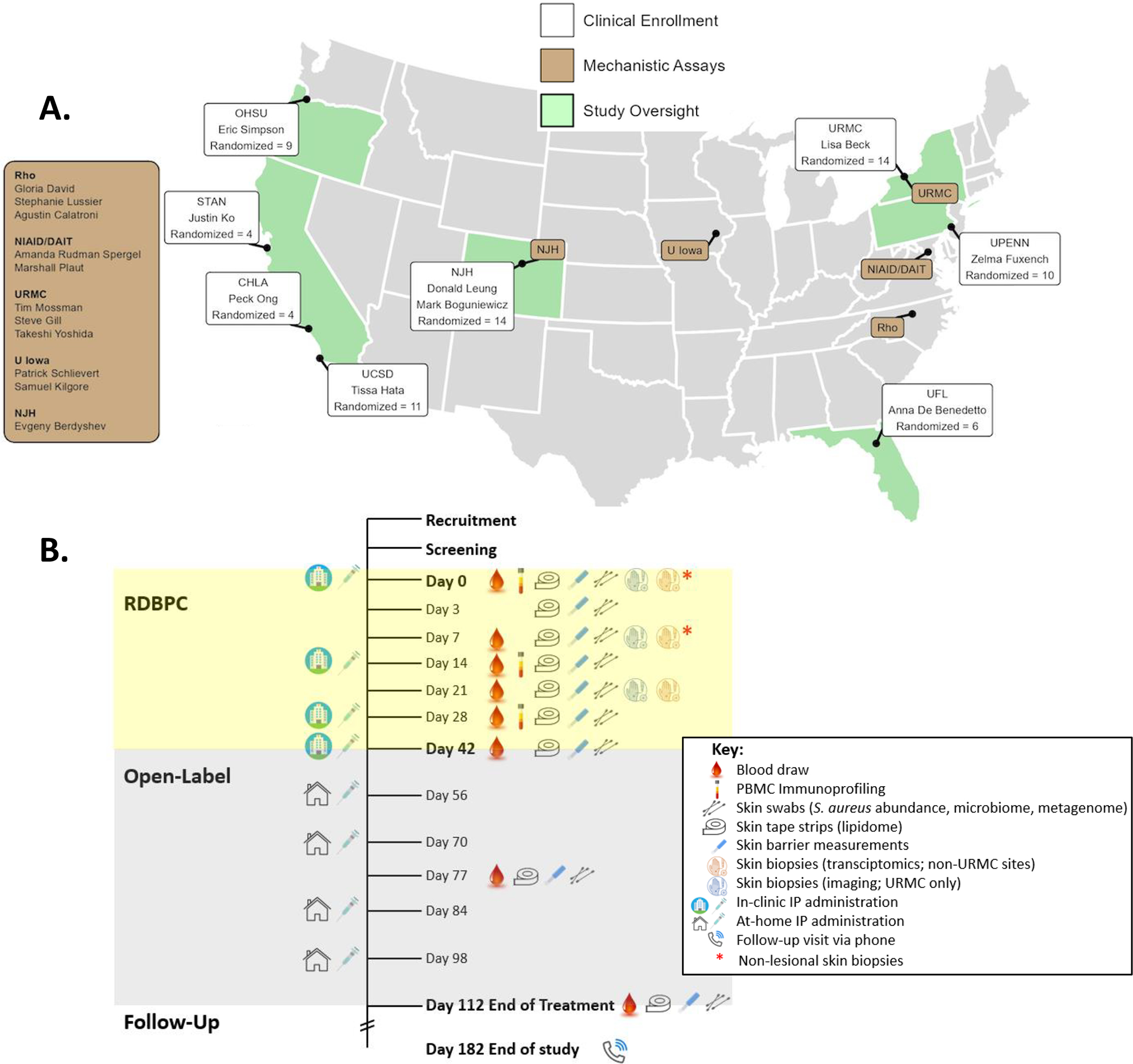
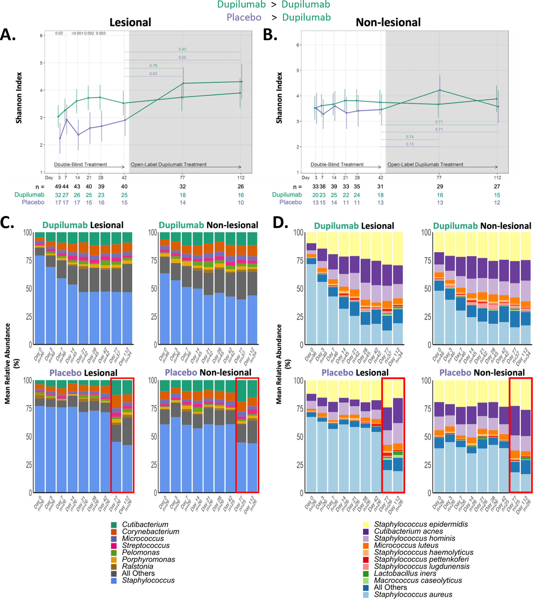
Methods: Participants (n = 71) with moderate-severe AD were enrolled in a randomized (dupilumab vs placebo; 2:1), double-blind study at Atopic Dermatitis Research Network centers. Bioassays were performed at multiple time points: S aureus and virulence factor quantification, 16s ribosomal RNA microbiome, serum biomarkers, skin transcriptomic analyses, and peripheral blood T-cell phenotyping.

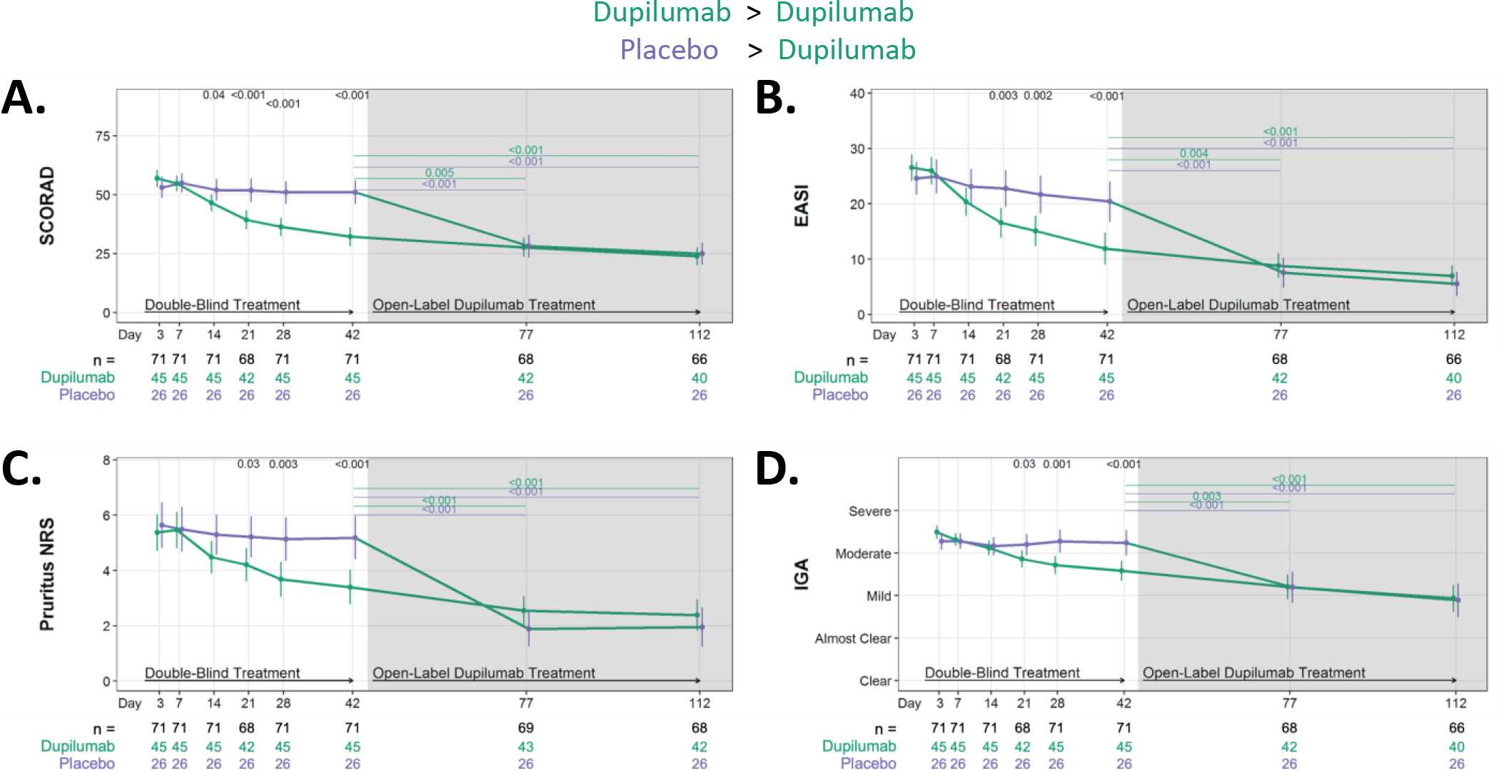
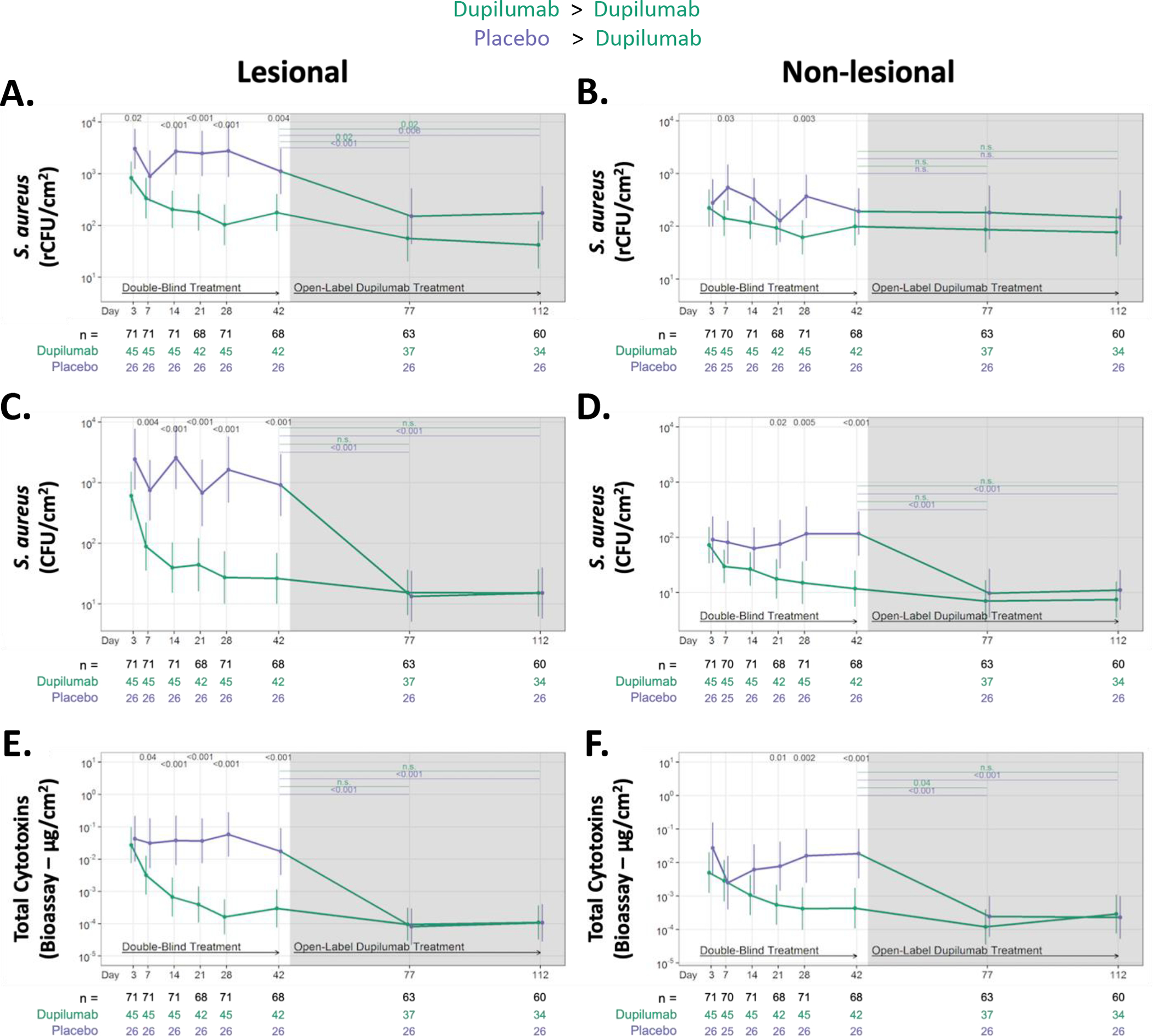
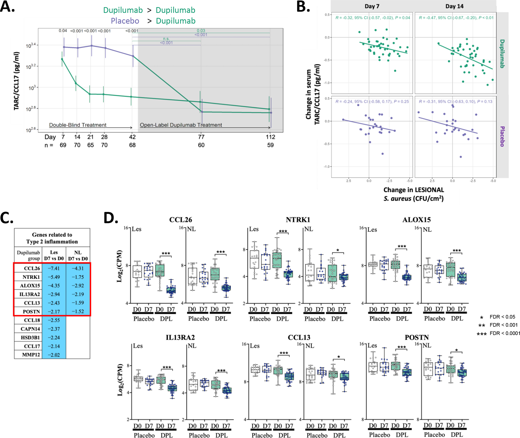
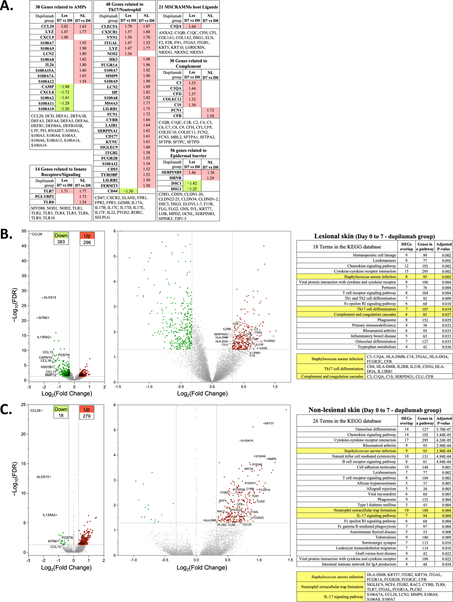
Results: At baseline, 100% of participants were S aureus colonized on the skin surface. Dupilumab treatment resulted in significant reductions in S aureus after only 3 days (compared to placebo), which was 11 days before clinical improvement. Participants with the greatest S aureus reductions had the best clinical outcomes, and these reductions correlated with reductions in serum CCL17 and disease severity. Reductions (10-fold) in S aureus cytotoxins (day 7), perturbations in TH17-cell subsets (day 14), and increased expression of genes relevant for IL-17, neutrophil, and complement pathways (day 7) were also observed.

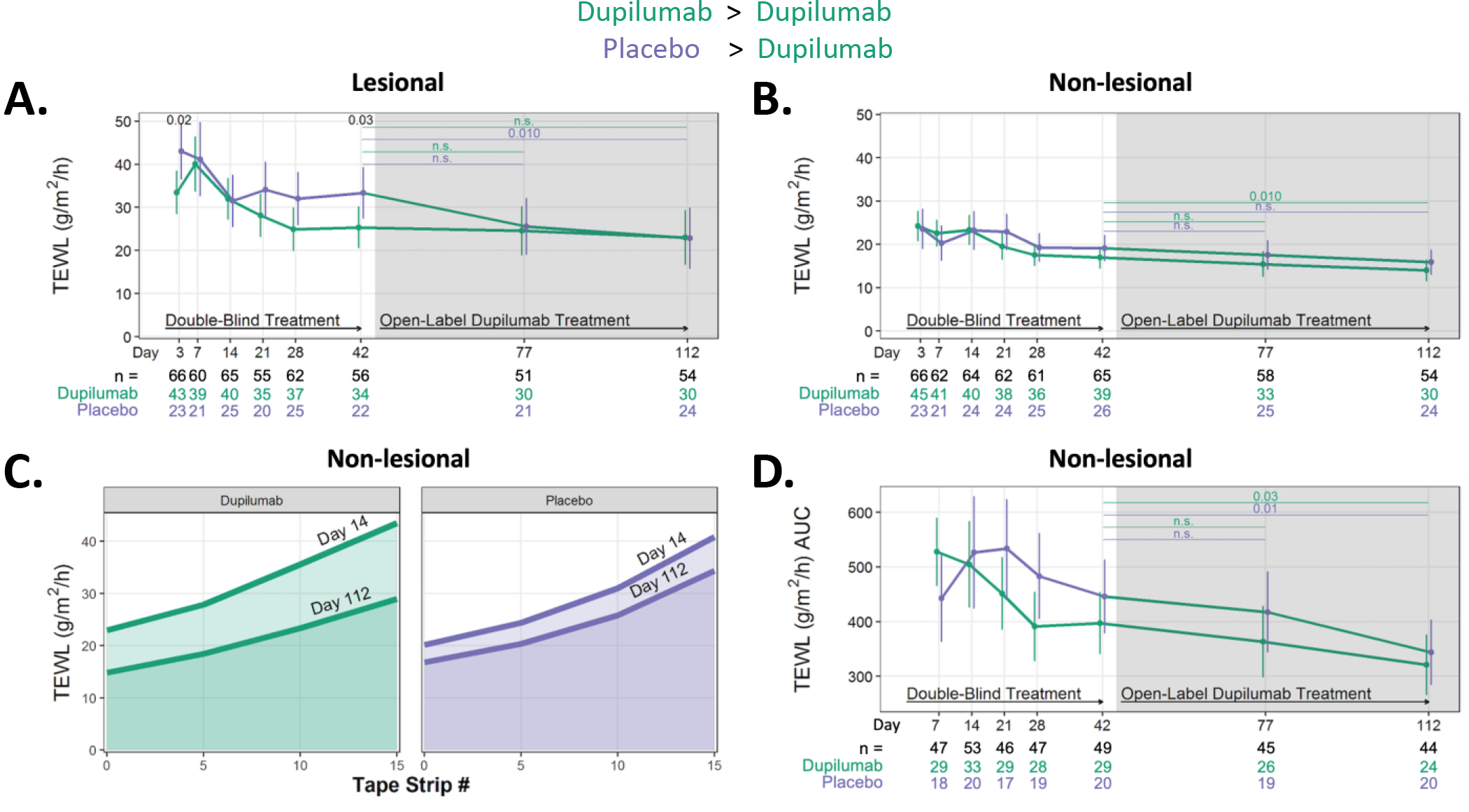
Conclusions: Blockade of IL-4 and IL-13 signaling, very rapidly (day 3) reduces S aureus abundance in subjects with AD, and this reduction correlates with reductions in the type 2 biomarker, CCL17, and measures of AD severity (excluding itch). Immunoprofiling and/or transcriptomics suggest a role for TH17 cells, neutrophils, and complement activation as potential mechanisms to explain these findings.

Keywords: Atopic dermatitis; IL-13; IL-17; IL-4; Staphylococcus aureus; barrier; cytotoxins; dupilumab; microbiome; type 2 immunity.


Conflict of interest statement

Conflicts of Interest: ADB – Consultant for dMed Biopharmaceutical Co, AbbVie, Sanofi-Aventis; Investigator for Kiniksa, Novartis, Incyte, and Pfizer. ELS – Consultant for AbbVie, Amgen, Arena Pharmaceuticals, Aslan Pharma, Boston Consulting Group, Collective Acumen, LLC (CA), Dermira, Eli Lilly, Evidera, Excerpta Medica, Forte Bio RX, Galderma, GlaxoSmithKline, Incyte, Janssen, Kyowa Kirin Pharmaceutical Development, Leo Pharm, Medscape LLC, Merck, Pfizer, Physicians World LLC, Regeneron, Roivant, Sanofi-Genzyme, Trevi therapeutics, Valeant, WebMD. Investigator for AbbVie, Amgen, Arcutis, Aslan, Castle Biosciences, Corevita, Dermavant, Dermira, Eli Lilly, Incyte, Kyowa Hakko Kirin, Leo Pharmaceuticals, Merck, Novartis, Pfizer, Regeneron, Sanofi, TARGET-DERM, Tioga, and Vanda. JK – Investigator and Consultant for Eli Lilly and Arena. Investigator for Abbvie and Regeneron. Consultant for Pfizer. MB - Investigator for Regeneron, Incyte. Consultant for Regeneron, Sanofi-Genzyme, Abbvie, Leo, Lilly, Pfizer, Janssen. PYO -Consultant for Incyte, Abbvie, Janssen; Investigator for Regeneron, Sanofi Genzyme, Leo, Incyte. DYML - Consultant for Genentech, LEO Pharma, and Incyte; Research grants from Sanofi Genzyme. LAB - Consultant for Allakos, Arena Pharmaceuticals, DermTech, Evelo Biosciences, Galderma, Incyte, Janssen, LEO Pharma, Merck, Numab Therapeutics, Pfizer, Rapt Therapeutics, Regeneron, Ribon Therapeutics, Sanofi/Genzyme, Sanofi-Aventis, Stealth BioTherapeutics, Trevi Therapeutics, Union Therapeutics and Xencor. DMC member – Novartis. Investigator for Abbvie, Astra-Zeneca, DermTech, Kiniksa, Pfizer, Regeneron, Ribon Therapeutics and Sanofi. TH - Investigator, Consultant for Eli Lilly and Arena. Investigator for Abbvie and Regeneron. Consultant for Pfizer. PMS, TY, SL, ZF AC, ALRS, MP, SAQ, SHK, LP, AG, GD, TM and SRG – No relevant conflicts of interest.

Figures

Figure 1.
Figure 1.. US academic centers involved in AD subject recruitment and Study Schematic for the Atopic Dermatitis Research Network (ADRN) 09 study (NCT03389893).
(A) A map of the US academic centers involved in clinical enrollment (white boxes) or mechanistic assays or study oversight (brown boxes). Each white box represents a single center with investigator(s) and enrollment totals. OHSU=Oregon Health & Science University, STAN=Stanford Medical Center, CHLA=Children’s Hospital of Los Angeles, UCSD=University of California San Diego, NJH=National Jewish Health, URMC=University of Rochester Medical Center, UPENN=University of Pennsylvania, UF=University of Florida. (B) High density sampling of the microbial, epithelial, and immune compartments was performed during the 6-week RDBPC phase (yellow) of the trial to characterize and quantify changes and their relationship to disease improvements, followed by a 10-week open-label extension (OLE; grey) phase with a safety assessment (by phone) 10 weeks after the OLE phase ended. ∗Timepoints when non-lesional skin biopsies were collected in addition to lesional biopsies for transcriptomics analysis.
Figure 2.
Figure 2.. Dupilumab treatment improves all measures of AD severity.
Absolute changes in AD severity measures during RDBPC phase (up to day 42 [6 weeks]) and during the OLE phase (day 42 – day 112 or [6 – 16 weeks]). (A) Absolute reductions in SCORing AD (SCORAD), (B) Eczema Area and Severity Index (EASI), (C) Pruritus Numerical Rating Scale (NRS) and (D) Investigator Global Assessment (IGA). Data for A, C, and D are shown as the means and 95% CIs adjusted for clinical site and EASI (≥ 21.1 or < 21.1), and the severity measure at day 0. Data for B is shown as the means and 95% CIs adjusted for clinical site and EASI at day 0. The number of participants with evaluable data at each timepoint are noted below the X axis (with the total population denoted in black, dupilumab-randomized participants in green and placebo-randomized in purple).
Figure 3.
Figure 3.. Dupilumab treatment rapidly reduces S. aureus abundance and cytotoxin production on the skin surface of AD participants.
Absolute changes in S. aureus abundance and cytotoxin production during RDBPC (up to day 42 [6 weeks]) and OLE phases (6 – 16 weeks) on lesional (left) and non-lesional (right) skin. S. aureus abundance was measured using qPCR for the femA gene (rCFU/cm2) in both lesional skin (A) and non-lesional skin (B) and by quantitative culture techniques (CFU/cm2) in both lesional skin (C) and non-lesional skin (D). Total S. aureus cytotoxin levels (μg/cm2), measured by bioassay from skin swabs of both lesional (E) and non-lesional skin (F), are shown. Data are shown as geometric means and 95% CIs and are adjusted for clinical site, disease severity at day 0, as measured by EASI ≥ 21.1 or < 21.1, and S. aureus abundance on lesional skin at day 0. The number of participants with evaluable data at each timepoint are noted below the X axis (with the total population denoted in black, dupilumab-randomized participants in green and placebo-randomized in purple). rCFU, relative colony-forming units
Figure 4.
Figure 4.. Measures of the cutaneous microbial community changed rapidly in response to dupilumab treatment.
(A) Longitudinal changes in Shannon diversity observed from 16S rRNA analysis of lesional and (B) non-lesional skin in the placebo and dupilumab-treated groups. The number of participants with evaluable data at each timepoint are noted below the X axis (with the total population denoted in black, dupilumab-randomized participants in green and placebo-randomized in purple). (C & D) Relative abundance of the most dominant genera (C) and species (D) in dupilumab and placebo randomized groups. Only the most abundant OTUs are presented. A decrease in the relative abundance of S. aureus is observed in the dupilumab treatment groups. The red box in the placebo-randomized group (C & D) illustrates that for those two timepoints (Day 77 and 112) participants originally randomized to placebo were now receiving dupilumab (as part of the OLE phase of the study). rRNA, ribosomal RNA; OTU, operational taxonomic unit
Figure 5.
Figure 5.. Reductions in type 2 serum and tissue biomarkers as a function of dupilumab treatment.
(A) Longitudinal changes in serum TARC/CCL17 during RDBPC and OLE phases of the study. The number of participants with evaluable data at each timepoint are noted below the X axis (with the total population denoted in black, dupilumab-randomized participants in green and placebo-randomized in purple). (B) Reductions in serum CCL17 significantly correlated with reductions in S. aureus lesional CFU abundance at both day 7 (p=0.04) and day 14 (p=0.01) in dupilumab-treated participants but there was no correlation observed at either timepoint in placebo-treated participants. (C) Differentially expressed genes (DEGs) related to type 2 inflammation (with a FDR of <0.05 and with a fold change > 2.0 in lesional skin) is shown comparing day 7 to day 0 in the dupilumab-treated group. Blue indicates reduced expression at day 7 in the dupilumab-treated group. (D) Graphical illustration of the genes reduced in both Les and NL skin (Red Box in [C]). Placebo data is shown in open box plots and dupilumab in shades of green. CFU, colony forming units; FDR, false discovery rate; les, lesional skin; NL, nonlesional skin
Figure 6.
Figure 6.. Transcriptomics changes in lesional and non-lesional skin in response to dupilumab treatment.
(A) DEGs with FDR of <0.05 and a log FC >1.25 in les and NL skin are shown. They are separated into functional groups: antimicrobial proteins (AMPs), Innate Receptors/Signaling, Th17/Neutrophil, microbial surface components recognizing adhesive matrix molecules (MSCRAMMs), complement and epidermal barrier. The genes listed at the bottom of each of these are those that are not significantly altered in their expression. (B) Volcano plot showing increased (n=296) or decreased (n=383) DEG from lesional skin of dupilumab treated subjects (Day 7 vs Day 0). The graphs show log2 FC in gene expression of lesional skin (Day 7) over lesional skin (Day 0) plotted against negative log10 FDR. (C) Volcano plot showing upregulated (n=279) or downregulated (n=18) DEGs from non-lesional skin of dupilumab treated subjects (Day 7 vs Day 0). Genes represented in red are upregulated by >1.25-fold in Day 7 lesional or non-lesional skin (FDR < 0.05). Genes represented in green are downregulated by >1.25-fold in Day 7 lesional or non-lesional skin (FDR < 0.05). The left most volcano plot is all of the DEG and the right most volcano plot reduces the y axis to enable labelling of some of the upregulated DEGs. The table lists the KEGG pathways with a few highlighted in yellow because of their potential relevance to dupilumab-induced S. aureus reductions. We list the DEGs in these pathways that are dysregulated in our samples. DEG, differentially expressed genes; FDR, false discovery rate; les, lesional skin; NL, nonlesional skin; KEGG, Kyoto Encyclopedia of Genes and Genomes
Figure 7.
Figure 7.. Dupilumab treatment modestly affects lesional and non-lesional barrier function at later timepoints.
(A) Changes in basal TEWL measurements in les and (B) NL skin sites. (C) The mean TEWL values obtained at NL skin site at Days 14 and 112 following five, ten and fifteen tape strips are shown for the dupilumab (green) and placebo (purple)-randomized populations to visualize how the AUC is calculated. (D) The longitudinal changes in TEWL AUC at NL skin sites. AUC, area under the curve; les, lesional skin; NL, nonlesional skin; TEWL, transepidermal water loss.

Similar articles

Cited by

References

    1. Boguniewicz M, Leung DY. Recent insights into atopic dermatitis and implications for management of infectious complications. J Allergy Clin Immunol. 2010;125(1):4–13; quiz 4–5. - PMC - PubMed
    1. Wang V, Boguniewicz J, Boguniewicz M, Ong PY. The infectious complications of atopic dermatitis. Ann Allergy Asthma Immunol. 2021;126(1):3–12. - PMC - PubMed
    1. Beck LA, Boguniewicz M, Hata T, Schneider LC, Hanifin J, Gallo R, et al. Phenotype of atopic dermatitis subjects with a history of eczema herpeticum. J Allergy Clin Immunol. 2009;124(2):260–9, 9.e1–7. - PMC - PubMed
    1. Leung DY, Gao PS, Grigoryev DN, Rafaels NM, Streib JE, Howell MD, et al. Human atopic dermatitis complicated by eczema herpeticum is associated with abnormalities in IFN-γ response. J Allergy Clin Immunol. 2011;127(4):965–73.e1–5. - PMC - PubMed
    1. Mathias RA, Weinberg A, Boguniewicz M, Zaccaro DJ, Armstrong B, Schneider LC, et al. Atopic dermatitis complicated by eczema herpeticum is associated with HLA B7 and reduced interferon-γ-producing CD8+ T cells. Br J Dermatol. 2013;169(3):700–3. - PMC - PubMed

Publication types

MeSH terms

Substances