. 2023 Aug:184:106196.
doi: 10.1016/j.nbd.2023.106196. Epub 2023 Jun 12.

A 14-day pulse of PLX5622 modifies α-synucleinopathy in preformed fibril-infused aged mice of both sexes

Affiliations

A 14-day pulse of PLX5622 modifies α-synucleinopathy in preformed fibril-infused aged mice of both sexes

Tarun N Bhatia et al. Neurobiol Dis. 2023 Aug.

Abstract

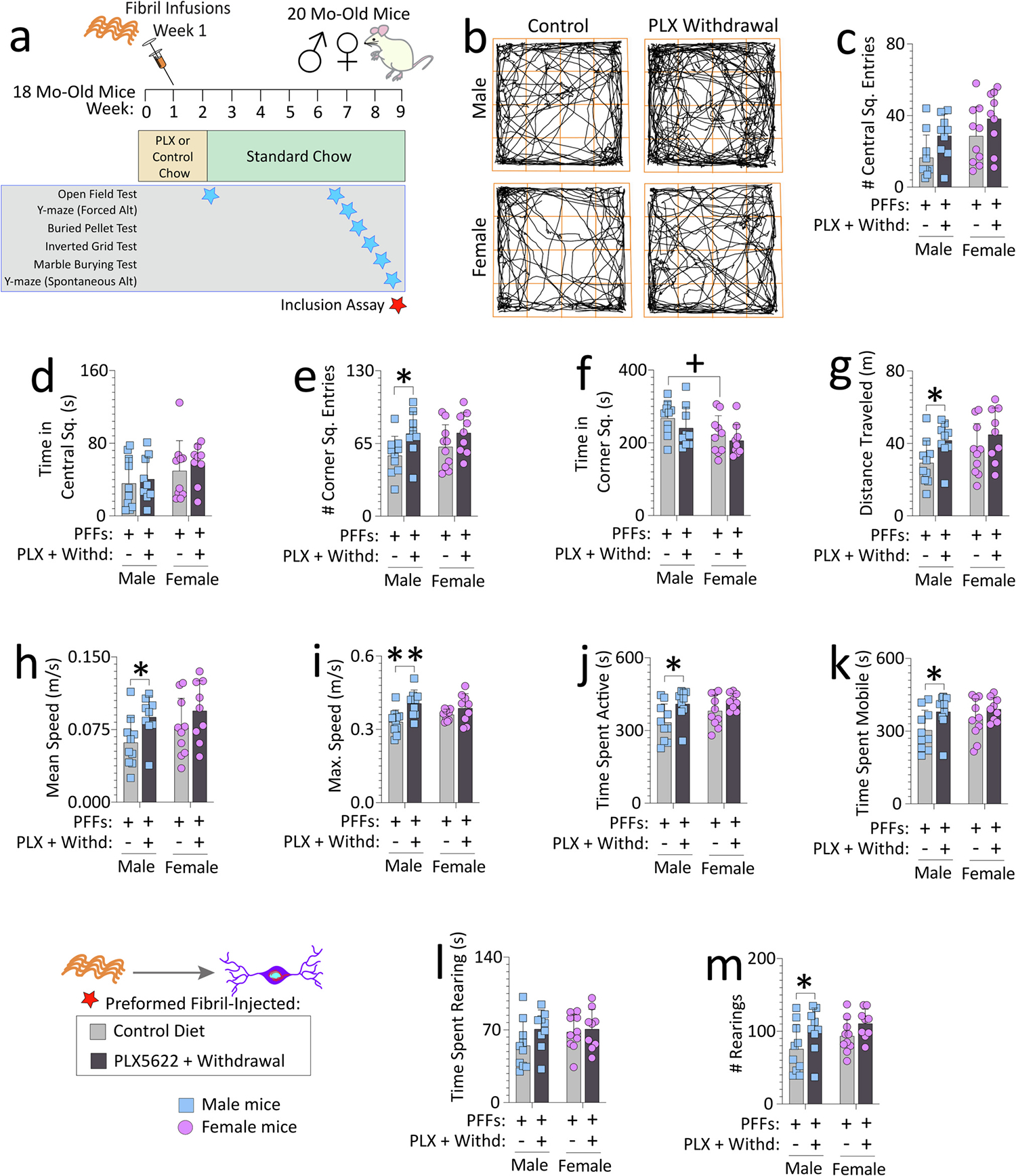
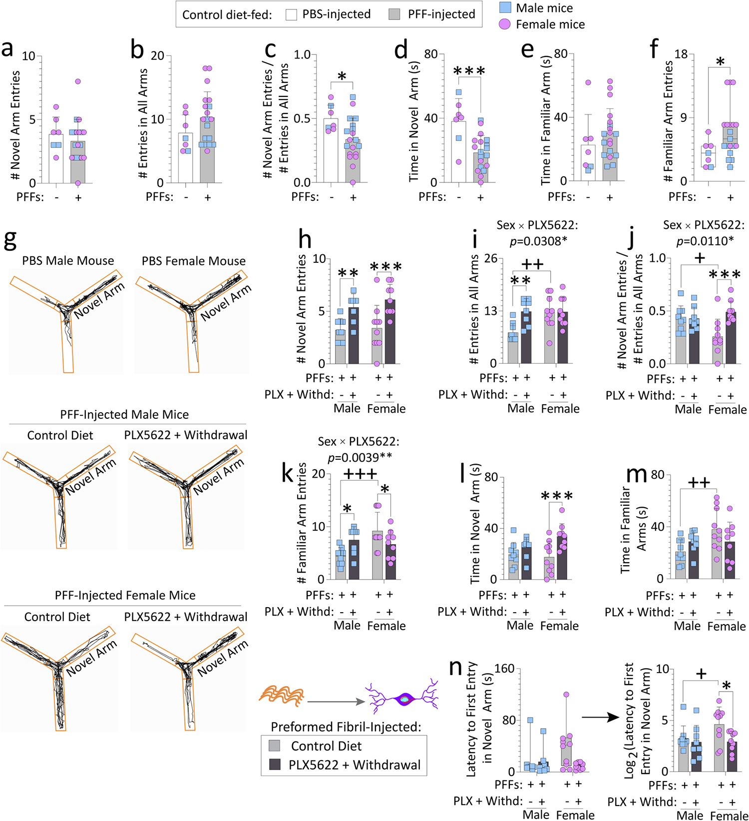
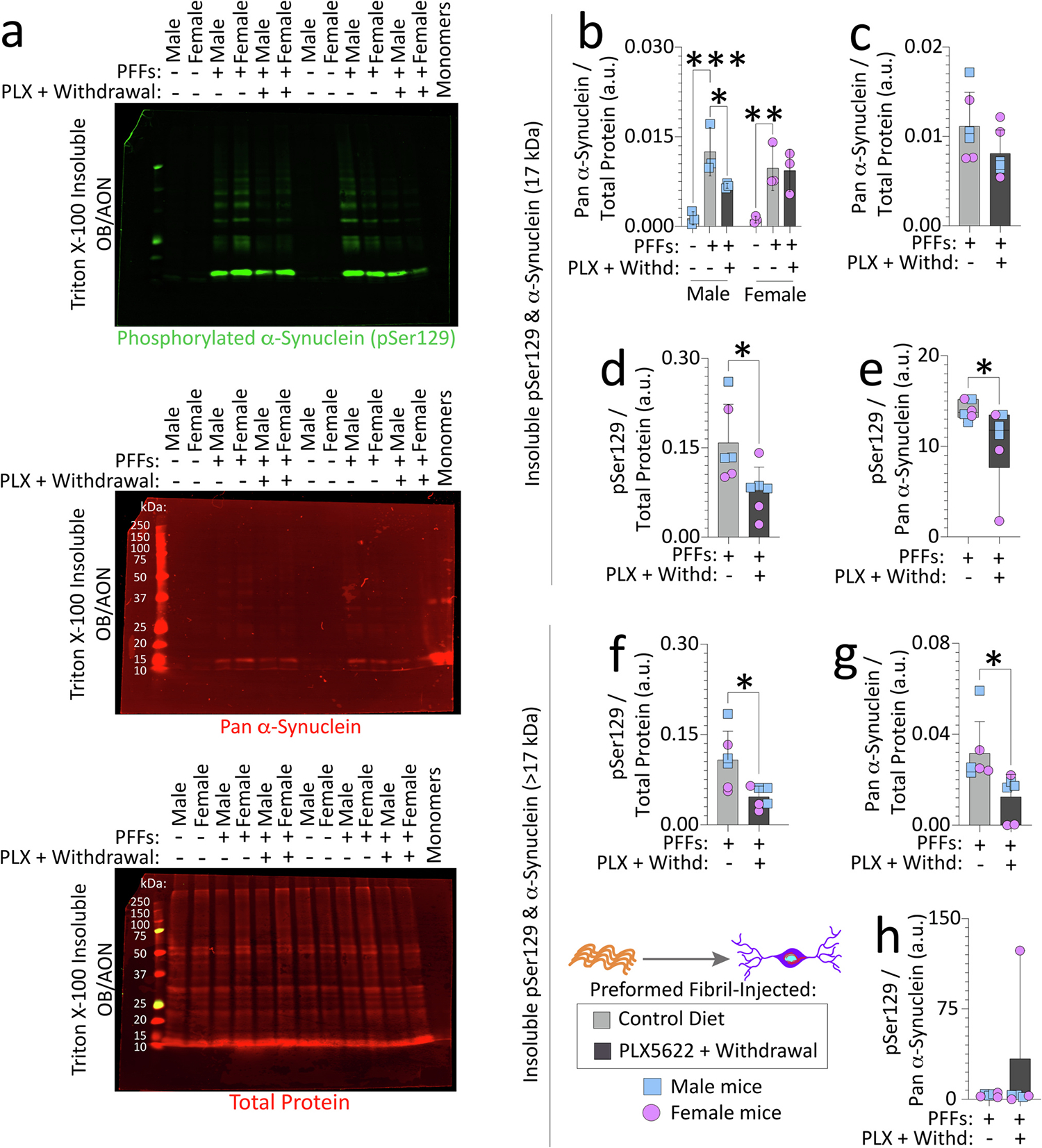
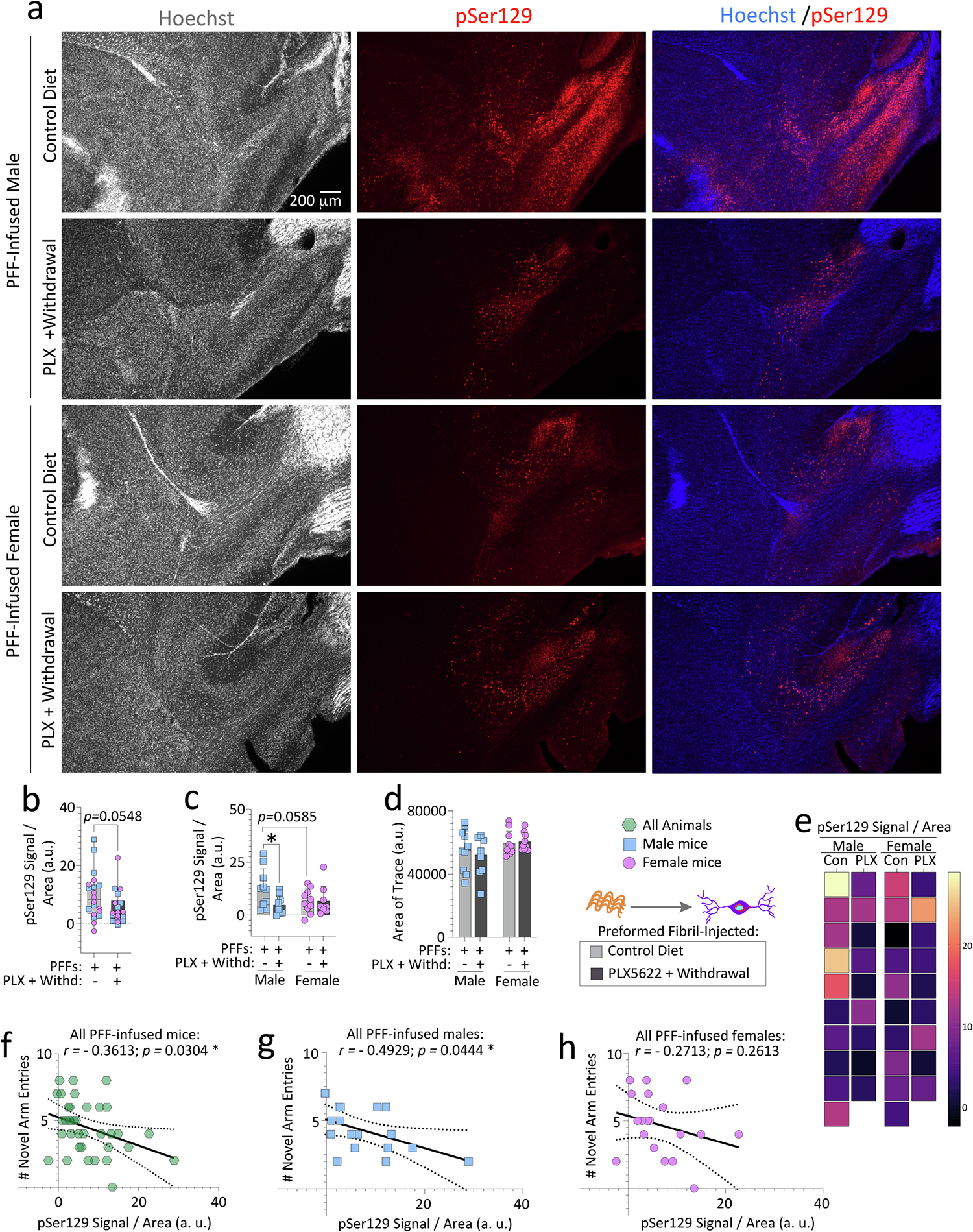
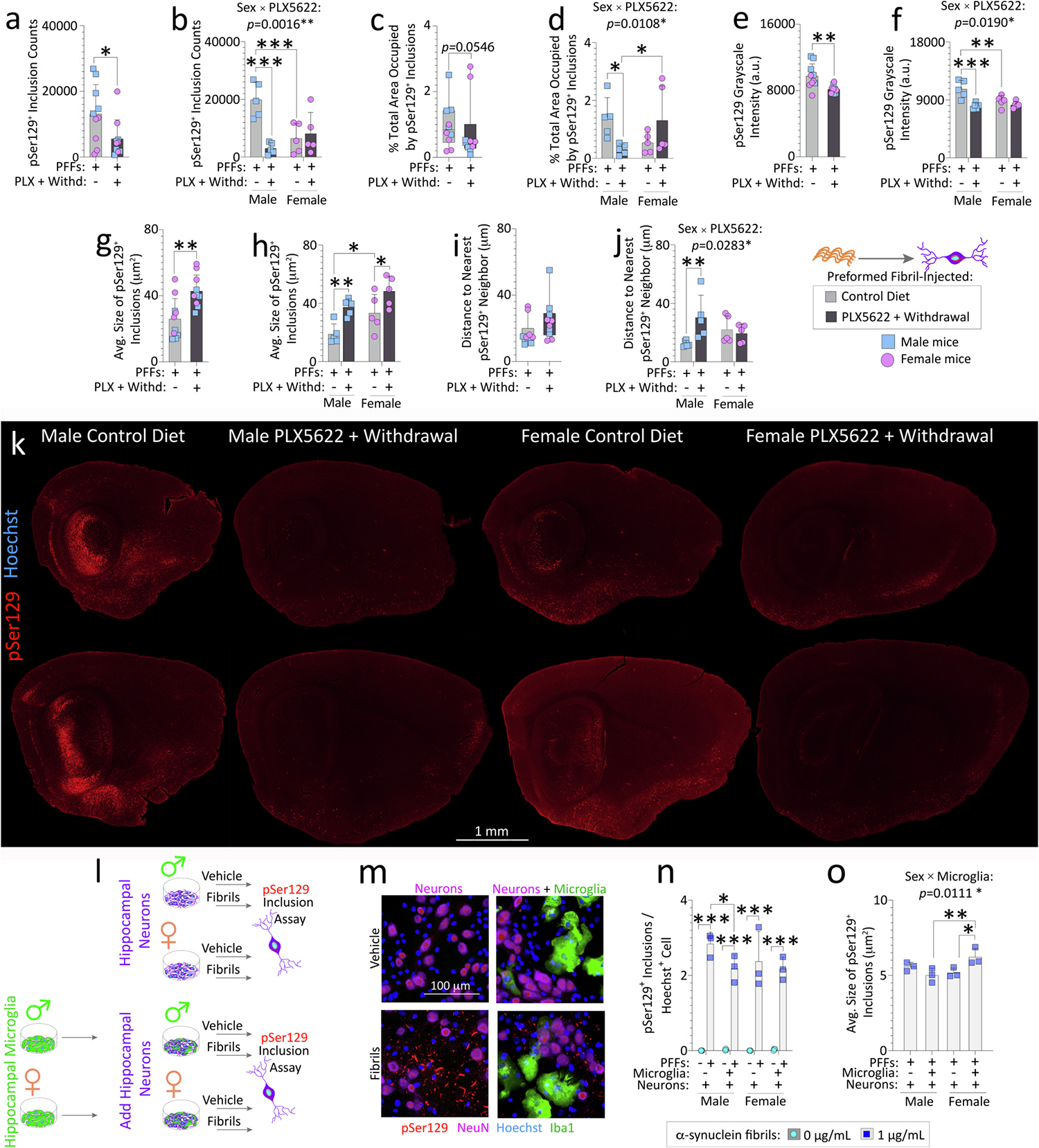
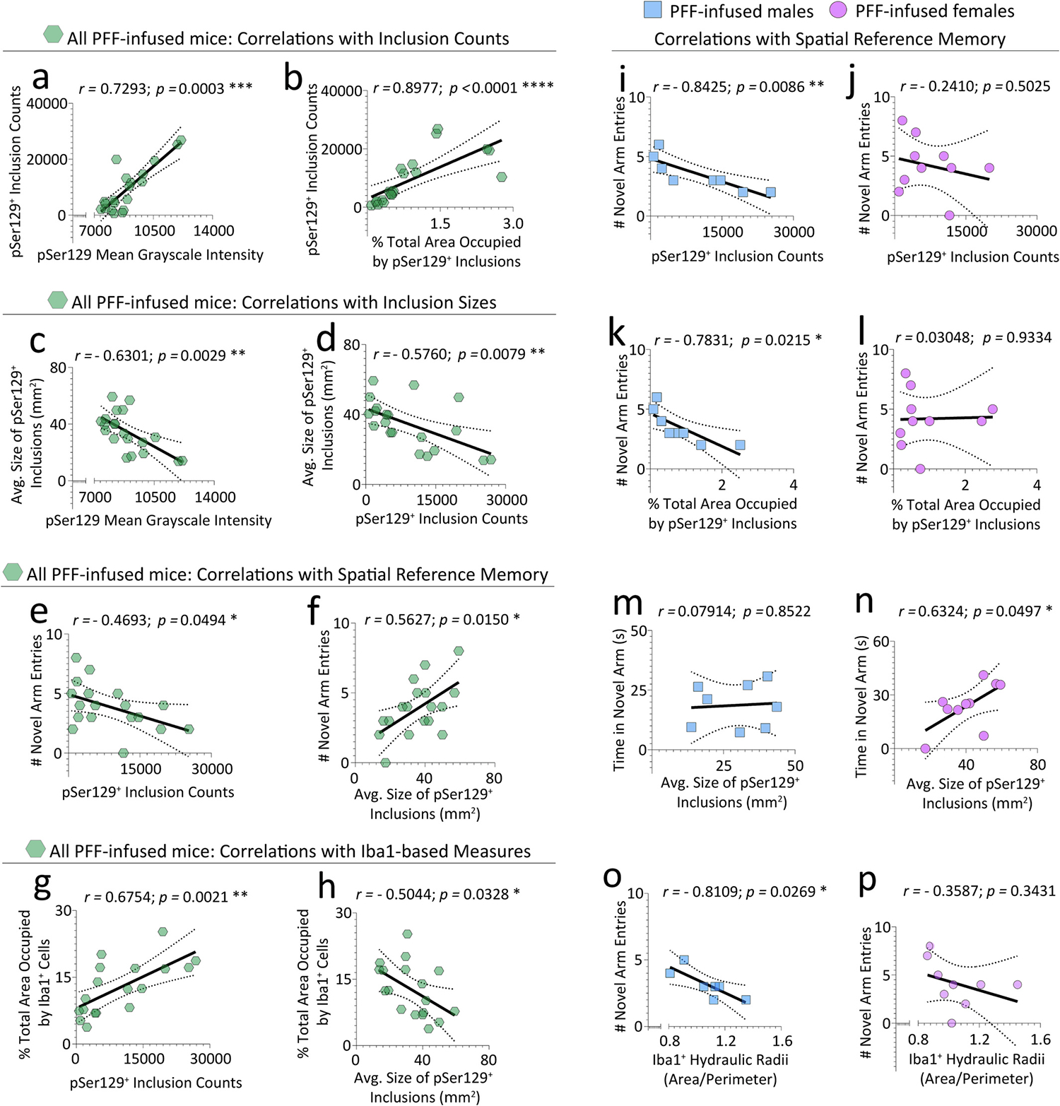
Reactive microglia are observed with aging and in Lewy body disorders, including within the olfactory bulb of men with Parkinson's disease. However, the functional impact of microglia in these disorders is still debated. Resetting these reactive cells by a brief dietary pulse of the colony-stimulating factor 1 receptor (CSF1R) inhibitor PLX5622 may hold therapeutic potential against Lewy-related pathologies. To our knowledge, withdrawal of PLX5622 after short-term exposure has not been tested in the preformed α-synuclein fibril (PFF) model, including in aged mice of both sexes. Compared to aged female mice, we report that aged males on the control diet showed higher numbers of phosphorylated α-synuclein+ inclusions in the limbic rhinencephalon after PFFs were injected in the posterior olfactory bulb. However, aged females displayed larger inclusion sizes compared to males. Short-term (14-day) dietary exposure to PLX5622 followed by control chow reduced inclusion numbers and levels of insoluble α-synuclein in aged males-but not females-and unexpectedly raised inclusion sizes in both sexes. Transient delivery of PLX5622 also improved spatial reference memory in PFF-infused aged mice, as evidenced by an increase in novel arm entries in a Y-maze. Superior memory was positively correlated with inclusion sizes but negatively correlated with inclusion numbers. Although we caution that PLX5622 delivery must be tested further in models of α-synucleinopathy, our data suggest that larger-sized-but fewer-α-synucleinopathic structures are associated with better neurological outcomes in PFF-infused aged mice.

Keywords: Dementia; Lewy body; Microglia; Neurodegeneration; Olfactory bulb; PLX5622; Parkinson's disease; Preformed fibril; Sex; Synuclein.


Conflict of interest statement

Declaration of Competing Interest The authors have no competing interests to declare.

Figures

Fig. 1.
Fig. 1.. Impact of transient exposure to the CSF1R antagonist PLX5622 (2-weeks “ON” and 7-weeks “OFF”) on open-field behaviors in preformed fibril-injected aged mice.
Aged, outbred CD-1 mice of both sexes were fed special chow with or without the CSF1R inhibitor, PLX5622, for two weeks, and then fed standard rodent chow for an additional 7 weeks. Preformed fibrils (PFFs; 5 μg in 1 μL) were injected into the bulbar anterior olfactory nucleus seven days after initiation of the PLX5622 diet (a). For evidence of PLX5622-driven depletion and repopulation of microglia/macrophages, see Figs. S4 and S10a. Mice were tested in an open-field arena after PLX5622 withdrawal (timeline in a; data for the open-field test during PLX5622 delivery are in Fig. S6). (b) Representative trackplots from ANY-maze. Numbers of central square entries (c) and the time spent in central squares (d) are shown. Numbers of corner square entries (e), time spent in corner squares (f), distance traveled (g), mean speeds (h), maximum speeds (i), time spent active (such as grooming) (j), time spent mobile (moving) (k), time spent rearing (l), and numbers of rears (m) are shown. Out of n = 40 PFF-infused mice, two mice were sacrificed early due to dermatitis and an abdominal abscess and thus excluded from the open field test (see Methods). Each mouse is illustrated as a colored dot on bar graphs with group means + SDs. *p ≤ 0.0500, **p ≤ 0.0100 for control diet vs. transient dietary PLX5622; +p ≤ 0.0500 for male vs. female two-way ANOVA/Bonferroni. All testing was two-tailed.
Fig. 2.
Fig. 2.. Transient exposure to PLX5622 (2-weeks “ON” and 7-weeks “OFF”) improves spatial reference memory in preformed fibril-injected aged female mice.
Aged, outbred CD-1 mice of both sexes were fed special chow with or without the CSF1R inhibitor, PLX5622, for two weeks, and then fed standard rodent chow for an additional 7 weeks (timeline in Fig. 1a). Preformed fibrils (PFFs; 5 μg in 1 μL) were injected into the bulbar anterior olfactory nucleus seven days after initiation of the PLX5622 diet. PFF-injected mice were tested on a Y-maze for spatial reference memory after PLX5622 was withdrawn from the diet (timeline in Fig. 1a). PBS-injected aged mice from each sex (total n = 7 across both sexes; remaining n = 2 PBS-infused aged mice had to be euthanized early due to sickness, see Methods) were included as controls to determine if PFF-infusions per se elicit loss of spatial reference memory (a-f). Numbers of novel arm entries are shown, expressed alone or as a fraction of total number of entries in all arms (a-c), to control for differences in activity. Time spent exploring the novel and familiar arms (d-e) and numbers of familiar arm entries (f) are shown, for the PBS versus PFF-infused aged mice. For control or transient PLX5622-diet fed, PFF-injected aged mice, numbers of novel arm entries (expressed alone in h or as a fraction of total number of entries in all arms in i-j), numbers of familiar arm entries (k), time spent exploring the novel arm (l), time spent exploring the familiar arms (m), and latencies to first entry in the novel arm (n) are shown. In g are representative ANY-maze trackplots. Out of the n = 40 PFF-infused mice, two mice were sacrificed early due to dermatitis and an abdominal abscess and were excluded from the Y-maze test (see Methods). Mice that made less than three arm entries during the first minute of the second Y-maze trial were also excluded (see SI Methods and (Wolf et al., 2016)). Data in n were non-Gaussian per the Shapiro-Wilk test and log-transformed (arrow) to assess statistical interactions between the two independent variables. Intervariable statistical interactions are shown above respective graphs. Each mouse is illustrated as a colored dot on bar graphs with group means + SDs or on box plots with interquartile ranges. *p ≤ 0.0500, ***p ≤ 0.0010 for PBS vs. PFFs; Mann-Whitney U (boxplot in f) or the unpaired t-test (a-e). *p ≤ 0.0500, **p ≤ 0.0100, ***p ≤ 0.0010 for control diet vs. transient dietary PLX5622; +p ≤ 0.0500, ++p ≤ 0.0100, +++p ≤ 0.0010 for male vs. female; Kruskal-Wallis (left panel of n) or two-way ANOVA/Bonferroni for h-m and right panel of n. All testing was two-tailed.
Fig. 3.
Fig. 3.. Transient exposure to PLX5622 (2-weeks “ON” and 7-weeks “OFF”) lowers nonionic detergent-insoluble phosphorylated and pan-α-synuclein in the OB/AON of preformed fibril-injected aged mice.
Aged, outbred CD-1 mice of both sexes were fed special chow with or without the CSF1R inhibitor, PLX5622, for two weeks, and then fed standard rodent chow for an additional 7 weeks (timeline in Fig. 1a). Preformed fibrils (PFFs; 5 μg in 1 μL) were injected into the bulbar anterior olfactory nucleus (OB/AON) seven days after initiation of the PLX5622 diet. OB/AON tissues from PFF-injected aged mice fed control chow or transiently exposed to the PLX5622 diet were sequentially extracted to obtain nonionic, detergent-insoluble fractions, and subjected to immunoblotting to probe for pSer129 and pan-α-synuclein (Volpicelli-Daley et al., 2014). OB/AON tissues from PBS-injected aged mice were included as controls and wild-type mouse α-synuclein monomers were loaded in the last lane to evaluate antibody specificities (a; higher-resolution image of the blot is in Fig. S8a). Note that monomeric α-synuclein migrates at ~16–17 kDa through denaturing gels, based on immunoblot in a and (Anderson et al., 2006; Sharon et al., 2001). Insoluble pSer129 and pan-α-synuclein bands at 17 kDa were quantified (b-e) separately from bands of higher molecular mass (> 17 kDa; f-h) and expressed as a fraction of the Total Protein Stain as a loading control. Levels of insoluble pSer129 were also expressed as a fraction of pan-α-synuclein in e and h. Additional data from these experiments are shown in Fig. S8. For studies in Figs. 3 and S8, three murine OB/AON samples had to be collapsed into one. Each group has three dots/statistical units, but data were generated from nine mice per group (see Methods). Shown are group means + SDs or box plots with interquartile ranges. *p ≤ 0.0500, **p ≤ 0.0100, ***p ≤ 0.0010; Mann-Whitney U (boxplots in e, h), unpaired t-test (c-d and f-g), or two-way ANOVA/Bonferroni (b). All testing was two-tailed.
Fig. 4.
Fig. 4.. Transient exposure to PLX5622 (2-weeks “ON” and 7-weeks “OFF”) reduces phosphorylated α-synuclein in the olfactory peduncle of preformed fibril-injected aged male but not female mice.
Aged, outbred CD-1 mice of both sexes were fed special chow with or without the CSF1R inhibitor, PLX5622, for two weeks, and then fed standard rodent chow for an additional 7 weeks (timeline in Fig. 1a). Preformed fibrils (PFFs; 5 μg in 1 μL) were injected into the bulbar anterior olfactory nucleus seven days after initiation of the PLX5622 diet. Brain sections were subjected to Tyramide Signal Amplification to improve the signal-to-noise ratio of the pSer129 immunolabel, scanned on an Odyssey Imager M at a resolution of 5 μm, and the olfactory peduncle (Brunjes et al., 2011) was outlined to obtain the pSer129 signal. pSer129 signal was then divided by the area of each trace (b-d). Representative photomicrographs of Hoechst-stained and pSer129-immunolabeled anterior olfactory nuclei are shown in a. Heatmap depicting the pSer129 signal per unit area of the olfactory peduncle from all mice is in e. Correlation analyses of the numbers of novel arm entries (from the Y-maze forced alternation test shown in Fig. 2) with the pSer129 signal in the olfactory peduncle of all PFF-infused mice (f) or of PFF-infused males vs. females (g-h). Note that two mice were excluded from the Y-maze and inclusion assays due to dermatitis and abdominal abscesses (see Methods). In addition, mice that made less than three arm entries during the first minute of the second Y-maze trial were also excluded from the behavior correlations (see SI Methods and (Wolf et al., 2016)). Each mouse is illustrated as a colored dot on bar graphs with group means + SDs, on box plots with interquartile ranges, or in scatterplots for correlations. *p ≤ 0.0500; Mann-Whitney U (boxplot in b) or two-way ANOVA/Bonferroni (c-d). Pearson correlation analyses are shown in f-h. All testing was two-tailed.
Fig. 5.
Fig. 5.. Sex-specific regulation of pSer129+inclusion numbers and sizes in PFF-injected aged mice after transient PLX5622 exposure (2-weeks “ON” and 7-weeks “OFF”).
Aged, outbred CD-1 mice of both sexes were fed special chow with or without the CSF1R inhibitor, PLX5622, for two weeks, and then fed standard rodent chow for an additional 7 weeks (timeline in Fig. 1a). Preformed fibrils (PFFs; 5 μg in 1 μL) were injected into the bulbar anterior olfactory nucleus seven days after initiation of the PLX5622 diet. Brain sections from PFF-injected aged mice (n = 5 randomized mice per group) were subjected to Tyramide Signal Amplification of the pSer129 signal and scanned on a VS200 Olympus imager (a-k). pSer129+ inclusions in the entire lateral limbic rhinencephalon were quantified via True AI/Deep Learning (Olympus) to identify the numbers of pSer129+ inclusions (a-b), % total area (area fraction) occupied by pSer129+ inclusions (c-d), mean grayscale intensity of pSer129+ inclusions (e-f), average sizes of the individual pSer129+ inclusions (g-h), and the distance to the nearest pSer129+ neighbor (i-j). Representative photomontage of the pSer129 staining (k; for higher resolution image and Hoechst labeling, please download Fig. S9). Young primary hippocampal neurons from male or female rat pups were cultured with or without sex-matched primary hippocampal microglia (l-o). Neurons and neuron/glia cocultures were exposed to vehicle or PFFs for 10 d. pSer129+ inclusions were expressed as a fraction of Hoechst+ cells (n). Average sizes of pSer129+ inclusions (o). Intervariable statistical interactions are shown above respective graphs. Each mouse (or each in vitro culture from one litter of pups) is illustrated as a colored dot on bar graphs with group means + SDs or on box plots with interquartile ranges. *p ≤ 0.0500, **p ≤ 0.0100, ***p ≤ 0.0010 for indicated comparisons; two or three-way ANOVA/Bonferroni (b, d, f, h, j, n-o), Mann-Whitney U (c, i), unpaired t-test (a, e, g). All testing was two-tailed.
Fig. 6.
Fig. 6.. Correlations between spatial reference memory, Iba1+cells, and pSer129+inclusions.
pSer129+ inclusion morphology data from Fig. 5 were tested for correlations to assess the consistency of the AI/Deep Learning, or with data from the Y-maze forced alternation test in Fig. 2. In addition, Iba1+ cell morphology data from Fig. S10 were tested for correlations with pSer129+ inclusion morphology data from Fig. 5 or with data from the Y-maze forced alternation test in Fig. 2. Inclusion counts were positively correlated with mean grayscale intensity of the pSer129 label (a) and with the total area occupied by the inclusions (i.e., area fraction; b). Average sizes of pSer129+ inclusions were negatively correlated with mean grayscale intensity of the pSer129 label (c) and with inclusion counts (d). Number of novel arm entries were negatively correlated with inclusion counts (e), but positively correlated with average inclusion sizes (f). The total area occupied by Iba1+ cells (i.e., area fraction) was positively correlated with pSer129+ inclusion counts (g), but negatively correlated with the average sizes of individual pSer129+ inclusions (h). The negative correlation between novel arm entries and inclusion numbers was mainly attributed to male (i) and not female (j) mice. The area fraction of the inclusions was also negatively correlated with novel arm entries in male (k), but not female (l) mice. Time spent in the novel arm was correlated with average sizes of pSer129+ inclusions in females but not males (m-n). Hydraulic radii of Iba1+ cells were negatively correlated with novel arm entries of PFF-infused male mice only (o-p). Note that additional correlation data are shown in Fig. S13. Each mouse is illustrated as a colored dot on the scatterplots. *p ≤ 0.0500, **p ≤ 0.0100, ***p ≤ 0.0010, ****p ≤ 0.0001; Pearson correlation analyses in d-p, and Spearman correlation analyses in a-c (i.e., failed the Shapiro-Wilk test for normality). All testing was two-tailed.

Similar articles

Cited by

References

    1. Aldinger KA, Sokoloff G, Rosenberg DM, Palmer AA, Millen KJ, 2009. Genetic variation and population substructure in outbred CD-1 mice: implications for genome-wide association studies. PLoS One 4, e4729. - PMC - PubMed
    1. Anderson JP, Walker DE, Goldstein JM, de Laat R, Banducci K, Caccavello RJ, Barbour R, Huang J, Kling K, Lee M, Diep L, Keim PS, Shen X, Chataway T, Schlossmacher MG, Seubert P, Schenk D, Sinha S, Gai WP, Chilcote TJ, 2006. Phosphorylation of Ser-129 is the dominant pathological modification of alpha-synuclein in familial and sporadic Lewy body disease. J. Biol. Chem. 281, 29739–29752. - PubMed
    1. Angelova DM, Brown DR, 2019. Microglia and the aging brain: are senescent microglia the key to neurodegeneration? J. Neurochem. 151, 676–688. - PubMed
    1. Attems J, Walker L, Jellinger KA, 2014. Olfactory bulb involvement in neurodegenerative diseases. Acta Neuropathol. 127, 459–475. - PubMed
    1. Attems J, Toledo JB, Walker L, Gelpi E, Gentleman S, Halliday G, Hortobagyi T, Jellinger K, Kovacs GG, Lee EB, Love S, McAleese KE, Nelson PT, Neumann M, Parkkinen L, Polvikoski T, Sikorska B, Smith C, Grinberg LT, Thal DR, Trojanowski JQ, McKeith IG, 2021. Neuropathological consensus criteria for the evaluation of Lewy pathology in post-mortem brains: a multi-Centre study. Acta Neuropathol. 141, 159–172. - PMC - PubMed

Publication types