. 2023 Nov;60(11):1092-1104.
doi: 10.1136/jmg-2022-109127. Epub 2023 Jun 14.

Dominant negative variants in IKZF2 cause ICHAD syndrome, a new disorder characterised by immunodysregulation, craniofacial anomalies, hearing impairment, athelia and developmental delay

Collaborators, Affiliations

Dominant negative variants in IKZF2 cause ICHAD syndrome, a new disorder characterised by immunodysregulation, craniofacial anomalies, hearing impairment, athelia and developmental delay

Arezoo Mohajeri et al. J Med Genet. 2023 Nov.

Abstract

Background: Helios (encoded by IKZF2), a member of the Ikaros family of transcription factors, is a zinc finger protein involved in embryogenesis and immune function. Although predominantly recognised for its role in the development and function of T lymphocytes, particularly the CD4+ regulatory T cells (Tregs), the expression and function of Helios extends beyond the immune system. During embryogenesis, Helios is expressed in a wide range of tissues, making genetic variants that disrupt the function of Helios strong candidates for causing widespread immune-related and developmental abnormalities in humans.

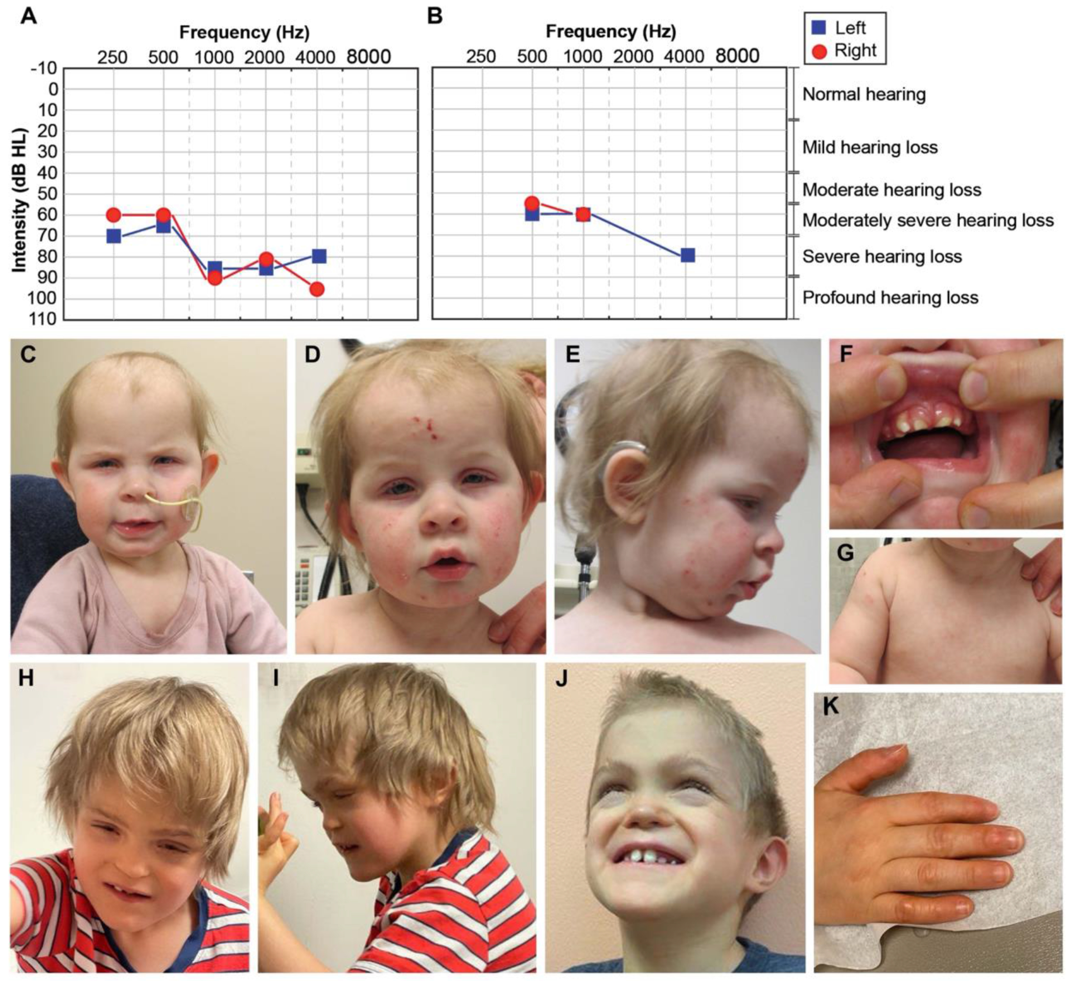
Methods: We performed detailed phenotypic, genomic and functional investigations on two unrelated individuals with a phenotype of immune dysregulation combined with syndromic features including craniofacial differences, sensorineural hearing loss and congenital abnormalities.

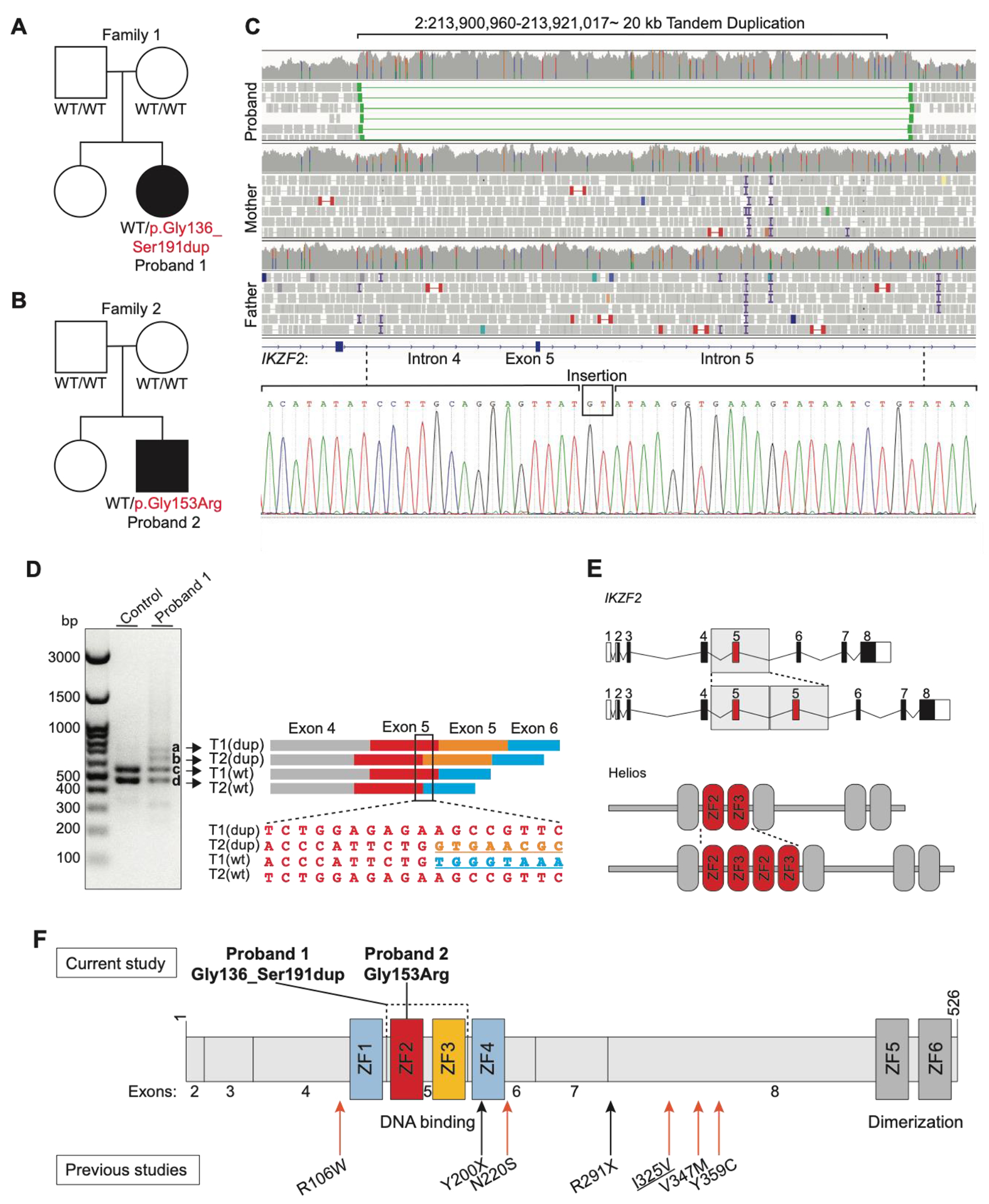
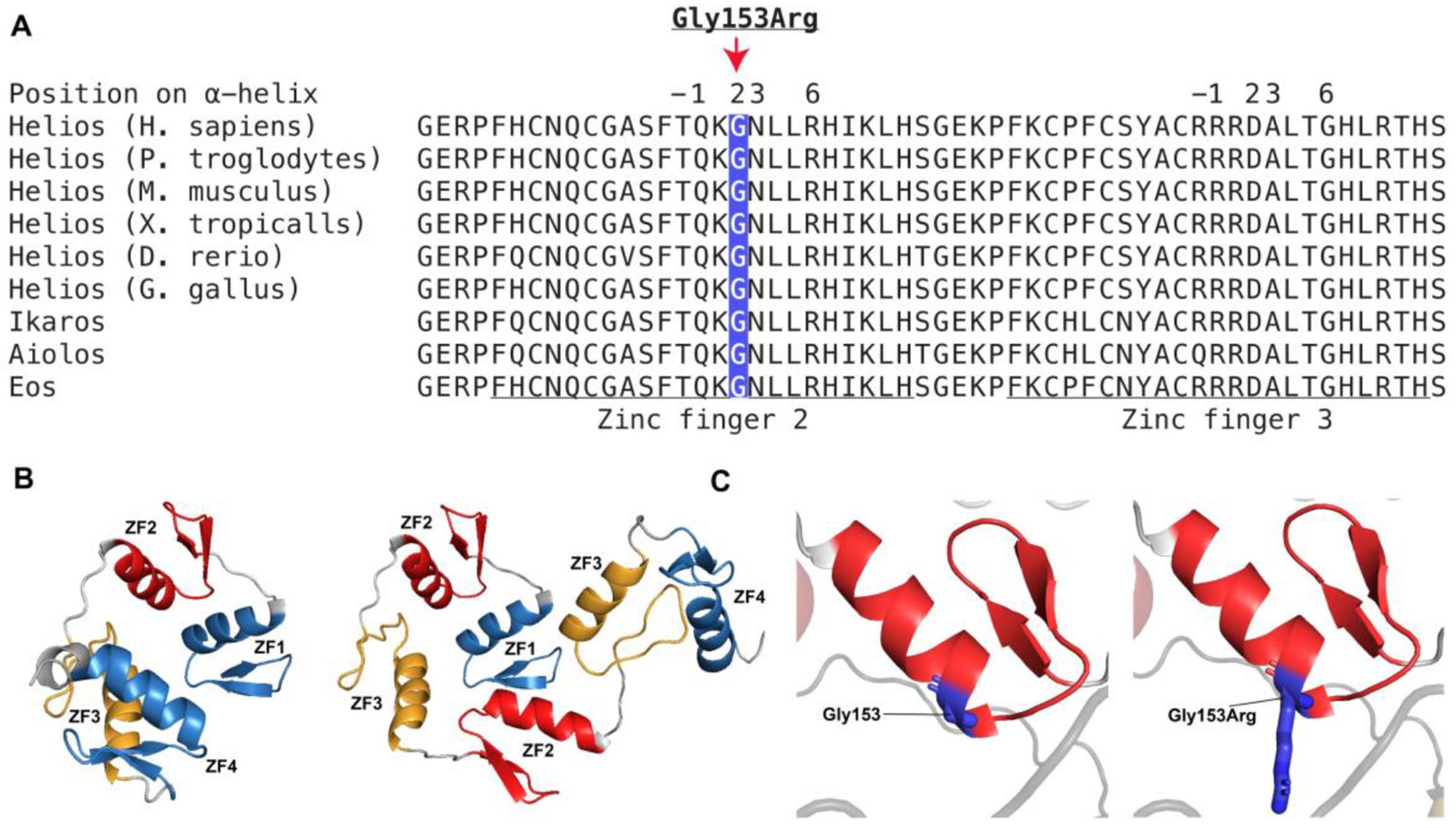
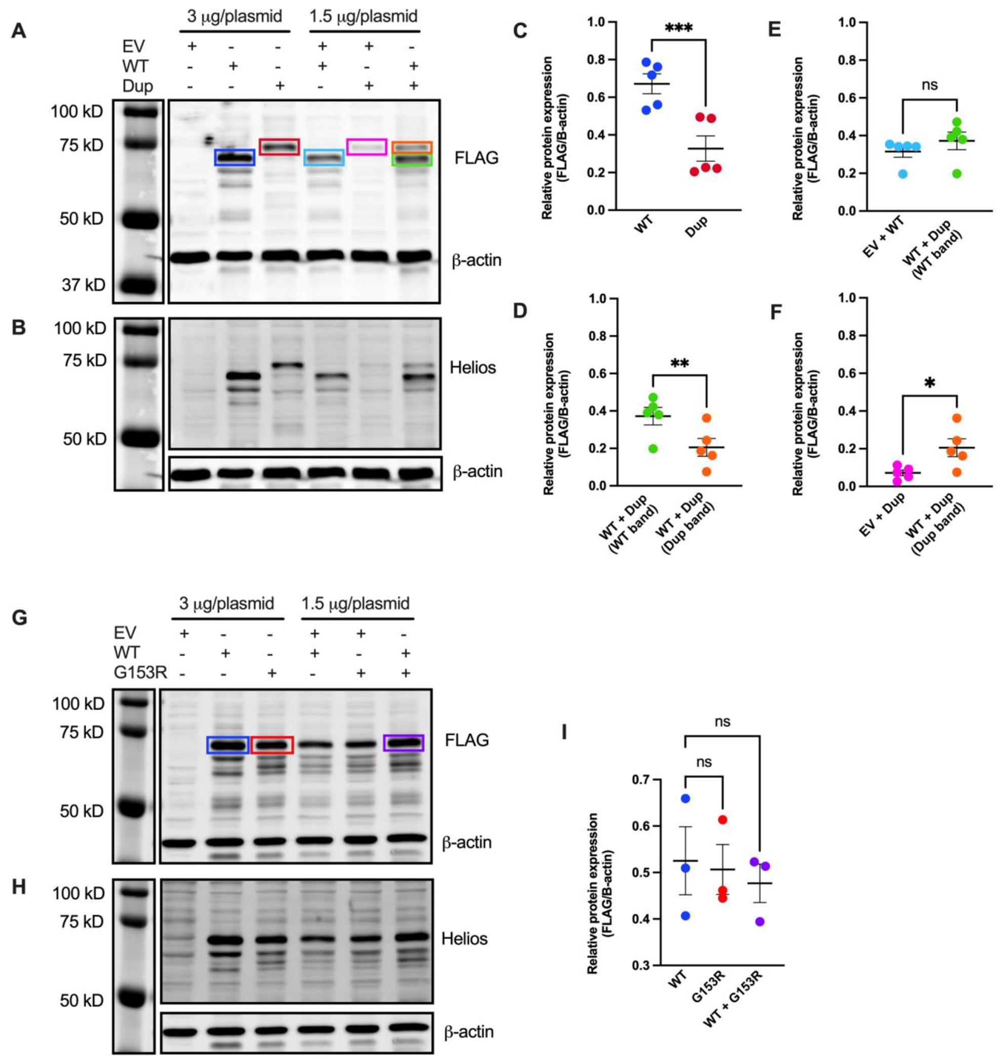
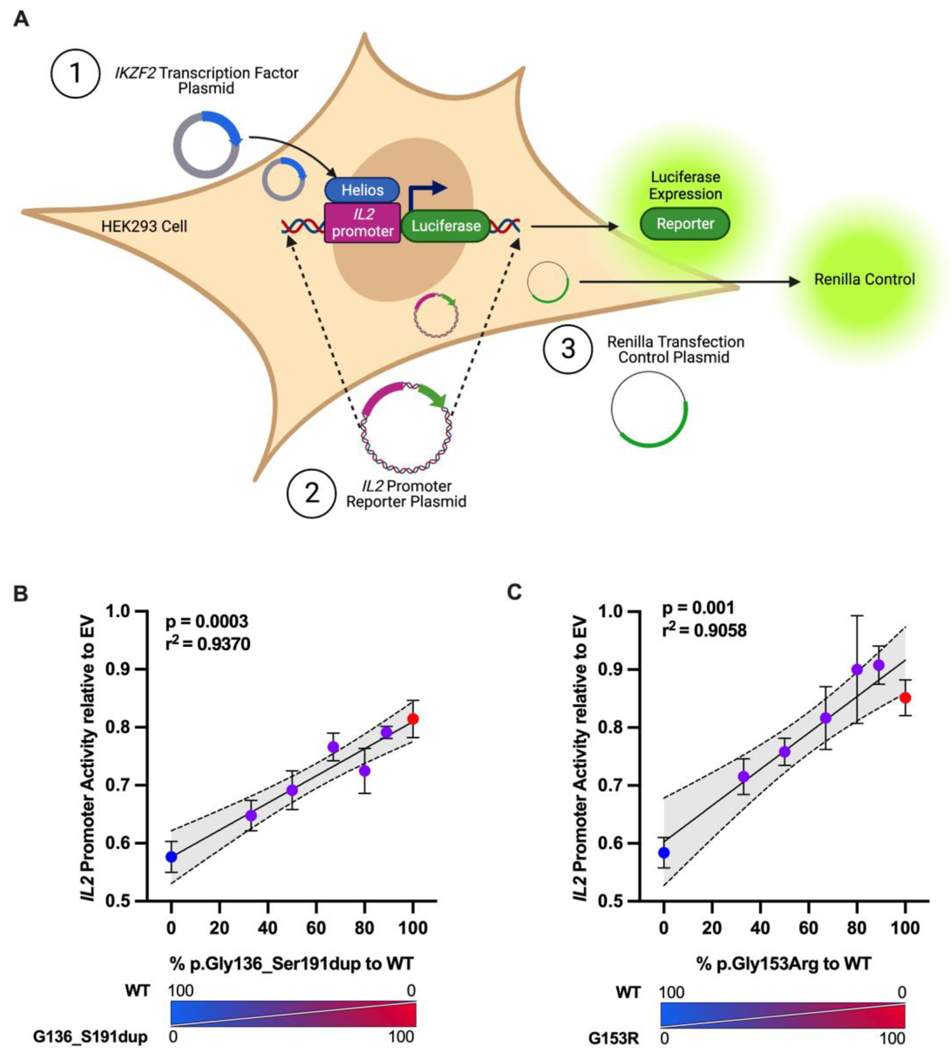
Results: Genome sequencing revealed de novo heterozygous variants that alter the critical DNA-binding zinc fingers (ZFs) of Helios. Proband 1 had a tandem duplication of ZFs 2 and 3 in the DNA-binding domain of Helios (p.Gly136_Ser191dup) and Proband 2 had a missense variant impacting one of the key residues for specific base recognition and DNA interaction in ZF2 of Helios (p.Gly153Arg). Functional studies confirmed that both these variant proteins are expressed and that they interfere with the ability of the wild-type Helios protein to perform its canonical function-repressing IL2 transcription activity-in a dominant negative manner.

Conclusion: This study is the first to describe dominant negative IKZF2 variants. These variants cause a novel genetic syndrome characterised by immunodysregulation, craniofacial anomalies, hearing impairment, athelia and developmental delay.

Keywords: Congenital, Hereditary, and Neonatal Diseases and Abnormalities; Genetics, Medical; Genomics; Immune System Diseases; Sequence Analysis, DNA.


Conflict of interest statement

Competing interests: The Department of Molecular and Human Genetics at Baylor College of Medicine receives revenue from clinical genetic testing completed at Baylor Genetics Laboratories.

Figures

Figure 1.
Figure 1.. Clinical phenotypes of Proband 1 and 2.
(A) Proband 1’s visual reinforcement audiometry (VRA) test completed at 17 months of age using insert headphones and warble tones. Air conduction thresholds from this test revealed moderately severe to profound hearing loss in the right ear, and moderately severe to severe hearing loss in the left ear. (B) Air conduction thresholds from Proband 2’s auditory brainstem response (ABR) test at 7 years of age indicated moderate to moderately severe hearing loss for the right ear and moderately severe to severe hearing loss for the left ear. (C-D) Face of Proband 1 at 10 months-1.5 years. In C and D note high forehead, macrotia, deep-set eyes, blepharophimosis, blepharitis (yellow crust on lashes), and bitemporal narrowing. In D and E note inflammation and excoriation marks from atopic dermatitis involving the whole face. In E note upturned nose and depressed nasal bridge. (F) Mouth of Proband 1. Note misaligned teeth and thickened gums. (G) Chest of Proband 1. Note athelia (absent nipples). (H-J) Face of Proband 2 at 6-7.5 years. Note brachycephaly, midface hypoplasia, narrow and long palpebral fissures, lagophthalmos (incomplete closure of eyelids), depressed nasal bridge, short philtrum, simplified ears, prominent chin, abnormal dentition, and misaligned teeth. (K) Hand of Proband 2. Note callused digits.
Figure 2.
Figure 2.. Family pedigrees, RT-PCR, Sanger results, and schematic of the IKZF2 variants on the gene and protein.
(A and B) The pedigrees of Proband 1 and 2. For individuals tested by genome and/or Sanger sequencing, the genotypes are noted. WT indicates a wild-type allele. Black boxes show individuals with ICHAD. Proband 1 harbors a de novo intragenic tandem duplication and Proband 2 harbors a de novo missense variant (C) The Integrative Genomics Viewer visualization of the duplicated region, with discordant read pairs and increased read depth providing evidence of the genomic duplication in Proband 1 (on top). Sanger sequencing chromatogram of the tandem duplication breakpoint junction on DNA and a GT insertion (at the bottom) (D) The gel image on the left shows the results of the RT-PCR. The two bottom bands (c and d) in the first and second lanes represent the amplicons from the two wild-type transcripts (T1, NM_016260.3; T2, NM_001079526.2; wt, wild-type), while the two top bands that are only visible in the second lane (a and b) correspond to the products from the two transcripts with exon 5 duplication (dup, exon 5 duplication). On the right side of the figure, a schematic of the RT-PCR products is shown alongside the Sanger trace from the region marked on the four RT-PCR products. The second sequence maps to the exon 5-exon 5 junction on altered T2. (E) Schematic of the wild-type and altered gene (top) and protein (bottom). (F) Helios protein and its functional domains (ZFs 1-6). The de novo tandem duplication (p.Gly136_Ser191dup) in Proband 1 and the de novo missense variant (p.Gly153Arg) in Proband 2 are shown on top of the protein, and the previously reported variants in the literature are shown at the bottom of the protein. Both variants presented in this study are located in the DNA binding domain of Helios. ZF, zinc finger; Refseq NM_016260.3; Underlined variants are homozygous variant; Orange arrow indicates missense variant; Black arrow indicates stop gain variant.
Figure 3.
Figure 3.. Multiple alignments of ZFs 2 and 3 and the linker sequences in orthologs and paralogs of IKZF2.
(A) The tandem duplication (p.Gly136_Ser191dup) affects ZFs 2 and 3 and linker sequences which are highly conserved across vertebrates and the paralogs, IKZF1, IKZF3, and IKZF4. The underlined sequence shows ZFs 2 and 3, and the remaining sequence represents the linkers. The missense variant (p.Gly153Arg) affects an amino acid which is situated at position 2 in reference to the start of α helix of ZF2 and has a critical role in DNA recognition and binding. (B) The Alphafold predicted structure of ZFs 1-4. The left structure shows four ZFs (ZFs 1-4), and the right structure shows six ZFs including the duplication (ZFs 1, 2, 3, 2, 3, 4). ZF, zinc finger. (C) The Alphafold predicted structure of human ZF2. ZF2 is shown in red and the amino acid Gly153 shown in dark blue. The left structure shows the wild-type residue, Gly153, and the right structure shows the altered residue, Gly153Arg.
Figure 4.
Figure 4.. Assessment of variant protein expression in HEK293 cells.
(A and B) Immunoblot of lysates obtained from HEK293 cells transfected with with pCMV6-XL4 plasmids expressing FLAG-tagged wild-type IKZF2 (WT), p.Gly136_Ser191dup variant (Dup), and/or empty vector control (EV). Protein was detected using anti-FLAG antibody in A and anti-Helios antibody in B. An anti-β actin antibody was used as loading control. A, B are representative blots from five independent experiments. (C, D, E and F) Band intensities of FLAG and β-actin fluorescence signals were quantified using Image Studio Lite Quantification Software (Licor). Data presented are mean ± SEM from five independent experiments. Paired two-tailed t-test was used for statistical analysis: *p≤ 0.05; **p ≤ 0.01; ***p ≤ 0.001. (G and H) Immunoblot of lysates obtained from HEK293 cells transfected with pCMV6-XL4 plasmids expressing FLAG-tagged WT IKZF2, p.Gly153Arg (G153R), and/or EV. Protein was detected using anti-FLAG antibody in G and anti-Helios antibody in H. An anti-β-actin antibody was used as loading control. G and H are representative blots from three independent experiments. (I) Quantification of band intensities of FLAG and β-actin fluorescence signals using Image Studio Lite. Data presented are mean ± SEM from three independent experiments. Paired two-tailed t-test was used for statistical analysis; ns, not significant.
Figure 5.
Figure 5.. Luciferase reporter assay to assess variants’ ability to repress IL2 promoter activity.
(A) Schematic representing the experiment. HEK293 cells were transfected with an empty pCMV6-XL4 (EV) plasmid or a plasmid encoding the wild-type (WT) and/or the indicated ratios of IKZF2 variants, a pGL4.14 firefly luciferase reporter plasmid carrying a region of the human IL2 promoter, and pGL4.74 renilla luciferase plasmid as transfection control. (B and C) After 24 hours, cells were lysed and measured for firefly luciferase and renilla luciferase activity. Data presented are mean ± SEM from three independent experiments, and each independent experiment is an average of three technical replicates. Dotted lines indicate 95% confidence intervals for Pearson’s Correlation test.

References

    1. Alsiö JM, Tarchini B, Cayouette M, et al. Ikaros promotes early-born neuronal fates in the cerebral cortex. PNAS 2013;110(8):E716–E25. doi: 10.1073/pnas.1215707110 - DOI - PMC - PubMed
    1. Chessum L, Matern MS, Kelly MC, et al. Helios is a key transcriptional regulator of outer hair cell maturation. Nature 2018;563(7733):696–700. doi: 10.1038/s41586-018-0728-4 - DOI - PMC - PubMed
    1. Javed A, Mattar P, Cui A, et al. Ikaros family proteins regulate developmental windows in the mouse retina through convergent and divergent transcriptional programs: Cold Spring Harbor Laboratory, 2021.
    1. Martín-Ibáñez R, Pardo M, Giralt A, et al. Helios expression coordinates the development of a subset of striatopallidal medium spiny neurons. Development 2017;144(8):1566–77. doi: 10.1242/dev.138248 - DOI - PMC - PubMed
    1. Sancho-Balsells A, Brito V, Fernandez B, et al. Lack of Helios During Neural Development Induces Adult Schizophrenia-Like Behaviors Associated With Aberrant Levels of the TRIF-Recruiter Protein WDFY1. Front Cell Neurosci 2020;14:93. doi: 10.3389/fncel.2020.00093 [published Online First: 2020/06/02] - DOI - PMC - PubMed

Publication types

Supplementary concepts