. 2023 Jun 15;25(1):32.
doi: 10.1186/s12968-023-00944-x.

Additional value of cardiac magnetic resonance feature tracking parameters for the evaluation of the arrhythmic risk in patients with mitral valve prolapse

Affiliations

Additional value of cardiac magnetic resonance feature tracking parameters for the evaluation of the arrhythmic risk in patients with mitral valve prolapse

Marco Guglielmo et al. J Cardiovasc Magn Reson. .

Abstract

Objectives: The identification of patients with mitral valve prolapse (MVP) presenting high arrhythmic risk remains challenging. Cardiovascular Magnetic Resonance (CMR) feature tracking (FT) may improve risk stratification. We analyzed the role of CMR-FT parameters in relation to the incidence of complex ventricular arrhythmias (cVA) in patients with MVP and mitral annular disjunction (MAD).

Methods: 42 patients with MVP and MAD who underwent 1.5 T CMR were classified as MAD-cVA (n = 23, 55%) in case of cVA diagnosed on a 24-h Holter monitoring and as MAD-noVA in the absence of cVA (n = 19, 45%). MAD length, late gadolinium enhancement (LGE), basal segments myocardial extracellular volume (ECV) and CMR-FT were assessed.

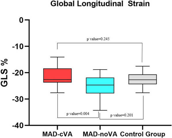
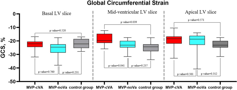
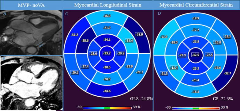
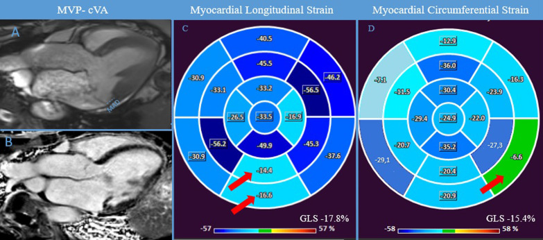
Results: LGE was more frequent in the MAD-cVA group in comparison with the MAD-noVA group (78% vs 42%, p = 0.002) while no difference was observed in terms of basal ECV. Global longitudinal strain (GLS) was reduced in MAD-cVA compared to MAD-noVA (- 18.2% ± 4.6% vs - 25.1% ± 3.1%, p = 0.004) as well as global circumferential strain (GCS) at the mid-ventricular level (- 17.5% ± 4.7% vs - 21.6% ± 3.1%, p = 0.041). Univariate analysis identified as predictors of the incidence of cVA: GCS, circumferential strain (CS) in the basal and mid infero-lateral wall, GLS, regional longitudinal strain (LS) in the basal and mid-ventricular inferolateral wall. Reduced GLS [Odd ratio (OR):1.56 (confidence interval (CI) 95%: 1.45-2.47; p < 0.001)] and regional LS in the basal inferolateral wall [OR: 1.62 (CI 95%: 1.22-2.13; p < 0.001)] remained independent prognostic factors in multivariate analysis.

Conclusion: In patients with MVP and MAD, CMR-FT parameters are correlated with the incidence of cVA and may be of interest in arrhythmic risk stratification.

Keywords: Cardiovascular magnetic resonance; Interstitial fibrosis; Mitral annular disjunction; Mitral valve prolapse; Strain.


Conflict of interest statement

The authors declare that they do not have any competing interests.

Figures

Fig. 1
Fig. 1
Box-plot representing GLS values for patients with MAD-cVA, MAD-noVA and control group. GLS Global Longitudinal Strain, MAD mitral annulus disjunction, cVA complex ventricular arrhythmias, no-VA no complex ventricular arrhythmias
Fig. 2
Fig. 2
Box-plot representing GCS values at basal LV, mid-ventricular LV and apical LV level in MAD-cVA, MAD-noVA and control group. GCS global circumferential strain, LV left ventricle, MAD mitral annulus disjunction, cVA complex ventricular arrhythmias, no-VA no complex ventricular arrhythmias
Fig. 3
Fig. 3
MAD-noVA. CMR examination of a patient known for known for bileaflet MVP with MAD (7.4 mm) without cVA at 24-h-Holter monitoring. Panel A shows 3 chamber view showing systolic MAD and the bileaflet MVP. Panel B shows the absence of macroscopic fibrosis in LGE and normal regional LS and GLS and GCS was finally evaluated in CMR-FT (Panel C-D). CMR cardiac magnetic resonance, FT feature-tracking, GCS global circumferential strain, GLS global longitudinal strain, LGE late gadolinium enhancement, LS longitudinal strain, MVP mitral valve prolapse, MAD mitral annulus disjunction
Fig. 4
Fig. 4
MAD-cVA. CMR examination of a patient known for bileaflet MVP with MAD (8.1 mm) with cVA at 24-h-Holter monitoring. Panel A shows 3 chamber view showing systolic MAD (blue lines) and the bileaflet MVP. Panel B, displays the presence of LGE in the inferolateral wall.  Abnormal GLS was found, with lower values of regional LS in the inferior wall (red arrow) (Panel C). Similarly, also GCS was reduced with lower values in the infero-lateral basal wall (red arrow) (Panel D). CMR cardiac magnetic resonance, cVA complex ventricular arrhythmias, FT feature-tracking, GCS global circumferential strain, GLS global longitudinal strain, LGE late gadolinium enhancement, LS longitudinal strain, MAD mitral annulus disjunction, MVP mitral valve prolapse

References

    1. Nordhues BD, Siontis KC, Scott CG, Nkomo VT, Ackerman MJ, Asirvatham SJ, Noseworthy PA. Bileaflet mitral valve prolapse and risk of ventricular dysrhythmias and death. J Cardiovasc Electrophysiol. 2016;27(4):463–468. doi: 10.1111/jce.12914. - DOI - PubMed
    1. Narayanan K, Uy-Evanado A, Teodorescu C, Reinier K, Nichols GA, Gunson K, Jui J, Chugh SS. Mitral valve prolapse and sudden cardiac arrest in the community. Heart Rhythm. 2016;13(2):498–503. doi: 10.1016/j.hrthm.2015.09.026. - DOI - PMC - PubMed
    1. Freed LA, Levy D, Levine RA, Larson MG, Evans JC, Fuller DL, Lehman B, Benjamin EJ. Prevalence and clinical outcome of mitral-valve prolapse. N Engl J Med. 1999;341(1):1–7. doi: 10.1056/NEJM199907013410101. - DOI - PubMed
    1. Jeresaty RM. Sudden death in the mitral valve prolapse-click syndrome. Am J Cardiol. 1976;37(2):317–318. doi: 10.1016/0002-9149(76)90328-3. - DOI - PubMed
    1. Oliveri F, Kakargias F, Panday P, Arcia Franchini AP, Iskander B, Anwer F, Hamid P. Arrhythmic mitral valve prolapse: diagnostic parameters for high-risk patients: a systematic review and meta-analysis. Pacing Clin Electrophysiol. 2021;44(10):1746–1755. doi: 10.1111/pace.14338. - DOI - PubMed