. 2023 Jun 14;14(1):160.
doi: 10.1186/s13287-023-03392-7.

Bioactivity of human adult stem cells and functional relevance of stem cell-derived extracellular matrix in chondrogenesis

Affiliations

Bioactivity of human adult stem cells and functional relevance of stem cell-derived extracellular matrix in chondrogenesis

Yangzi Jiang et al. Stem Cell Res Ther. .

Abstract

Background: Autologous chondrocyte implantation (ACI) has been used to treat articular cartilage defects for over two decades. Adult stem cells have been proposed as a solution to inadequate donor cell numbers often encountered in ACI. Multipotent stem/progenitor cells isolated from adipose, bone marrow, and cartilage are the most promising cell therapy candidates. However, different essential growth factors are required to induce these tissue-specific stem cells to initiate chondrogenic differentiation and subsequent deposition of extracellular matrix (ECM) to form cartilage-like tissue. Upon transplantation into cartilage defects in vivo, the levels of growth factors in the host tissue are likely to be inadequate to support chondrogenesis of these cells in situ. The contribution of stem/progenitor cells to cartilage repair and the quality of ECM produced by the implanted cells required for cartilage repair remain largely unknown. Here, we evaluated the bioactivity and chondrogenic induction ability of the ECM produced by different adult stem cells.

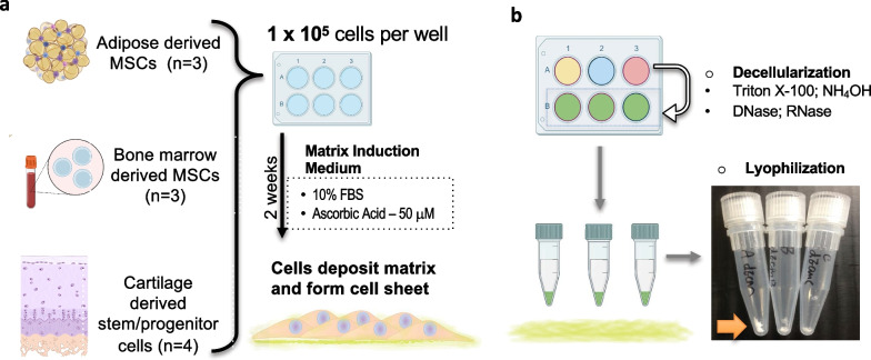
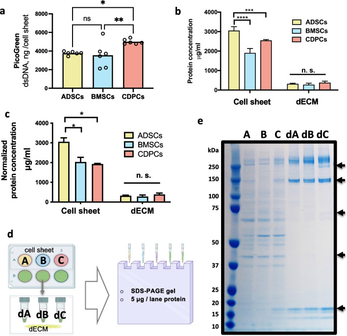
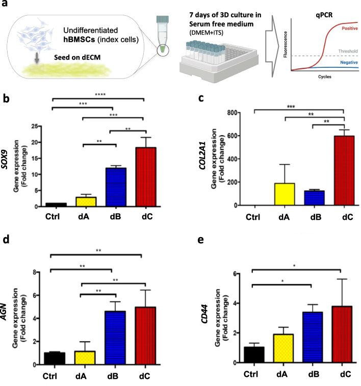
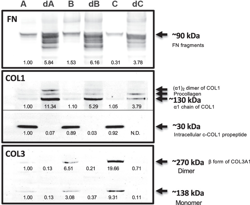
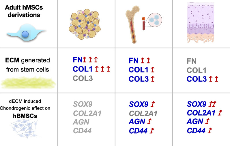
Methods: Adult stem/progenitor cells were isolated from human adipose (hADSCs), bone marrow (hBMSCs), and articular cartilage (hCDPCs) and cultured for 14 days in monolayer in mesenchymal stromal cell (MSC)-ECM induction medium to allow matrix deposition and cell sheet formation. The cell sheets were then decellularized, and the protein composition of the decellularized ECM (dECM) was analyzed by BCA assay, SDS-PAGE, and immunoblotting for fibronectin (FN), collagen types I (COL1) and III (COL3). The chondrogenic induction ability of the dECM was examined by seeding undifferentiated hBMSCs onto the respective freeze-dried solid dECM followed by culturing in serum-free medium for 7 days. The expression levels of chondrogenic genes SOX9, COL2, AGN, and CD44 were analyzed by q-PCR.

Results: hADSCs, hBMSCs, and hCDPCs generated different ECM protein profiles and exhibited significantly different chondrogenic effects. hADSCs produced 20-60% more proteins than hBMSCs and hCDPCs and showed a fibrillar-like ECM pattern (FNhigh, COL1high). hCDPCs produced more COL3 and deposited less FN and COL1 than the other cell types. The dECM derived from hBMSCs and hCDPCs induced spontaneous chondrogenic gene expression in hBMSCs.

Conclusions: These findings provide new insights on application of adult stem cells and stem cell-derived ECM to enhance cartilage regeneration.

Keywords: Adipose-derived stem cells; Adult stem cells; Bone marrow-derived stem cells; Cartilage repair; Cartilage-derived stem/progenitor cells; Chondrogenesis; Extracellular matrix.


Conflict of interest statement

YJ, none. RST serves as one of the Editors-in-Chiefs of Stem Cell Research & Therapy. Rocky S Tuan was not involved in the peer review or decision making of this article.

Figures

Fig. 1
Fig. 1
Schematic diagram of stem cell-derived cell sheet and dECM sample preparation. a. Adult human adult stem/progenitor cells were isolated from adipose (hADSC, lipoaspirate, donors n = 3), bone marrow (hBMSC, hip joint replacement, donors n = 3), and articular cartilage tissue (hCDPC, knee joint replacement, donors n = 4) with Institutional Review Board approval. The cells were pooled and expanded to Passage 2. hADSCs, hBMSCs, and hCDPCs were seeded into 6-well plates (1 × 105 cells/well) and cultured in regular MSC–ECM induction medium (growth medium with 50 µM ascorbic acid) for 14 days to allow matrix deposition and cell sheet formation. b. The cell sheets were decellularized by treatment with Triton X-100, NH4OH, DNase, and RNase. The decellularized cell sheets were then freeze-dried using a lyophilizer and stored at 4 °C until further usage. The bottom right shows the morphology of freeze-dried decellularized cell sheets in 1.5-mL tubes for each group, and the arrow indicates the dECM derived from 1 × 105 hADSCs, hBMSCs, and hCDPCs (left to right). hADSCs, human adipose-derived stem cells; hBMSCs, human bone marrow-derived stem cells; hCDPCs, human cartilage-derived progenitor cells; ECM, extracellular matrix; dECM, decellularized ECM; MSC, mesenchymal stromal cell. The schematic diagrams were created with BioRender.com by the authors with license obtained
Fig. 2
Fig. 2
ECM proteins produced by hADSCs, hBMSCs, and hCDPCs. The protein content and components of the cell sheets and dECM were determined by the BCA assay and SDS-PAGE. a. PicoGreen readouts of double-stranded DNA (dsDNA) amount of the cell sheets generated from different stem cells. b. hADSCs produced significantly higher amounts of protein than hBMSCs and hCDPCs. Group A: 3054.3 ± 196.3 μg/mL; group B: 1909.3 ± 221.9 μg/mL; group C: 2552.7 ± 37.9 μg/mL; ****p < 0.0001; ***p = 0.0005. Group dA, 321.0 ± 27.8 μg/mL; group dB, 278.5 ± 71.7 μg/mL; group dC, 381.8 ± 74.3 μg/mL; not significant between the dECM groups, p > 0.3. The experiment was repeated three times, with at least three technical replicates for each batch. One representative batch is shown, and the data are presented as mean ± SD. A two-way ANOVA, followed by LSD, was used for statistical analysis, and p < 0.05 was considered statistically significant. c. The BCA protein amount readouts of cell sheet and dECM groups, the cell sheet groups were normalized to PicoGreen dsDNA readouts, *p < 0.01; d. Diagram of the loading strategy used for SDS-PAGE. The samples were normalized to the protein concentration, and 5 μg protein was loaded into each lane of the gel. e. The SDS-PAGE results indicate the different ECM protein profiles (specifically at the ~ 10–20, 60, 75, 140, 230, and 260 kDa zones) of hADSCs, hBMSCs, and hCDPCs. The experiment was repeated at least three times, with at least two technical replicates for each trial. A representative trial is presented. A, cell sheet formed by hADSCs; dA, dECM from hADSC cell sheet; B, cell sheet formed by hBMSCs; dsDNA, double-stranded DNA; dB, dECM from hBMSC cell sheet; C, cell sheet formed by hCDPCs; dC, dECM from hCDPC cell sheet. hADSCs, human adipose-derived stem cells; hBMSCs, human bone marrow-derived stem cells; hCDPCs, human cartilage-derived progenitor cells; ECM, extracellular matrix; dECM, decellularized ECM; BCA, bicinchoninic acid, SD, standard deviation; ANOVA, analysis of variance; LSD, least significant difference; SDS-PAGE, sodium dodecyl sulfate–polyacrylamide gel electrophoresis. Figure 2d was created with BioRender.com by the authors with license obtained
Fig. 3
Fig. 3
Chondrogenic induction ability of the dECM generated by hADSCs, hBMSCs, and hCDPCs. a. Experimental set up. To examine the chondrogenic induction ability of the dECM, undifferentiated hBMSCs were used as index cells and seeded atop the dECM derived from different cell sources. 1 × 105 hBMSCs (n = 3, pooled from three donors other than the matrix donor) grown in serum-free medium were seeded onto freeze-dried dECM. The attached BMSC–dECM constructs were then moved to new tubes and cultured in serum-free growth medium (DMEM-high glucose and ITS) without the addition of any other growth factors for 7 days, followed by q-PCR examination. be. The expression levels of chondrogenic/cartilage matrix-related genes (SOX9, COL2A1, AGN, CD44, HKG:18SrRNA, and RPL13a; Control: monolayer-cultured BMSCs). The experiment was repeated at least three times, with three technical replicates (cell–dECM constructs) for each trial. The mRNA extracted from each cell–dECM construct was examined with three technical replicates in the q-PCR assay. Data are presented as mean fold changes ± SD from a representative trial. A one-way ANOVA, followed by LSD, was used for statistical analysis, and p < 0.05 was considered statistically significant. Ctrl, monolayer cultures of index BMSCs; dA, index BMSCs seeded onto the hADSC–dECM; dB, index BMSCs seeded onto the hBMSC–dECM; dC, index BMSCs seeded onto the hCDPC–dECM. SOX9, SRY-box transcription factor 9; COL2A1, pro-alpha1(II) chain of type II collagen; AGN, aggrecan; CD44, cell surface glycoprotein, receptor of hyaluronic acid; 18SrRNA, 18S ribosomal RNA; RPL13a, ribosomal protein L13a. hADSCs, human adipose-derived stem cells; hBMSCs, human bone marrow-derived stem cells; hCDPCs, human cartilage-derived progenitor cells; ECM, extracellular matrix; dECM, decellularized ECM; q-PCR, quantitative polymerase chain reaction; DMEM, Dulbecco’s modified Eagle medium; ITS, insulin–transferrin–selenium; SD, standard deviation; ANOVA, analysis of variance; LSD, least significant difference. Figure 3a was created with BioRender.com by the authors with license obtained
Fig. 4
Fig. 4
Fibrillary components of FN, COL1, and COL3 in the stem cell-generated ECM. Western blot results of FN, COL1, and COL3 in the cell sheets and dECM generated by hADSCs, hBMSCs, and hCDPCs. A, cell sheet formed by hADSCs; dA, dECM from hADSC cell sheet; B, cell sheet formed by hBMSCs; dB, dECM from hBMSC cell sheet; C, cell sheet formed by hCDPCs; dC, dECM from CDPC cell sheet. FN: fibronectin fragments, ~ 90 kDa (bands at ~ 263 kDa under non-reducing conditions can be found in Additional file 1: Fig. S1); COL1, collagen type I, intracellular c-terminal-COL1 propeptide can be found at ~ 30 kDa; mature ECM COL1 at ~ 130 kDa (arrows from bottom to top indicate the α1 chain of COL1, procollagen, and (α1)2 dimer of COL1, respectively); COL3, collagen type III, monomer at ~ 138 kDa, and dimer at ~ 270 kDa. The same amounts of protein (5 μg protein/lane) were loaded into each well, and SDS-PAGE was performed under reducing conditions. The experiment was repeated at least three times, with at least two technical replicates for each trial. The data were quantified using Image J (NIH, USA), normalized with group A, and a representative trial is presented. Full-length blots/gels are presented in Additional file 1: Figs. S2–S4. hADSCs, human adipose-derived stem cells; hBMSCs, human bone marrow-derived stem cells; hCDPCs, human cartilage-derived progenitor cells; ECM, extracellular matrix; dECM, decellularized ECM; SDS-PAGE, sodium dodecyl sulfate polyacrylamide gel electrophoresis
Fig. 5
Fig. 5
Schematic of the study. Schematic of the study. In this study, we characterized the bioactivity, particularly the chondrogenic induction ability, of the ECM produced by adult stem/progenitor cells derived from human adipose, bone marrow, and articular cartilage tissue. Our results suggest the presence of different in vivo cartilage regenerative potencies and patterns of adult stem cells derived from adipose, bone marrow, and cartilage tissue. FN: fibronectin; COL1, collagen type I; COL3, collagen type III; SOX9, SRY-box transcription factor 9; COL2A1, pro-alpha1(II) chain of type II collagen; AGN, aggrecan; CD44, cell surface glycoprotein, receptor of hyaluronic acid; ECM; extracellular matrix. Figure 5 was created with BioRender.com by the authors with license obtained

Similar articles

Cited by

References

    1. Jiang Y, Zhang S, Qi Y, Wang L, Ouyang H. Cell transplantation for articular cartilage defects: principles of past, present, and future practice. Cell Transplant. 2011;20(5):593–607. doi: 10.3727/096368910X532738. - DOI - PubMed
    1. Jiang Y, Cai Y, Zhang W, Yin Z, Hu C, Tong T, Lu P, Zhang S, Neculai D, Tuan RS, et al. Human cartilage-derived progenitor cells from committed chondrocytes for efficient cartilage repair and regeneration. Stem Cells Transl Med. 2016;5(6):733–744. doi: 10.5966/sctm.2015-0192. - DOI - PMC - PubMed
    1. Jiang Y, Tuan RS. Origin and function of cartilage stem/progenitor cells in osteoarthritis. Nat Rev Rheumatol. 2015;11(4):206–212. doi: 10.1038/nrrheum.2014.200. - DOI - PMC - PubMed
    1. Handorf AM, Li WJ. Induction of mesenchymal stem cell chondrogenesis through sequential administration of growth factors within specific temporal windows. J Cell Physiol. 2014;229(2):162–171. doi: 10.1002/jcp.24428. - DOI - PubMed
    1. Correa D, Somoza RA, Lin P, Greenberg S, Rom E, Duesler L, Welter JF, Yayon A, Caplan AI. Sequential exposure to fibroblast growth factors (FGF) 2, 9 and 18 enhances hMSC chondrogenic differentiation. Osteoarthr Cartil. 2015;23(3):443–453. doi: 10.1016/j.joca.2014.11.013. - DOI - PMC - PubMed

Publication types

Substances