

Clinical presentation, diagnosis, treatment, and outcome in 8 dogs and 2 cats with global hypoxic-ischemic brain injury (2010-2022)
- PMID: 37316975
- PMCID: PMC10365066
- DOI: 10.1111/jvim.16790
Clinical presentation, diagnosis, treatment, and outcome in 8 dogs and 2 cats with global hypoxic-ischemic brain injury (2010-2022)
Abstract
Background: Global hypoxic-ischemic brain injury (GHIBI) results in variable degrees of neurological dysfunction. Limited data exists to guide prognostication on likelihood of functional recovery.
Hypothesis: Prolonged duration of hypoxic-ischemic insult and absence of neurological improvement in the first 72 hours are negative prognostic indicators.
Animals: Ten clinical cases with GHIBI.
Methods: Retrospective case series describing 8 dogs and 2 cats with GHIBI, including clinical signs, treatment, and outcome.
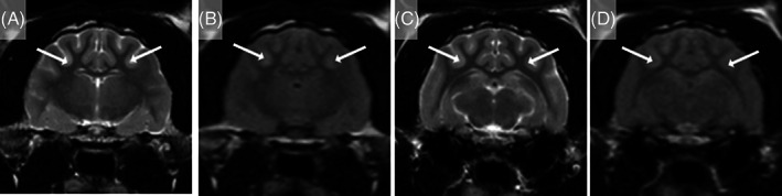
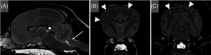
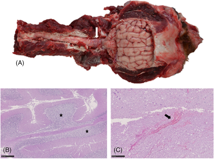
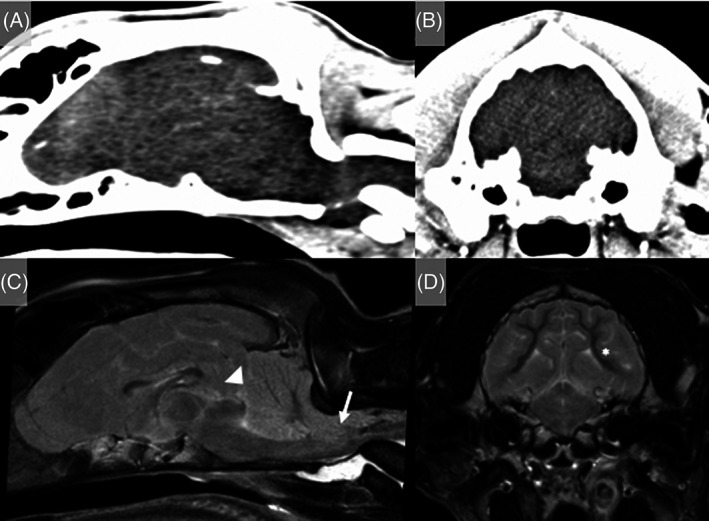
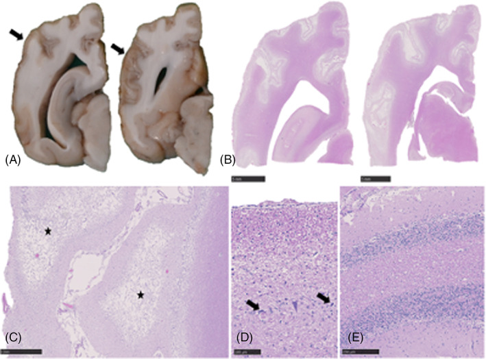
Results: Six dogs and 2 cats experienced cardiopulmonary arrest or anesthetic complication in a veterinary hospital and were promptly resuscitated. Seven showed progressive neurological improvement within 72 hours of the hypoxic-ischemic insult. Four fully recovered and 3 had residual neurological deficits. One dog presented comatose after resuscitation at the primary care practice. Magnetic resonance imaging confirmed diffuse cerebral cortical swelling and severe brainstem compression and the dog was euthanized. Two dogs suffered out-of-hospital cardiopulmonary arrest, secondary to a road traffic accident in 1 and laryngeal obstruction in the other. The first dog was euthanized after MRI that identified diffuse cerebral cortical swelling with severe brainstem compression. In the other dog, spontaneous circulation was recovered after 22 minutes of cardiopulmonary resuscitation. However, the dog remained blind, disorientated, and ambulatory tetraparetic with vestibular ataxia and was euthanized 58 days after presentation. Histopathological examination of the brain confirmed severe diffuse cerebral and cerebellar cortical necrosis.
Conclusions and clinical importance: Duration of hypoxic-ischemic insult, diffuse brainstem involvement, MRI features, and rate of neurological recovery could provide indications of the likelihood of functional recovery after GHIBI.
Keywords: canine; cerebral; feline; hypoxia; ischemic.
© 2023 The Authors. Journal of Veterinary Internal Medicine published by Wiley Periodicals LLC on behalf of American College of Veterinary Internal Medicine.
Conflict of interest statement
Authors declare no conflict of interest.
Figures
Similar articles
-
Successful resuscitation and neurological monitoring of a dog with out-of-hospital cardiopulmonary arrest due to pentobarbital overdose.J Vet Emerg Crit Care (San Antonio). 2023 May-Jun;33(3):393-400. doi: 10.1111/vec.13283. Epub 2023 Feb 23. J Vet Emerg Crit Care (San Antonio). 2023. PMID: 36815742
-
Prognostic indicators for dogs and cats with cardiopulmonary arrest treated by cardiopulmonary cerebral resuscitation at a university teaching hospital.J Am Vet Med Assoc. 2009 Jul 1;235(1):50-7. doi: 10.2460/javma.235.1.50. J Am Vet Med Assoc. 2009. PMID: 19566454
-
Assessment of cardiopulmonary resuscitation in 121 dogs and 30 cats at a university teaching hospital (2009-2012).J Vet Emerg Crit Care (San Antonio). 2014 Nov-Dec;24(6):693-704. doi: 10.1111/vec.12250. J Vet Emerg Crit Care (San Antonio). 2014. PMID: 25471644
-
Cardiopulmonary cerebral resuscitation.Vet Clin North Am Small Anim Pract. 2001 Nov;31(6):1253-64, vii. doi: 10.1016/s0195-5616(01)50102-3. Vet Clin North Am Small Anim Pract. 2001. PMID: 11727336 Review.
-
[Recent insights into the possibilities of resuscitation of dogs and cats].Tijdschr Diergeneeskd. 1998 Aug 15;123(16):464-70. Tijdschr Diergeneeskd. 1998. PMID: 9728386 Review. Dutch.
Cited by
-
Beyond the surface: how ex-vivo diffusion-weighted imaging reveals large animal brain microstructure and connectivity.Front Neurosci. 2024 Jun 26;18:1411982. doi: 10.3389/fnins.2024.1411982. eCollection 2024. Front Neurosci. 2024. PMID: 38988768 Free PMC article. Review.
References
-
- Erbsloh F, Bernsmeier A, Hillesheim H. The glucose consumption of the brain & its dependence on the liver. Arch Psychiatr Nervenkr Z Gesamte Neurol Psychiatr. 1958;196:611‐626. - PubMed
-
- Panarello GL, Dewey CW, Barone G, Stefanacci JD. Magnetic resonance imaging of two suspected cases of global brain ischemia. J Vet Emerg Crit Car. 2004;14:269‐277.
-
- Collard CD, Gelman S. Pathophysiology, clinical manifestations, and prevention of ischemia‐reperfusion injury. Anesthesiology. 2001;94:1133‐1138. - PubMed
-
- Busl KM, Greer DM. Hypoxic‐ischemic brain injury: pathophysiology, neuropathology and mechanisms. NeuroRehabilitation. 2010;26:5‐13. - PubMed
MeSH terms
LinkOut - more resources
Full Text Sources
Medical
Miscellaneous
