. 2023 May 1;11(5):1192.
doi: 10.3390/microorganisms11051192.

Cloning and Molecular Characterization of the Recombinant CVB4E2 Immunogenic Viral Protein (rVP1), as a Potential Subunit Protein for Vaccine and Immunodiagnostic Reagent Candidate

Affiliations

Cloning and Molecular Characterization of the Recombinant CVB4E2 Immunogenic Viral Protein (rVP1), as a Potential Subunit Protein for Vaccine and Immunodiagnostic Reagent Candidate

Ikbel Hadj Hassine et al. Microorganisms. .

Abstract

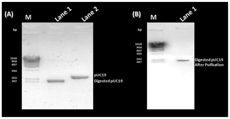
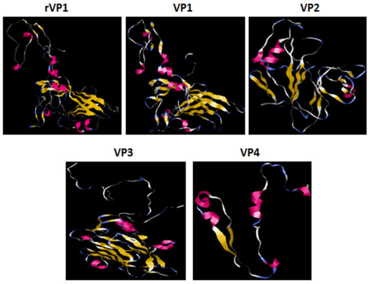
The aim of the present study was, first, to clone the VP1 gene of the human coxsackievirus B4 strain E2 (CVB4E2) in the prokaryotic pUC19 plasmid expression vector then to compare it with the structural capsid proteins of the same strain using bioinformatic tools. PCR colony amplification followed through a restriction digestion analysis and sequencing process which affirmed the success of the cloning process. SDS-PAGE and Western Blotting were used to characterize the purified recombinant viral protein expressed in bacteria cells. The BLASTN tool revealed that the nucleotide sequence of the recombinant VP1 (rVP1) expressed by pUC19 highly matched the target nucleotide sequence of the diabetogenic CVB4E2 strain. Secondary structure and three-dimension structure prediction suggested that rVP1, such as wild-type VP1, is chiefly composed of random coils and a high percentage of exposed amino acids. Linear B-cell epitope prediction showed that several antigenic epitopes are likely present in rVP1 and CVB4E2 VP1 capsid protein. Additionally, phosphorylation site prediction revealed that both proteins may affect the signal transduction of host cells and can be involved in virus virulence. The present work highlights the usefulness of cloning and bioinformatics characterizations for gene investigation. Furthermore, the collected data are helpful for future experimental research related to the development of immunodiagnostic reagents and subunit vaccines based on the expression of immunogenic viral capsid proteins.

Keywords: bioinformatics; coxsackievirus B4; molecular characterization; recombinant rVP1.


Conflict of interest statement

The authors declare no conflict of interest.

Figures

Figure 1
Figure 1
PCR amplification and Purification of the VP1 gene. (A) Lane M: GeneRuler 100 bp DNA Ladder (ThermoFisher, Waltham, MD, USA); Lane 1: negative control; Lane 2: PCR amplification product of the VP1 gene; Lane 3: PCR amplification product of VP1 with primers containing EcoRI and BamHI recognition sites. (B) Lane M: GeneRuler 100 bp DNA Ladder (ThermoFisher); Lanes 1 and 3: negative control; Lanes 2 and 4: Purification products of VP1 and VP1 flanked by the restriction enzyme sites, respectively.
Figure 2
Figure 2
Preparation of pUC19 for the cloning process. (A) Lane M: Lambda DNA/HindIII Marker (ThermoFisher); Lane 1: Digestion product of purified pUC19; Lane 2: Purified pUC19 extracted from transformed bacterial cells. (B) Lane M: Lambda DNA/HindIII Marker (ThermoFisher); Lane 1: Purification product of the digested pUC19.
Figure 3
Figure 3
Confirmation of the cloning process. (A) Lane M: GeneRuler 100 bp DNA Ladder (ThermoFisher); Lane 1: Negative control; Lanes 2 to 6: PCR colony of Non-transformed colonies; Lane 7: PCR colony of a transformed colony. (B) Lane M: Lambda DNA/HindIII Marker (ThermoFisher); Lane 1: The extracted DNA of the same colony confirmed the cloning of VP1 in pUC19. (C) Lane M: Lambda DNA/HindIII Marker (ThermoFisher); Lane 1: The restriction analysis showing two bands corresponding to pUC19 and VP1.
Figure 4
Figure 4
Molecular characterization of the recombinant CVB4 E2 rVP1 subunit viral protein. (A) SDS-PAGE analysis of the recombinant rVP1 protein. MW: Protein Weight Marker; 1: Control DH5α cells; 2–3: Transformed DH5α cells expressing VP1 gene. (B) Purification of recombinant rVP1 subunit protein using Ni-NTA affinity chromatography. 1: Clarified Bacteria cells expressing VP1; 2–3: Wash fractions; 4–6: Elution fractions. (C) Western Blotting analysis of the recombinant rVP1 protein. 1: Negative control; 2–3: Non-transformed bacteria cell lysates; 4: Transformed DH5α cell lysate expressing VP1 gene.
Figure 5
Figure 5
Prediction of the conserved domain, signal peptides sequence, and phosphorylation sites of the rVP1. The conserved domain, Signal peptide sequence, and phosphorylation site of rVP1 were analyzed using NCBI Conserved Domains search tool (A), SignalP-4.0 Server (B), and NetPhos 2.0 program (C), respectively.
Figure 6
Figure 6
Prediction of the tertiary structure of rVP1 and CVB4E2 capsid proteins. The colored ribbons show the tertiary structure of the viral proteins. The red ones show the α helices, the yellows are the β-folding, and the white ones correspond to other residues.
Figure 7
Figure 7
The possible B-epitope regions of the proteins and their presentation in tertiary structures. RasMol generated the tertiary ribbon and molecular surface representations. B cell epitopes are shown in red, blue, green, gold, purple, orange, and light green based on the order: AA 18–63, 83–93, 130–140, 155–168, 202–213, 258–273 of rVP1; AA 5–31, 33–50, 82–96, 127–138, 152–165, 199–209, 251–280 of VP1; AA 5–27, 38–60, 66–92, 130–165, 223–244 of VP2; AA 7–39, AA 55–67 and AA 139–150 of VP3; and AA 5–19 and AA 29–54 of VP4.

References

    1. Tapparel C., Siegrist F., Petty T.J., Kaiser L. Picornavirus and enterovirus diversity with associated human diseases. Infect. Genet. Evol. 2013;14:282–293. doi: 10.1016/j.meegid.2012.10.016. - DOI - PubMed
    1. Zell R., Delwart E., Gorbalenya A.E., Hovi T., King A.M.Q., Knowles N.J., Lindberg A.M., Pallansch M.A., Palmenberg A.C., Reuter G., et al. ICTV virus taxonomy profile: Picornaviridae. J. Gen. Virol. 2017;98:2421–2422. doi: 10.1099/jgv.0.000911. - DOI - PMC - PubMed
    1. Solomon T., Lewthwaite P., DaCardosa M.J., McMinn P., Ooi M.H. Virology, epidemiology, pathogenesis, and control of enterovirus 71. Lancet Infect. Dis. 2010;10:778–790. doi: 10.1016/S1473-3099(10)70194-8. - DOI - PubMed
    1. Hassine I.H., Gharbi J., Hamrita B., Almalki M.A., Rodríguez J.F., Ben M’hadheb M. Characterization of Coxsackievirus B4 virus-like particles VLP produced by the recombinant baculovirus-insect cell system expressing the major capsid protein. Mol. Biol. Rep. 2020;47:2835–2843. doi: 10.1007/s11033-020-05333-6. - DOI - PubMed
    1. Ben M’hadheb M., Souii A., Harrabi M., Jrad-Battikh N., Gharbi J. In Vitro-reduced translation efficiency of coxsackievirus B3 Sabin3-like strain is correlated to impaired binding of cellular initiation factors to viral IRES RNA. Curr. Microbiol. 2015;70:756–761. doi: 10.1007/s00284-015-0784-z. - DOI - PubMed

LinkOut - more resources