

When a dermatopathologist encounters the ultra-rare: A case series of superficial soft tissue/cutaneous myxopapillary ependymomas
- PMID: 37317818
- PMCID: PMC10721733
- DOI: 10.1111/cup.14475
When a dermatopathologist encounters the ultra-rare: A case series of superficial soft tissue/cutaneous myxopapillary ependymomas
Abstract
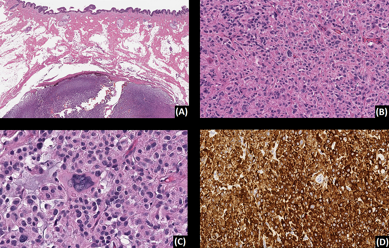
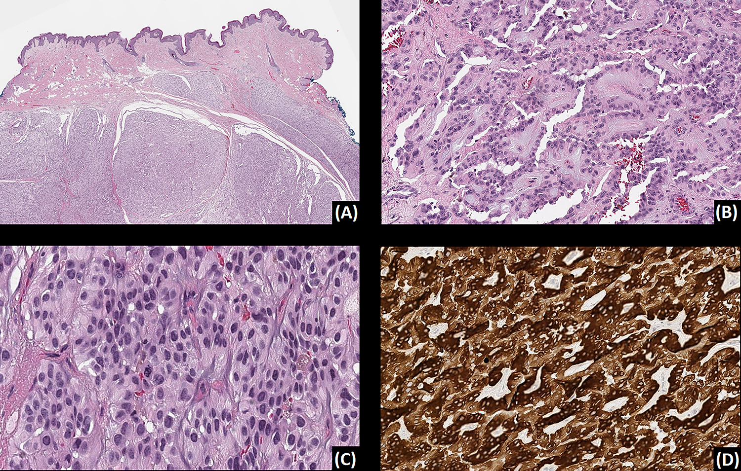
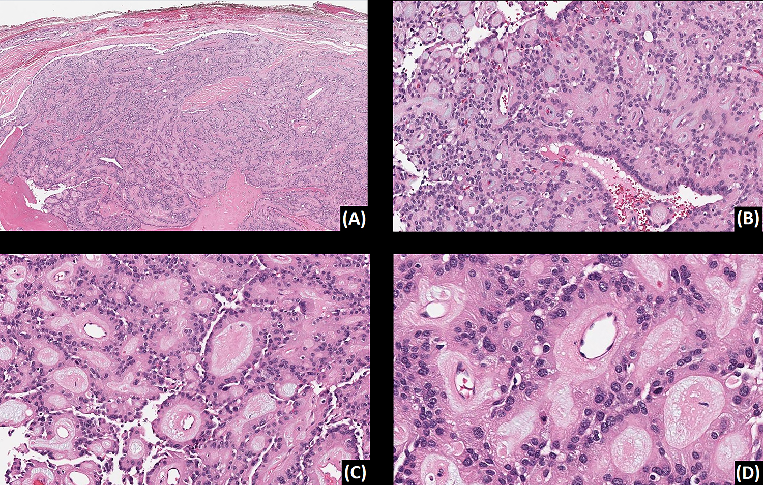
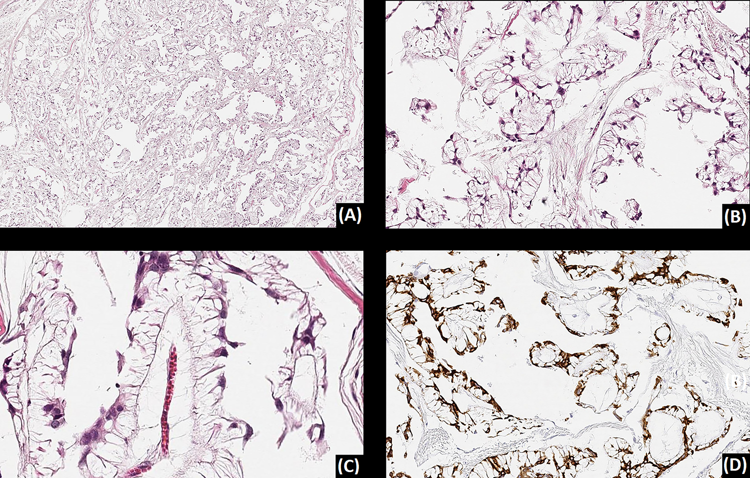
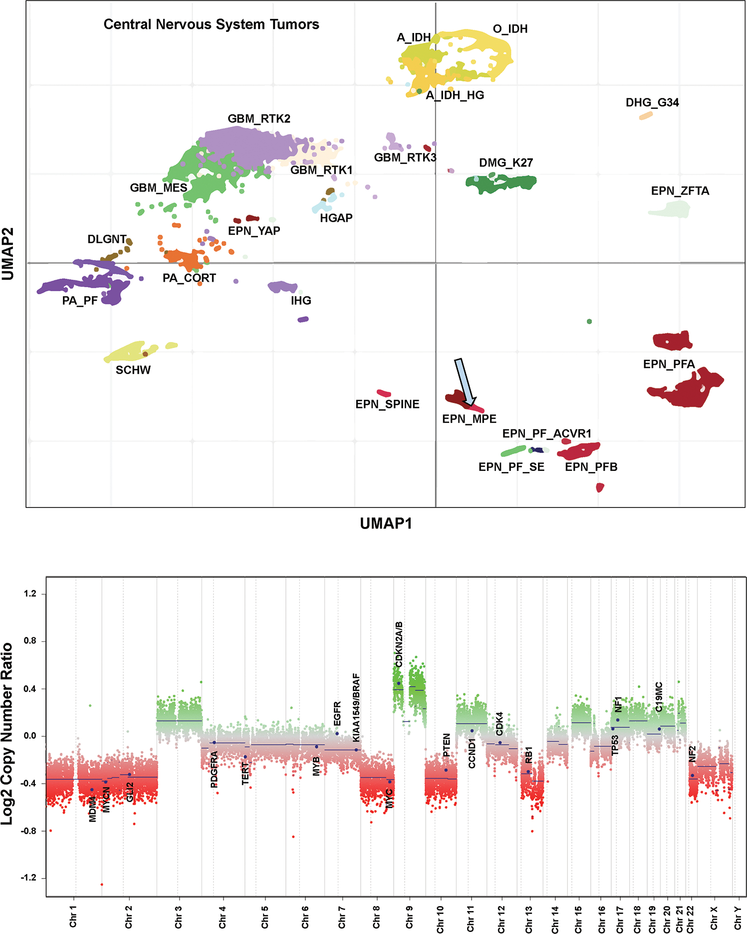
Myxopapillary ependymoma (MPE) is an uncommon variant of ependymoma, almost exclusively seen in conus medullaris or filum terminale. MPE can be diagnostically challenging, especially when arising extra-axially. Here we report 5 cases of superficial soft tissue/cutaneous MPE, identified across three tertiary institutions. All patients were female and three of them (3/5, 60%) were children (median age 11 years, range 6-58 years). The tumors presented as slow-growing masses of the sacrococcygeal subcutaneous soft tissues, occasionally identified after minor trauma and clinically favored to be pilonidal sinuses. Imaging showed no neuraxis connection. Macroscopically, tumors were well-circumscribed, lobulated, and solid and microscopically they exhibited typical histopathology of MPE, at least focally. Two of the tumors (2/5, 40%) showed predominantly solid or trabecular architecture with greater cellular pleomorphism, scattered giant cells, and increased mitotic activity. All tumors (5/5, 100%) showed strong diffuse immunohistochemical expression of GFAP. One tumor clustered at the category "ependymoma, myxopapillary" by methylome analysis. Two patients (2/5, 40%) had local recurrence at 8 and 30 months after the initial surgery. No patients developed metastases during the follow-up period (median 60 months, range 6-116 months). Since a subset of extra-axial MPEs behaves more aggressively, timely and accurate diagnosis is of paramount importance.
Keywords: GFAP; cutaneous; methylation; myxopapillary ependymoma; superficial.
© 2023 John Wiley & Sons A/S. Published by John Wiley & Sons Ltd.
Conflict of interest statement
Figures

References
-
- Rosenblum MK, Pietsch T, Korshunov A et al. Myxopapillary ependymoma. In: WHO classification of tumors editorial board. Central nervous system tumours. Lyon (France): International Agency for Research on Cancer; 2021:183–185. (WHO classification of tumors series, 5th ed.; vol. 6). https://publication.iarc.fr/601
-
- Goldblum JR, Weiss SW, Folpe AL. Extraspinal (soft tissue) ependymoma. In: Goldblum JR, Folpe AL eds. Enzinger and Weiss’s soft tissue tumors. 7th ed. Amsterdam, Netherlands: Elsevier; 2019:985–987.
-
- Cimino PJ, Ketchum C, Turakulov R, et al. Expanded analysis of high-grade astrocytoma with piloid features identifies an epigenetically and clinically distinct subtype associated with neurofibromatosis type 1. Acta Neuropathol. 2023;145(1):71–82. doi: 10.1007/s00401-022-02513-5. - DOI - PMC - PubMed
Publication types
MeSH terms
Grants and funding
LinkOut - more resources
Full Text Sources
Miscellaneous
