. 2023 Aug 1;325(2):F164-F176.
doi: 10.1152/ajprenal.00321.2022. Epub 2023 Jun 15.

Renal, but not platelet or skin, extracellular vesicles decrease oxidative stress, enhance nascent peptide synthesis, and protect from ischemic renal injury

Affiliations

Renal, but not platelet or skin, extracellular vesicles decrease oxidative stress, enhance nascent peptide synthesis, and protect from ischemic renal injury

Jesus H Dominguez et al. Am J Physiol Renal Physiol. .

Abstract

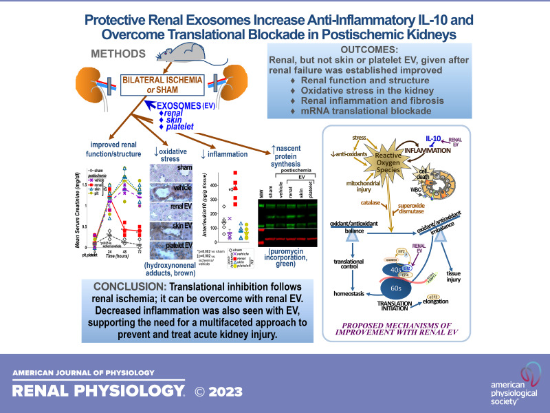
Acute kidney injury (AKI) is deadly and expensive, and specific, effective therapy remains a large unmet need. We have demonstrated the beneficial effects of transplanted adult tubular cells and extracellular vesicles (EVs; exosomes) derived from those renal cells on experimental ischemic AKI, even when administered after renal failure is established. To further examine the mechanisms of benefit with renal EVs, we tested the hypothesis that EVs from other epithelia or platelets (a rich source of EVs) might be protective, using a well-characterized ischemia-reperfusion model. When given after renal failure was present, renal EVs, but not those from skin or platelets, markedly improved renal function and histology. The differential effects allowed us to examine the mechanisms of benefit with renal EVs. We found significant decreases in oxidative stress postischemia in the renal EV-treated group with preservation of renal superoxide dismutase and catalase as well as increases in anti-inflammatory interleukin-10. In addition, we propose a novel mechanism of benefit: renal EVs enhanced nascent peptide synthesis following hypoxia in cells and in postischemic kidneys. Although EVs have been used therapeutically, these results serve as "proof of principle" to examine the mechanisms of injury and protection.NEW & NOTEWORTHY Acute kidney injury is common and deadly, yet the only approved treatment is dialysis. Thus, a better understanding of injury mechanisms and potential therapies is needed. We found that organ-specific, but not extrarenal, extracellular vesicles improved renal function and structure postischemia when given after renal failure occurred. Oxidative stress was decreased and anti-inflammatory interleukin-10 increased with renal, but not skin or platelet, exosomes. We also propose enhanced nascent peptide synthesis as a novel protective mechanism.

Keywords: acute kidney injury; exosomes; inflammation; protein biosynthesis; reactive oxygen species.


Conflict of interest statement

No conflicts of interest, financial or otherwise, are declared by the authors.

Figures

None
Graphical abstract
Figure 1.
Figure 1.
Extracellular vesicle (EV) characterization and uptake. The median diameter of the EVs derived from renal tubular and skin epithelial cells and platelets is consistent with EVs (A and D). The yield was nonsignificantly greater from platelets than epithelial cells (A). EVs were fluorescently labeled (red) to assess uptake into cultured renal tubular epithelial cells (B). The uptake of an equivalent number of EVs was not significantly different among the three groups of EVs examined. Nuclei were counterstained with Hoechst (blue). The different groups of EVs expressed the marker CD63 (with a representative immunoblot shown in C) at similar levels. D: the size distribution of EVs by nanotracker. Values are means ± SE. Statistical analysis used ANOVA. n = 4/group. Scale bar = 30 µm. Original magnification: ×200. MW, molecular weight marker.
Figure 2.
Figure 2.
Hypoxia and reoxygenation in renal tubular cells. To assess the protective effects of extracellular vesicles (EVs), we first subjected renal tubular cells to hypoxia (<1% O2, 24 h) followed by reoxygenation (24 h) with or without EVs. Live cells were labeled with calcein (green) and dead cells were labeled with propidium (red; A). Quantification showed no significant cell death with normoxia and large increases following hypoxia and reoxygenation, which were significantly limited when renal EVs were added at the time of reoxygenation. Although readily accessible, neither skin epithelial nor platelet EVs provided benefit (B). To assess the mechanism of protection, reactive oxygen species (ROS) were quantified (as fluorescence of the oxidant product 2′,7′-dichlorfluorescein) in cultured renal tubular cells. The ROS generation seen following hypoxia was largely prevented with renal but not skin or platelet EVs (C). We also found that renal EVs contain more catalase (cat) and superoxide dismutase (sod) than platelet or skin EVs (with a representative immunoblot shown in D), as quantified in E. In this and subsequent figures, individual values are presented with means and SEs shown as horizontal and vertical bars, respectively. For cell death and ROS, *P < 0.001 vs. normoxia and §P < 0.005 vs. hypoxia/vehicle. For immunoblots, †P < 0.02 vs. renal EVs. Statistical analysis used ANOVA. n = 9 for cell culture experiments and n = 5 for immunoblots. Scale bar = 50 µm. Original magnification: ×200. MW, molecular weight marker.
Figure 3.
Figure 3.
Renal function and structure. A: serum creatinine levels were stable in sham rats but increased rapidly in rats after bilateral renal artery occlusion for 50 min [ischemia (Isch); time 0] and remained elevated for at least another 24 h (48 h). Rapid improvement was seen in the group that received renal extracellular vesicles (EVs) 24 h following ischemia when renal failure was established. Although skin and platelet (plt) EVs are readily accessible, they did not provide the benefits of renal EVs. Renal tubular architecture was also preserved in the renal EV group (B). C−G: representative periodic acid Schiff-stained sections. Extensive injury with tubular casts (†) and dilation/simplification (‡) was seen postischemia (D). This was much less severe in the renal EV group (E). *P < 0.05 vs. ischemia/vehicle. Statistical analysis used ANOVA. n = 5/group. Scale bar = 100 µm. Original magnification: ×200.
Figure 4.
Figure 4.
Oxidative stress in the kidney. Oxidative stress has been shown to be one mechanism of injury in acute kidney injury. Representative images of renal 4-hydroxynonenal (HNE) adducts and quantification as well as levels of catalase and superoxide dismutase in the kidney are shown. Catalase and superoxide dismutase were significantly decreased following untreated renal ischemia (Isch) and improved in renal but not platelet (plt) or skin extracellular vesicles (EV)-treated groups (A). HNE adducts (brown; quantified in B) were undetectable after sham surgery but were extensive in untreated postischemic kidneys and were seen in both areas of tubular dilation (†) and intact epithelium (‡). Renal EVs prevented HNE adduct formation. With skin or platelet EVs, the level of HNE adducts was not significantly different than in the untreated postischemia group (C−G). *P < 0.002 vs. sham; §P < 0.05 vs. ischemia/vehicle. Statistical analysis used ANOVA. n = 5/group. Scale bar = 100 µm. Original magnification: ×200.
Figure 5.
Figure 5.
Fibrosis. To examine the effect of extracellular vesicles (EVs) on the kidney following resolution of renal failure, sections were stained with trichrome to examine fibrosis. Six days following untreated renal ischemia (Isch; C), fibrosis (blue, examples at *) was extensive, replacing normal (red) tissue, compared with sham surgery (B). In the renal EV group, there was significantly less fibrosis (D), consistent with a sustained benefit of EVs. In contrast, there were no significant differences in fibrosis among the untreated ischemia and skin (E) and platelet (plt; F) EV groups. Quantification is shown in the graph in A. *P < 0.001 vs. sham; §P < 0.001 vs. ischemia/vehicle (veh). Statistical analysis used ANOVA. n = 5/group. Scale bar = 50 µm. Original magnification: ×200.
Figure 6.
Figure 6.
Renal inflammation. Representative kidney sections stained for neutrophils (brown; AE) and complement component C3 (red; FJ) are shown. Nuclei were counterstained with hematoxylin and Hoechst, respectively. Arrows indicate neutrophils, arrowheads indicate C3 in tubules, and double arrows indicate C3 in glomeruli. Quantification is shown in K and L. Tissue myeloperoxidase (MPO) activity (M), interleukin-10 (N), and intercellular adhesion molecule-1 (ICAM) transcript levels (O) were also quantified. *P < 0.002 and †P < 0.04 vs. sham; ‡P < 0.002 and §P < 0.03 vs. ischemia (Isch)/vehicle (veh). Statistical analysis used ANOVA. n = 5/group. Scale bar = 100 µm. Original magnification: ×200. plt, platelet; hpf, high-power field.
Figure 7.
Figure 7.
Nascent peptide synthesis. To test the hypothesis that extracellular vesicles (EVs) would enhance nascent protein synthesis needed for renal recovery, incorporation of administered puromycin was assessed using anti-puromycin antibody (green). In cultured renal tubular cells, puromycin was incorporated into nascent peptides at a basal level in normoxic cells (A). Nascent peptide synthesis decreased following hypoxia and reoxygenation (B) and increased in the renal EV group (C). Puromycin incorporation was not improved with skin EVs (D) and was slightly higher with platelet (plt) EVs (E), but the difference did not reach statistical significance (L). To further assess the role of new peptide synthesis, cells were incubated with the protein synthesis inhibitor cycloheximide (cyclohex) for 0 min (FH), 30 min (I), 60 min (J), or 120 min (K) minutes under normoxic conditions (F), hypoxic conditions (G), or hypoxia with renal EV (H−K) conditions with cell death (red) quantified in M. Representative immunoblot (N, puromycin, green; actin, red) and quantification (O) of new peptide synthesis in kidney tissue showed similar results as in cells. Molecular analyses of translation initiation [eukaryotic initiation factor (eIF)2 and eiF4, growth arrest and DNA damage-inducible 34 (GADD34), and polyadenylate binding protein cytosolic 1 (PABPC1)] and elongation [eukaryotic elongation factor 2 (eEF2)] transcripts are shown in P, and a proposed schema for the influence of postischemia inflammation and oxidative stress on protein homeostasis is shown in Q. For puromycin fluorescence in cells, *P < 0.04 vs. normoxia and †P < 0.01 vs. all other groups. For cell death, *P < 0.04 vs. normoxia, †P < 0.03 vs. hypoxia, ‡P < 0.008 vs. hypoxia/renal EVs, and §P < 0.04 vs. hypoxia/renal EVs/cycloheximide (30 min). For kidneys, *P < 0.0002 vs. all other groups. Statistical analysis used ANOVA. n = 8/group for cells and n = 5/group for immunoblots. Scale bar = 50 µm. Original magnification: ×200. Isch, ischemia; MW, molecular weight markers; 40S and 60S, ribosomal subunits.
Figure A1.
Figure A1.
Origin of renal tubular cells. Cultured renal cells were probed for the tubule segment markers organic anion transporter-1, aquaporin 2, and Dolichos biflorus lectin as well as the epithelial cell marker pan-cytokeratin (all red).
Figure A2.
Figure A2.
Additional functional measures. Quantification of creatinine clearance and albuminuria in the 5 groups is shown. *P < 0.02 vs. sham and †P < 0.03 vs. ischemia/vehicle. Statistical analysis used ANOVA. n = 5/group. EVs, extracellular vesicles; plt, platelet.

Similar articles

References

    1. Faubel S, Chawla LS, Chertow GM, Goldstein SL, Jaber BL, Liu KD; Acute Kidney Injury Advisory Group of the American Society of Nephrology. Ongoing clinical trials in AKI. Clin J Am Soc Nephrol 7: 861–873, 2012. doi:10.2215/CJN.12191111. - DOI - PubMed
    1. Hukriede NA, Soranno DE, Sander V, Perreau T, Starr MC, Yuen PST, Siskind LJ, Hutchens MP, Davidson AJ, Burmeister DM, Faubel S, de Caestecker MP. Experimental models of acute kidney injury for translational research. Nat Rev Nephrol 18: 277–293, 2022. doi:10.1038/s41581-022-00539-2. - DOI - PubMed
    1. Hoste EAJ, Kellum JA, Selby NM, Zarbock A, Palevsky PM, Bagshaw SM, Goldstein SL, Cerdá J, Chawla LS. Global epidemiology and outcomes of acute kidney injury. Nat Rev Nephrol 14: 607–625, 2018. doi:10.1038/s41581-018-0052-0. - DOI - PubMed
    1. Susantitaphong P, Cruz DN, Cerda J, Abulfaraj M, Alqahtani F, Koulouridis I, Jaber BL; Acute Kidney Injury Advisory Group of the American Society of Nephrology. World incidence of AKI: a meta-analysis. Clin J Am Soc Nephrol 8: 1482–1493, 2013. [Erratum in Clin J Am Soc Nephrol 9: 1148, 2014]. doi:10.2215/CJN.00710113. - DOI - PMC - PubMed
    1. Saran R, Li Y, Robinson B, Abbott KC, Agodoa LY, Ayanian J, et al. . US Renal Data System 2015 annual data report: epidemiology of kidney disease in the United States. Am J Kidney Dis 67, Suppl 1: S1–S305, 2016. doi:10.1053/j.ajkd.2015.12.014. - DOI - PMC - PubMed

Publication types

LinkOut - more resources