. 2023 Sep 1;41(25):4164-4177.
doi: 10.1200/JCO.23.00570. Epub 2023 Jun 15.

Relapse Timing Is Associated With Distinct Evolutionary Dynamics in Diffuse Large B-Cell Lymphoma

Affiliations

Relapse Timing Is Associated With Distinct Evolutionary Dynamics in Diffuse Large B-Cell Lymphoma

Laura K Hilton et al. J Clin Oncol. .

Abstract

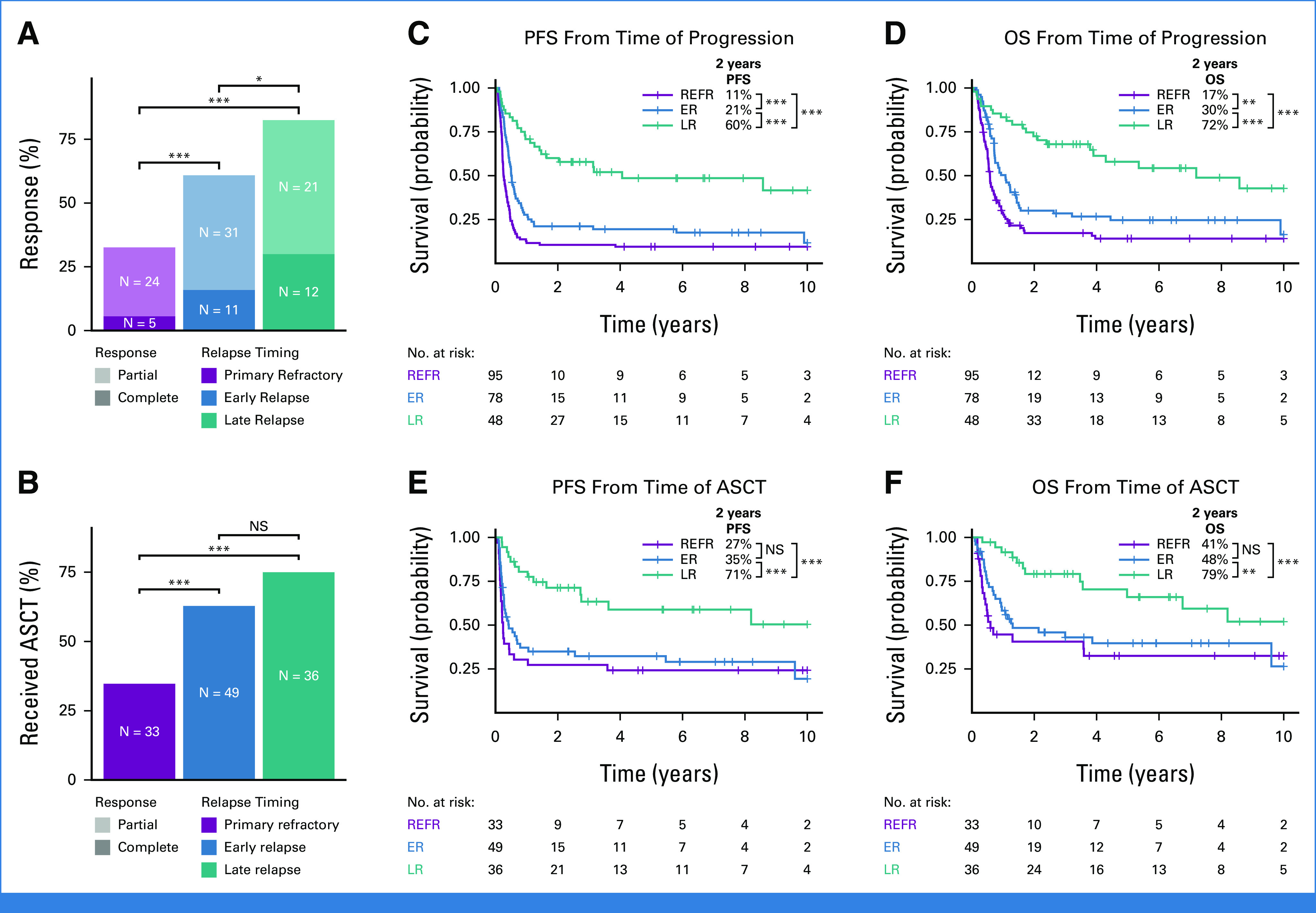
Purpose: Diffuse large B-cell lymphoma (DLBCL) is cured in more than 60% of patients, but outcomes remain poor for patients experiencing disease progression or relapse (refractory or relapsed DLBCL [rrDLBCL]), particularly if these events occur early. Although previous studies examining cohorts of rrDLBCL have identified features that are enriched at relapse, few have directly compared serial biopsies to uncover biological and evolutionary dynamics driving rrDLBCL. Here, we sought to confirm the relationship between relapse timing and outcomes after second-line (immuno)chemotherapy and determine the evolutionary dynamics that underpin that relationship.

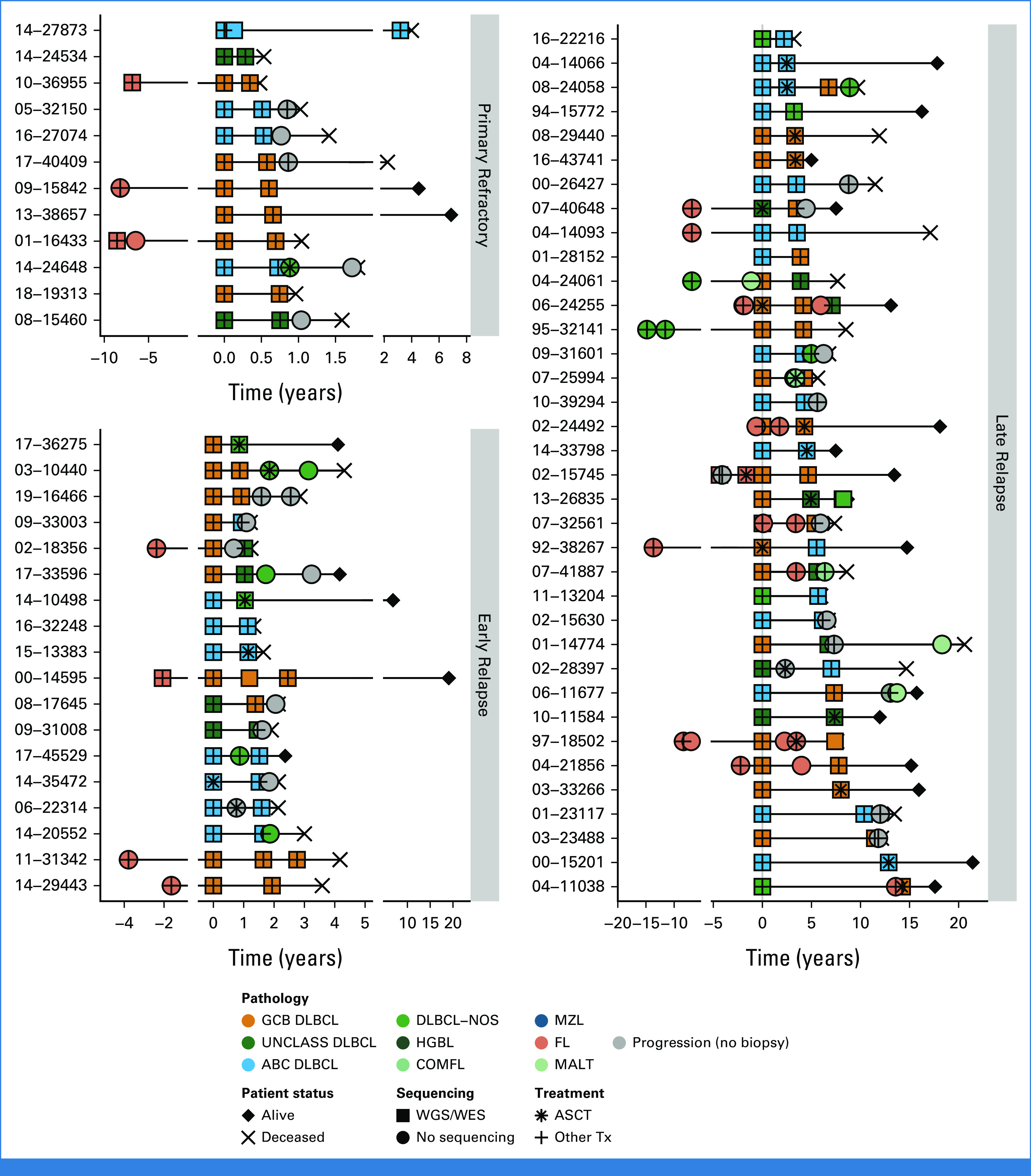
Patients and methods: Outcomes were examined in a population-based cohort of 221 patients with DLBCL who experienced progression/relapse after frontline treatment and were treated with second-line (immuno)chemotherapy with an intention-to-treat with autologous stem-cell transplantation (ASCT). Serial DLBCL biopsies from a partially overlapping cohort of 129 patients underwent molecular characterization, including whole-genome or whole-exome sequencing in 73 patients.

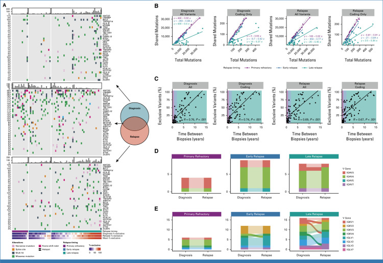
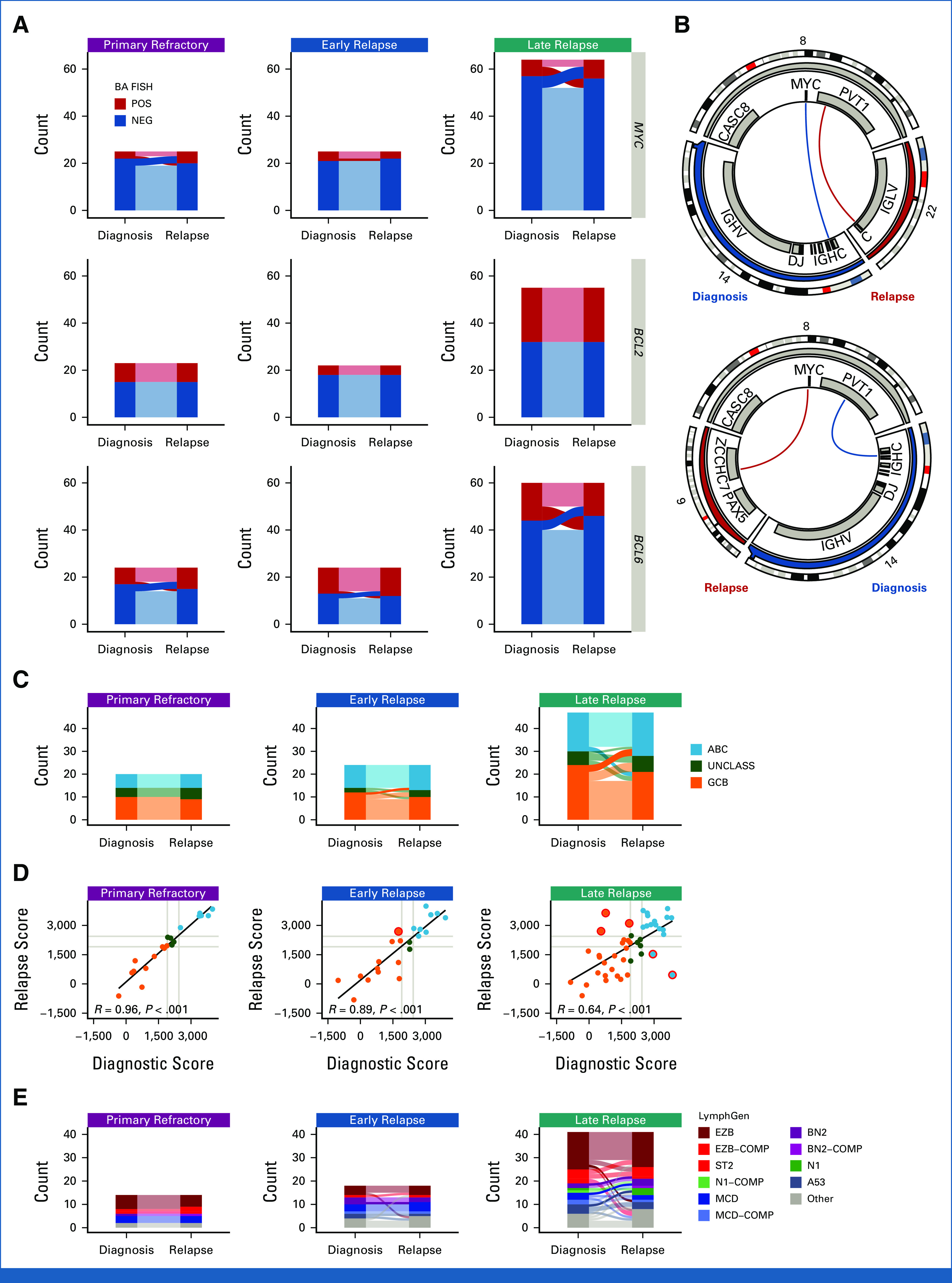
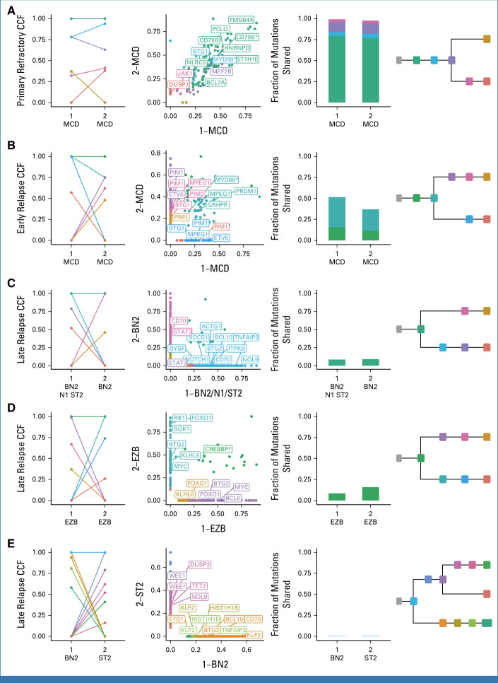
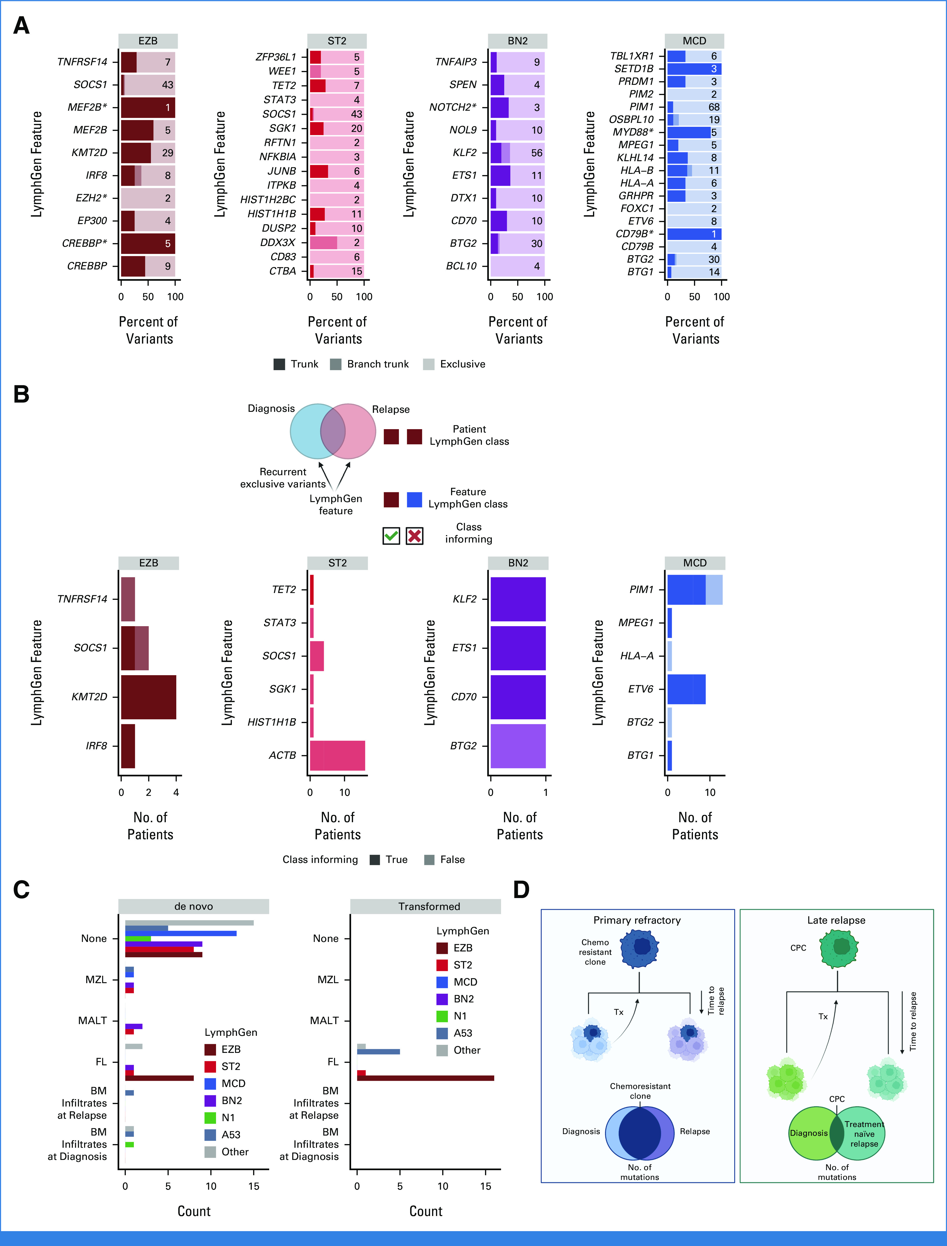
Results: Outcomes to second-line therapy and ASCT are superior for late relapse (>2 years postdiagnosis) versus primary refractory (<9 months) or early relapse (9-24 months). Diagnostic and relapse biopsies were mostly concordant for cell-of-origin classification and genetics-based subgroup. Despite this concordance, the number of mutations exclusive to each biopsy increased with time since diagnosis, and late relapses shared few mutations with their diagnostic counterpart, demonstrating a branching evolution pattern. In patients with highly divergent tumors, many of the same genes acquired new mutations independently in each tumor, suggesting that the earliest mutations in a shared precursor cell constrain tumor evolution toward the same genetics-based subgroups at both diagnosis and relapse.

Conclusion: These results suggest that late relapses commonly represent genetically distinct and chemotherapy-naïve disease and have implications for optimal patient management.


Conflict of interest statement

The following represents disclosure information provided by authors of this manuscript. All relationships are considered compensated unless otherwise noted. Relationships are self-held unless noted. I = Immediate Family Member, Inst = My Institution. Relationships may not relate to the subject matter of this manuscript. For more information about ASCO's conflict of interest policy, please refer to www.asco.org/rwc or ascopubs.org/jco/authors/author-center.

Open Payments is a public database containing information reported by companies about payments made to US-licensed physicians (Open Payments).

Figures

FIG 1.
FIG 1.
Relationship between relapse timing and outcomes to second-line therapy. (A and B) The percent of patients in each relapse timing category (A) whose relapse responded to second-line therapy and (B) who received ASCT. Groups were compared with pairwise Fisher's exact tests. (C-F) Kaplan-Meier curves showing PFS and OS from the time of progression or ASCT. P values were determined with a log-rank test. *P < .05; **P < .01; ***P < .001. ASCT, autologous stem-cell transplantation; ER, early relapse; LR, late relapse; NS, not significant; OS, overall survival; PFS, progression-free survival; REFR, primary refractory.
FIG 2.
FIG 2.
Sequencing cohort patient histories. Disease and treatment histories for all known biopsies and progression time points for patients for which WGS/WES data were generated, distributed according to relapse timing categories. Five patients were omitted because of incomplete histories. DLBCL tumors are colored according to NanoString cell-of-origin where available or are otherwise labeled DLBCL-NOS. ABC, activated B-cell-like DLBCL; ASCT, autologous stem-cell transplantation; COMFL, composite lymphoma with areas of DLBCL and FL morphology; DLBCL, diffuse large B-cell lymphoma; FL, follicular lymphoma; GCB, germinal center B-cell-like DLBCL; HGBL, high-grade B-cell lymphoma; MALT, extranodal MZL of mucosa-associated lymphoid tissue; MZL, marginal zone lymphoma; NOS, not otherwise specified; PROG, clinical progression without a biopsy; Tx, treatment; UNCLASS, unclassified DLBCL; WES, whole-exome sequencing; WGS, whole-genome sequencing.
FIG 3.
FIG 3.
Patterns of evolution in diagnostic and relapse tumor pairs. (A) Oncoplots of variants identified exclusively at diagnosis (top), relapse (bottom), or shared between biopsies (middle), highlighting the most frequently mutated genes involved in LymphGen classification. Patients are stratified by relapse timing and ordered by the mean percentage of exclusive variants. Barplots indicate the number of coding mutations present per patient in each mutation subset. (B) The relationship between total variants (all or coding only) at diagnosis or relapse versus the number of mutations shared between tumors. The dashed gray line represents the line of unity. (C) The percent of variants exclusive to either diagnostic or relapse tumors as a function of time between biopsies. R represents the Pearson correlation coefficient. (D) Concordance of heavy chain and (E) light chain V gene usage derived from RNAseq data for tumor pairs colored by V gene subgroup. Light chain rearrangements were more frequently discordant, which may suggest ongoing receptor editing. In all plots, alluvia connecting each tumor pair are opaque for discordant pairs and translucent for concordant pairs. N.B. Where V gene usage was discordant but both V genes belong to the same subgroup, the color is consistent across time points.
FIG 4.
FIG 4.
Comparison of structural variants and gene expression profiling and genetic classifications between biopsies. (A) Concordance of BA-FISH results between diagnosis and first relapse for MYC, BCL2, and BCL6 translocations. (B) Circos plots showing discordant MYC translocations in two patients who experienced late relapse. Top: a tumor pair that was positive for BA-FISH at both timepoints; bottom: a tumor pair that was BA-FISH positive at diagnosis and negative at relapse. (C) Alluvial comparison of COO classifications in diagnostic/relapse pairs stratified by relapse timing. Frank discordance (ABC to GCB or vice versa) is indicated by opaque alluvia. (D) A scatter plot comparing DLBCL90 COO scores across tumor pairs. Red circles highlight frank discordance in COO classification. R values indicate Pearson correlation coefficient. (E) Comparison of LymphGen classifications between tumor pairs. Frank discordance (a switch between two mutually exclusive non-other classifications) is emphasized with opaque alluvia. ABC, activated B-cell-like DLBCL; BA, break-apart; COMP, composite; COO, cell-of-origin; FISH, fluorescence in situ hybridization; GCB, germinal center B-cell-like DLBCL; NEG, negative; POS, positive; UNCLASS, unclassified DLBCL.
FIG 5.
FIG 5.
Representative phylogenetic reconstructions. (A-E) Each row of plots displays data for a single patient. Tumors are labeled according to order of occurrence and LymphGen classification. Subclones are colored consistently across all plots for each patient. From left to right: CCF of subclones estimated by PhyClone; VAF of each variant as a scatter plot with the diagnostic tumor on the x-axis and relapse tumor on the y-axis with selected genes labeled; the fraction of mutations shared between both tumors (ie, all mutations in a cluster with a CCF > 0.1); and the inferred phylogenetic relationship between tumors. Hotspot mutations at MYD88 L265P and CD79B Y179 and missense mutations in the CREBBP lysine acetyltransferase (KAT) domain are indicated with an asterisk. The VAF scatter plots without gene labels are also presented in the Data Supplement (Supplemental Fig 9). CCF, cancer cell fraction; VAF, variant allele frequency.
FIG 6.
FIG 6.
Classification features in divergent tumor pairs. (A) LymphGen classification features that were mutated in two or more tumors from the same patient. Trunk variants (darkest) are identical in all tumors from the same patient; branch trunk are variants not shared across all tumors but are common between at least two; and exclusive are those found in only one tumor. Numbers on each bar represent the total number of variants considered in each feature. (B) LymphGen classification features that acquired exclusive variants in two or more tumors from the same patient. Class informing indicates that the mutations arose in patients in which LymphGen classification matched the class association of the acquired variants. (C) Patient LymphGen classifications stratified according to associated low-grade lymphoma entities. Transformed indicates low-grade disease preceded the first DLBCL diagnosis while de novo indicates that the low-grade diagnosis was made after DLBCL diagnosis. (D) A model of the relationship between relapse timing, evolutionary patterns, and outcomes. BM, bone marrow; CPC, common precursor cell; DLBCL, diffuse large B-cell lymphoma; FL, follicular lymphoma; MALT, extranodal MZL of mucosa-associated lymphoid tissue; MZL, marginal zone lymphoma; Tx, treatment. Created with BioRender.com.

Update of

Similar articles

Cited by

References

    1. Sehn LH, Salles G. Diffuse large B-cell lymphoma. N Engl J Med. 2021;384:842–858. - PMC - PubMed
    1. Rovira J, Valera A, Colomo L, et al. Prognosis of patients with diffuse large B cell lymphoma not reaching complete response or relapsing after frontline chemotherapy or immunochemotherapy. Ann Hematol. 2015;94:803–812. - PMC - PubMed
    1. Crump M, Neelapu SS, Farooq U, et al. Outcomes in refractory diffuse large B-cell lymphoma: Results from the international SCHOLAR-1 study. Blood. 2017;130:1800–1808. - PMC - PubMed
    1. Morin RD, Mendez-Lago M, Mungall AJ, et al. Frequent mutation of histone-modifying genes in non-Hodgkin lymphoma. Nature. 2011;476:298–303. - PMC - PubMed
    1. Pasqualucci L, Trifonov V, Fabbri G, et al. Analysis of the coding genome of diffuse large B-cell lymphoma. Nat Genet. 2011;43:830–837. - PMC - PubMed

Publication types

MeSH terms

LinkOut - more resources