

Free-electron interactions with van der Waals heterostructures: a source of focused X-ray radiation
- PMID: 37321995
- PMCID: PMC10272160
- DOI: 10.1038/s41377-023-01141-2
Free-electron interactions with van der Waals heterostructures: a source of focused X-ray radiation
Abstract
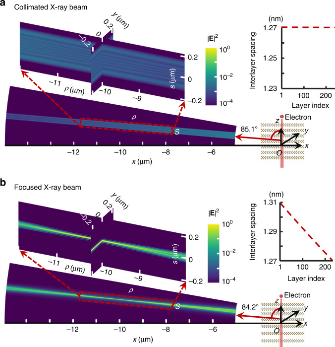
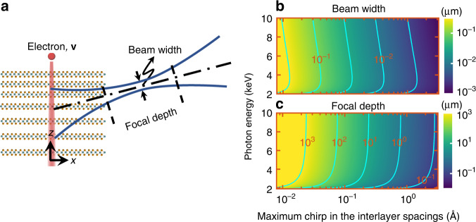
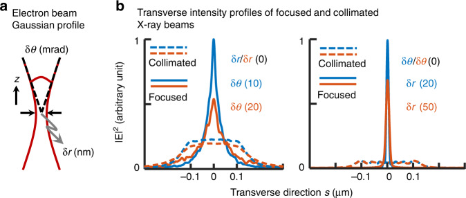
The science and technology of X-ray optics have come far, enabling the focusing of X-rays for applications in high-resolution X-ray spectroscopy, imaging, and irradiation. In spite of this, many forms of tailoring waves that had substantial impact on applications in the optical regime have remained out of reach in the X-ray regime. This disparity fundamentally arises from the tendency of refractive indices of all materials to approach unity at high frequencies, making X-ray-optical components such as lenses and mirrors much harder to create and often less efficient. Here, we propose a new concept for X-ray focusing based on inducing a curved wavefront into the X-ray generation process, resulting in the intrinsic focusing of X-ray waves. This concept can be seen as effectively integrating the optics to be part of the emission mechanism, thus bypassing the efficiency limits imposed by X-ray optical components, enabling the creation of nanobeams with nanoscale focal spot sizes and micrometer-scale focal lengths. Specifically, we implement this concept by designing aperiodic vdW heterostructures that shape X-rays when driven by free electrons. The parameters of the focused hotspot, such as lateral size and focal depth, are tunable as a function of an interlayer spacing chirp and electron energy. Looking forward, ongoing advances in the creation of many-layer vdW heterostructures open unprecedented horizons of focusing and arbitrary shaping of X-ray nanobeams.
© 2023. The Author(s).
Conflict of interest statement
The authors declare no competing interests.
Figures


Similar articles
-
Multicolor x-rays from free electron-driven van der Waals heterostructures.Sci Adv. 2023 Dec;9(48):eadj8584. doi: 10.1126/sciadv.adj8584. Epub 2023 Dec 1. Sci Adv. 2023. PMID: 38039369 Free PMC article.
-
Enhanced Versatility of Table-Top X-Rays from Van der Waals Structures.Adv Sci (Weinh). 2022 May;9(16):e2105401. doi: 10.1002/advs.202105401. Epub 2022 Mar 31. Adv Sci (Weinh). 2022. PMID: 35355443 Free PMC article.
-
Predicting Van der Waals Heterostructures by a Combined Machine Learning and Density Functional Theory Approach.ACS Appl Mater Interfaces. 2022 Jun 8;14(22):25907-25919. doi: 10.1021/acsami.2c04403. Epub 2022 May 27. ACS Appl Mater Interfaces. 2022. PMID: 35622945
-
Recent Advances on Tuning the Interlayer Coupling and Properties in van der Waals Heterostructures.Small. 2022 Apr;18(15):e2105877. doi: 10.1002/smll.202105877. Epub 2022 Jan 19. Small. 2022. PMID: 35044721 Review.
-
Unconventional van der Waals heterostructures beyond stacking.iScience. 2021 Aug 28;24(9):103050. doi: 10.1016/j.isci.2021.103050. eCollection 2021 Sep 24. iScience. 2021. PMID: 34553135 Free PMC article. Review.
Cited by
-
Shaping free-electron radiation via van der Waals heterostructures.Light Sci Appl. 2023 Jul 28;12(1):187. doi: 10.1038/s41377-023-01221-3. Light Sci Appl. 2023. PMID: 37507415 Free PMC article.
-
Transverse recoil imprinted on free-electron radiation.Nat Commun. 2024 Sep 6;15(1):7803. doi: 10.1038/s41467-024-52050-w. Nat Commun. 2024. PMID: 39242627 Free PMC article.
-
Multicolor x-rays from free electron-driven van der Waals heterostructures.Sci Adv. 2023 Dec;9(48):eadj8584. doi: 10.1126/sciadv.adj8584. Epub 2023 Dec 1. Sci Adv. 2023. PMID: 38039369 Free PMC article.
References
-
- Sakdinawat A, Attwood D. Nanoscale X-ray imaging. Nat. Photonics. 2010;4:840–848. doi: 10.1038/nphoton.2010.267. - DOI
-
- Bostwick A, Rotenberg E, Avila J, Asensio MC. Zooming in on electronic structure: nanoARPES at SOLEIL and ALS. Synchrotron Radiat. N. 2012;25:19–25. doi: 10.1080/08940886.2012.720162. - DOI
-
- Avila J, Asensio MC. First NanoARPES user facility available at SOLEIL: an innovative and powerful tool for studying advanced materials. Synchrotron Radiat. N. 2014;27:24–30. doi: 10.1080/08940886.2014.889549. - DOI
Grants and funding
- 851780/EC | Horizon 2020 Framework Programme (EU Framework Programme for Research and Innovation H2020)
- 2018288/United States-Israel Binational Science Foundation (BSF)
- NRF2020- NRF-ISF004-3525/National Research Foundation Singapore (National Research Foundation-Prime Minister's office, Republic of Singapore)
LinkOut - more resources
Full Text Sources
