. 2023 Jun 15;14(1):3568.
doi: 10.1038/s41467-023-39335-2.

Structural insight into the human SID1 transmembrane family member 2 reveals its lipid hydrolytic activity

Affiliations

Structural insight into the human SID1 transmembrane family member 2 reveals its lipid hydrolytic activity

Dandan Qian et al. Nat Commun. .

Abstract

The systemic RNAi-defective (SID) transmembrane family member 2 (SIDT2) is a putative nucleic acid channel or transporter that plays essential roles in nucleic acid transport and lipid metabolism. Here, we report the cryo-electron microscopy (EM) structures of human SIDT2, which forms a tightly packed dimer with extensive interactions mediated by two previously uncharacterized extracellular/luminal β-strand-rich domains and the unique transmembrane domain (TMD). The TMD of each SIDT2 protomer contains eleven transmembrane helices (TMs), and no discernible nucleic acid conduction pathway has been identified within the TMD, suggesting that it may act as a transporter. Intriguingly, TM3-6 and TM9-11 form a large cavity with a putative catalytic zinc atom coordinated by three conserved histidine residues and one aspartate residue lying approximately 6 Å from the extracellular/luminal surface of the membrane. Notably, SIDT2 can hydrolyze C18 ceramide into sphingosine and fatty acid with a slow rate. The information presented advances the understanding of the structure-function relationships in the SID1 family proteins.


Conflict of interest statement

The authors declare no competing interests.

Figures
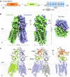
Fig. 1
Fig. 1. Overall structure of human SIDT2.
a A schematic diagram showing the domain organization of SIDT2. SP signal peptide, BRD β-sheet rich domain, TMD transmembrane domain, CTD cytoplasmic domain. The dashed line indicates the unresolved region in the structure. b The overall EM density map of the human SIDT2. SIDT2 is a homodimer, and the protomers A and B are shown in lemon and light blue, respectively. The map was contoured at 0.3 and visualized in ChimeraX (www.cgl.ucsf.edu/chimerax). c Overall structure of SIDT2. The N-linked glycans are displayed as black sticks. All structural figures were prepared in PyMOL (www.pymol.org).
Fig. 2
Fig. 2. Structural features of each domain.
a Topological diagram and overall structure showing one SIDT2 protomer. The CTD is invisible in the structure, reflecting its flexibility. b BRD1 contains eleven β-sheet folds as two lamellar structures facing each other. One disulfide bond is formed between C117 of BRD1 and C207 of BRD2, and five glycosylation sites are observed in BRD1. c BRD2 contains eight β-sheet folded into two lamellar structures facing each other. One glycosylation site and one disulfide bond are present. d The TMD contains eleven transmembrane helices containing a putative Zn2+-binding site formed by three histidine residues, one aspartate residue, and one serine residue. The putative Zn2+ is located approximately 6 Å from the extracellular/luminal surface of the membrane. Two pairs of disulfide bonds are formed in the extracellular/luminal loops. Except for TM2, the TMs are arranged counterclockwise in an orderly manner from the extracellular/luminal view. loop 2–3 indicates the loop between the TM2 and TM3, loop 10-11 indicates that the loop between the TM10 and TM11.
Fig. 3
Fig. 3. Dimer interface of SIDT2.
a The dimer interface of SIDT2 can be divided into three regions. b The first region is formed by the β1, β3, β10, β5, and β6 of the two BRD1 molecules through extensive hydrophobic interactions and one hydrogen bond. The contact hydrophobic residues are indicated. The black dashed line indicates a hydrogen bond. c The second region is formed between loop 4-5 of one protomer with that of the opposing protomer and loop 7-8 of BRD1 of the opposing protomer. The blue dashed lines indicate ionic interactions, and the gray dashed lines indicate cation-π interactions. d The third region is formed between TM2 of one protomer and TM6 and the extracellular/luminal end of TM5 of the opposing protomer.
Fig. 4
Fig. 4. SIDT2 has a Zn2+-dependent catalytic core similar to that of ACER3 and ADIPOR2.
a TM3-6 and TM9-10 of SIDT2 are aligned with TM1-7s of ACER3 and ADIPOR2. Superimposing the TMD of SITD2 with that of ACER3 and ADIPOR2 relative to the Zn2+-binding sites. b A large but distinct cavity around the Zn2+-binding site was observed in all the structures of ACER3, ADIPOR2, and SIDT2. The binding of the lipids in the cavity of the ACER3 and ADIPOR2 structures are indicated as black sticks.
Fig. 5
Fig. 5. SIDT2 exhibits ceramidase activity.
a The ceramidase activity was detected in the wild-type SIDT2 sample but not in the H796A-H800A mutant sample. The SIDT2 alone condition and the reaction buffer plus ceramide (d18:1/18:0) (C18) condition serve as controls. Detected sphingosine values are represented as the mean ± s.d. (standard deviations) of three independent measurements (n = 3). Source data are provided as a Source Data file. b Michaelis-Menten analysis of the SIDT2 ceramidase activity. Detected sphingosine values are represented as the mean ± s.d. of three independent measurements (n = 3). Source data are provided as a Source Data file. c Schematic illustration of the hydrolysis of ceramide by SIDT2.

References

    1. Fire A, et al. Potent and specific genetic interference by double-stranded RNA in Caenorhabditis elegans. Nature. 1998;391:806–811. doi: 10.1038/35888. - DOI - PubMed
    1. Tabara H, Grishok A, Mello CC. RNAi in C. elegans: soaking in the genome sequence. Science. 1998;282:430–431. doi: 10.1126/science.282.5388.430. - DOI - PubMed
    1. Timmons, L. & Fire, A. Specific interference by ingested dsRNA. Nature395, 854 (1998). - PubMed
    1. Winston WM, Molodowitch C, Hunter CP. Systemic RNAi in C. elegans requires the putative transmembrane protein SID-1. Science. 2002;295:2456–2459. doi: 10.1126/science.1068836. - DOI - PubMed
    1. Feinberg EH, Hunter CP. Transport of dsRNA into cells by the transmembrane protein SID-1. Science. 2003;301:1545–1547. doi: 10.1126/science.1087117. - DOI - PubMed

Publication types

LinkOut - more resources