

Bacteria evolve macroscopic multicellularity by the genetic assimilation of phenotypically plastic cell clustering
- PMID: 37322016
- PMCID: PMC10272148
- DOI: 10.1038/s41467-023-39320-9
Bacteria evolve macroscopic multicellularity by the genetic assimilation of phenotypically plastic cell clustering
Abstract
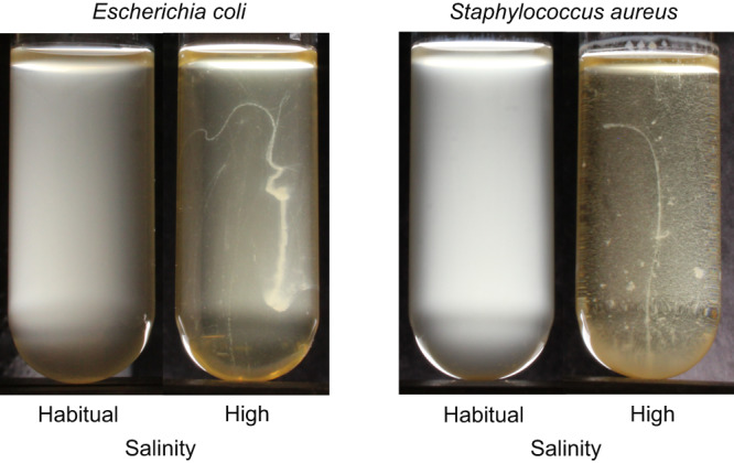
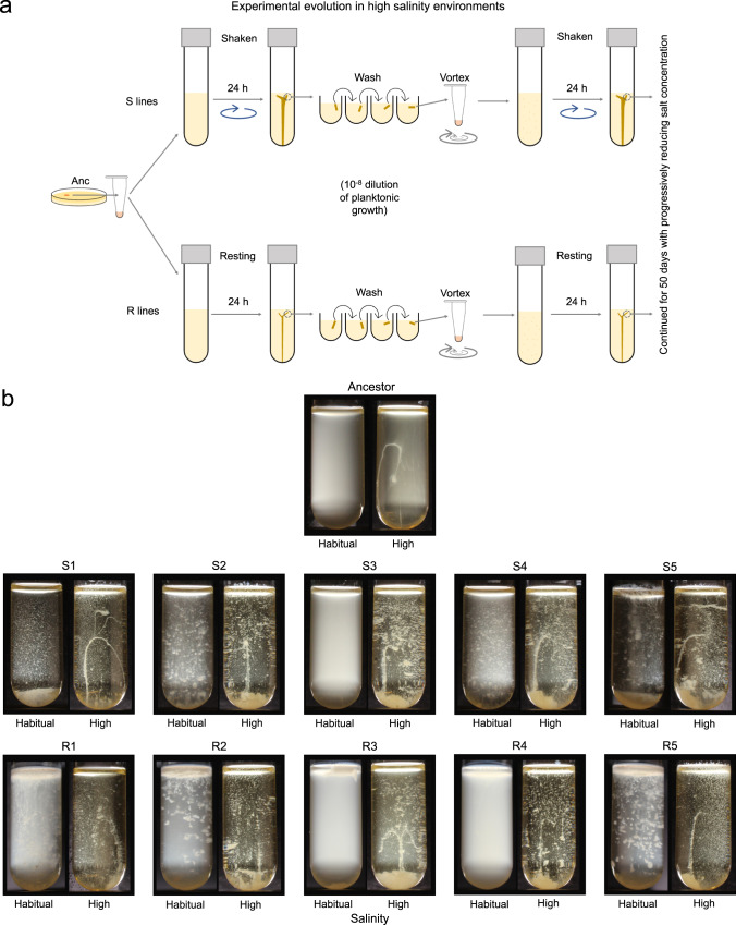
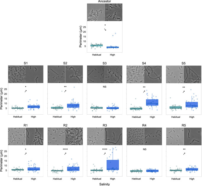
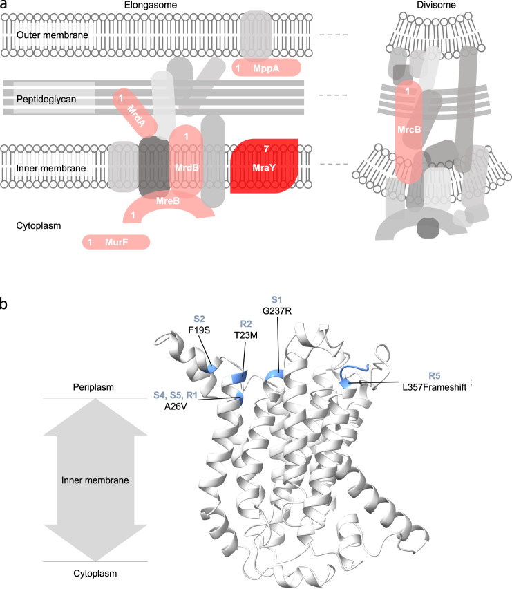
The evolutionary transition from unicellularity to multicellularity was a key innovation in the history of life. Experimental evolution is an important tool to study the formation of undifferentiated cellular clusters, the likely first step of this transition. Although multicellularity first evolved in bacteria, previous experimental evolution research has primarily used eukaryotes. Moreover, it focuses on mutationally driven (and not environmentally induced) phenotypes. Here we show that both Gram-negative and Gram-positive bacteria exhibit phenotypically plastic (i.e., environmentally induced) cell clustering. Under high salinity, they form elongated clusters of ~ 2 cm. However, under habitual salinity, the clusters disintegrate and grow planktonically. We used experimental evolution with Escherichia coli to show that such clustering can be assimilated genetically: the evolved bacteria inherently grow as macroscopic multicellular clusters, even without environmental induction. Highly parallel mutations in genes linked to cell wall assembly formed the genomic basis of assimilated multicellularity. While the wildtype also showed cell shape plasticity across high versus low salinity, it was either assimilated or reversed after evolution. Interestingly, a single mutation could genetically assimilate multicellularity by modulating plasticity at multiple levels of organization. Taken together, we show that phenotypic plasticity can prime bacteria for evolving undifferentiated macroscopic multicellularity.
© 2023. The Author(s).
Conflict of interest statement
The authors declare no competing interests.
Figures
Similar articles
-
Stabilizing multicellularity through ratcheting.Philos Trans R Soc Lond B Biol Sci. 2016 Aug 19;371(1701):20150444. doi: 10.1098/rstb.2015.0444. Philos Trans R Soc Lond B Biol Sci. 2016. PMID: 27431522 Free PMC article.
-
Adaptive evolutionary trajectories in complexity: Transitions between unicellularity and facultative differentiated multicellularity.Proc Natl Acad Sci U S A. 2025 Jan 28;122(4):e2411692122. doi: 10.1073/pnas.2411692122. Epub 2025 Jan 22. Proc Natl Acad Sci U S A. 2025. PMID: 39841150 Free PMC article.
-
A nonadaptive explanation for macroevolutionary patterns in the evolution of complex multicellularity.Proc Natl Acad Sci U S A. 2024 Feb 13;121(7):e2319840121. doi: 10.1073/pnas.2319840121. Epub 2024 Feb 5. Proc Natl Acad Sci U S A. 2024. PMID: 38315855 Free PMC article.
-
Why have aggregative multicellular organisms stayed simple?Curr Genet. 2021 Dec;67(6):871-876. doi: 10.1007/s00294-021-01193-0. Epub 2021 Jun 10. Curr Genet. 2021. PMID: 34114051 Review.
-
Diversity of 'simple' multicellular eukaryotes: 45 independent cases and six types of multicellularity.Biol Rev Camb Philos Soc. 2023 Dec;98(6):2188-2209. doi: 10.1111/brv.13001. Epub 2023 Jul 20. Biol Rev Camb Philos Soc. 2023. PMID: 37475165 Review.
Cited by
-
Nutrient-rich spatial refuges buffer against extinction and promote evolutionary rescue in evolving microbial populations.Proc Biol Sci. 2024 Dec;291(2036):20242197. doi: 10.1098/rspb.2024.2197. Epub 2024 Dec 11. Proc Biol Sci. 2024. PMID: 39657803 Free PMC article.
-
Metabolically driven flows enable exponential growth in macroscopic multicellular yeast.Sci Adv. 2025 Jun 20;11(25):eadr6399. doi: 10.1126/sciadv.adr6399. Epub 2025 Jun 20. Sci Adv. 2025. PMID: 40540574 Free PMC article.
-
Evolutionary dynamics of nascent multicellular lineages.Proc Biol Sci. 2025 Apr;292(2045):20241195. doi: 10.1098/rspb.2024.1195. Epub 2025 Apr 30. Proc Biol Sci. 2025. PMID: 40300626 Free PMC article.
-
Metabolically-driven flows enable exponential growth in macroscopic multicellular yeast.bioRxiv [Preprint]. 2024 Jun 22:2024.06.19.599734. doi: 10.1101/2024.06.19.599734. bioRxiv. 2024. Update in: Sci Adv. 2025 Jun 20;11(25):eadr6399. doi: 10.1126/sciadv.adr6399. PMID: 38948761 Free PMC article. Updated. Preprint.
-
Multicellular magnetotactic bacteria are genetically heterogeneous consortia with metabolically differentiated cells.PLoS Biol. 2024 Jul 11;22(7):e3002638. doi: 10.1371/journal.pbio.3002638. eCollection 2024 Jul. PLoS Biol. 2024. PMID: 38990824 Free PMC article.
References
-
- Maynard Smith, J. & Szathmáry, E. The major transitions in evolution (Freeman, 1995).
-
- Michod, R. E. Darwinian dynamics: evolutionary transitions in fitness and individuality (Princeton University Press, 1999).
-
- Grosberg RK, Strathmann RR. The evolution of multicellularity: a minor major transition? Annu. Rev. Ecol. Evol. Syst. 2007;38:621–654. doi: 10.1146/annurev.ecolsys.36.102403.114735. - DOI
Publication types
MeSH terms
LinkOut - more resources
Full Text Sources
Molecular Biology Databases
