. 2023 Jun 15;14(1):3560.
doi: 10.1038/s41467-023-39281-z.

CNK2 promotes cancer cell motility by mediating ARF6 activation downstream of AXL signalling

Affiliations

CNK2 promotes cancer cell motility by mediating ARF6 activation downstream of AXL signalling

Guillaume Serwe et al. Nat Commun. .

Abstract

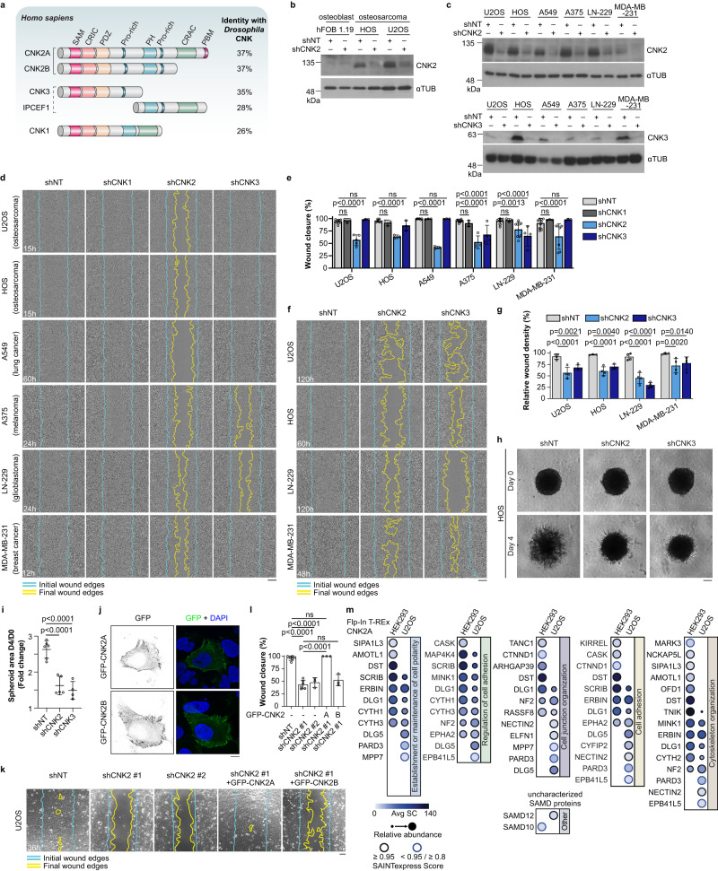
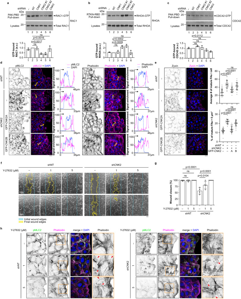
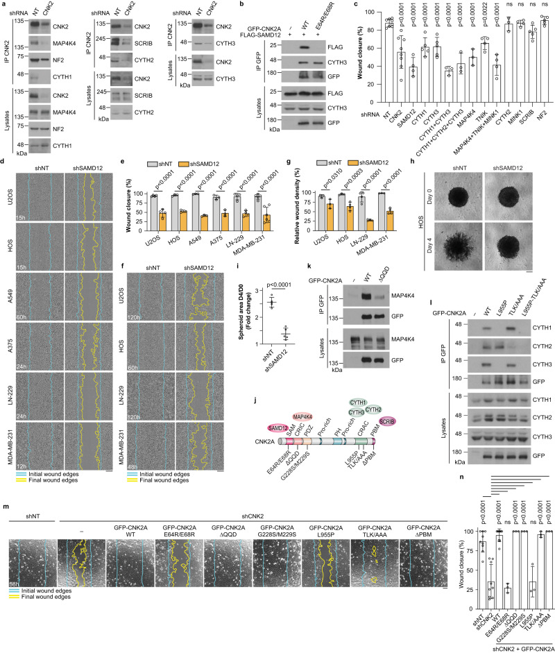
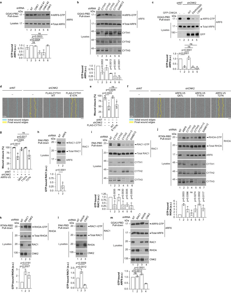
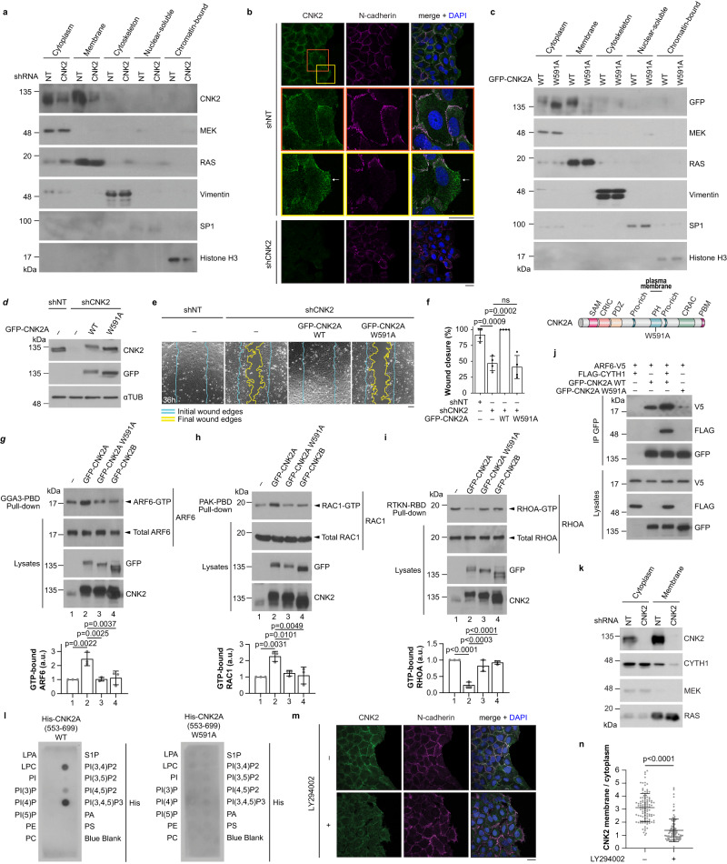
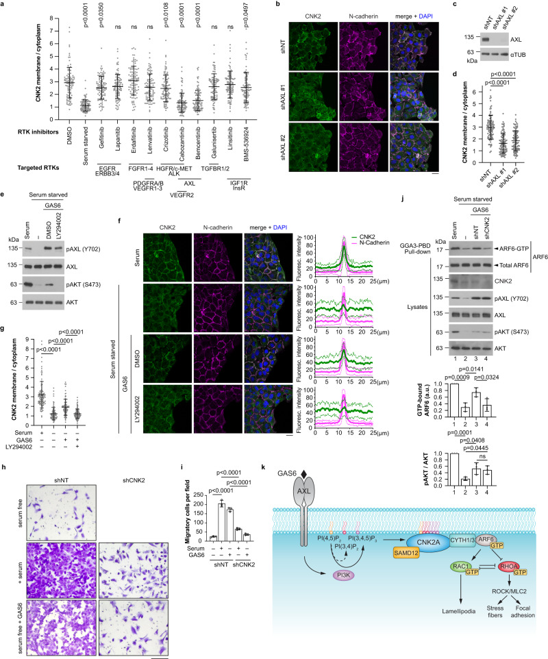
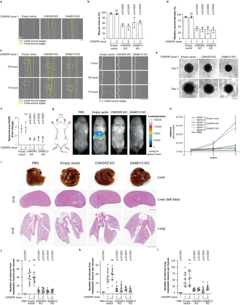
Cell motility is a critical feature of invasive tumour cells that is governed by complex signal transduction events. Particularly, the underlying mechanisms that bridge extracellular stimuli to the molecular machinery driving motility remain partially understood. Here, we show that the scaffold protein CNK2 promotes cancer cell migration by coupling the pro-metastatic receptor tyrosine kinase AXL to downstream activation of ARF6 GTPase. Mechanistically, AXL signalling induces PI3K-dependent recruitment of CNK2 to the plasma membrane. In turn, CNK2 stimulates ARF6 by associating with cytohesin ARF GEFs and with a novel adaptor protein called SAMD12. ARF6-GTP then controls motile forces by coordinating the respective activation and inhibition of RAC1 and RHOA GTPases. Significantly, genetic ablation of CNK2 or SAMD12 reduces metastasis in a mouse xenograft model. Together, this work identifies CNK2 and its partner SAMD12 as key components of a novel pro-motility pathway in cancer cells, which could be targeted in metastasis.


Conflict of interest statement

The authors declare no competing interests.

Figures

Fig. 1
Fig. 1. CNK2 knockdown delays cell migration and invasion of multiple cancer cell lines.
a Schematic representation of human CNK proteins. CNK homologs are depicted with their conserved domains or regions (see text for description). Bracketed CNK2 proteins indicate two splicing variants encoded by the CNKSR2 gene. CNK2A contains a PDZ domain-binding motif (PBM) at its C-terminus. The dashed bracket denotes that CNK3 and IPCEF1 might be encoded by the same gene. CNK2 is the closest ortholog to Drosophila CNK based on sequence identity. b CNK2 expression is detected in two osteosarcoma cell lines (U2OS, HOS) but not in osteoblast cells (hFOB 1.19). shRNA-mediated depletion of CNK2 confirmed the specificity of our custom-made antibody. Levels of α-tubulin (αTUB) were used as loading controls. c Immunoblots confirming shRNA-mediated depletion of endogenous CNK2 and CNK3 in six cancer cell lines. d shRNA-mediated depletion of CNK2 delayed migration of six cancer cell lines and knockdown of CNK3 reduced migration of A375 and LN-229 in wound healing assays. Blue lines delineate the initial wound edges and yellow lines delineate the migration or invasion front at the indicated time point. The same colour-coded lines were used for all subsequent wound healing assays. e Quantification of the migration assays shown in d at the indicated time points for each cell line. f Depletion of CNK2 or CNK3 delayed invasion of the indicated cell lines used in Matrigel-based wound healing assays. g Quantification of the invasion assays shown in (f) at the indicated time points for each cell line. h Depletion of CNK2 or CNK3 by shRNA reduced 3D invasion of HOS cell spheroids into Matrigel. i Quantification of the invasion assays shown in (h). j GFP-CNK2A and GFP-CNK2B are mainly detected at the plasma membrane in U2OS cells. k Expression of shRNA-insensitive GFP-CNK2A but not GFP-CNK2B rescued the migration delay observed in CNK2-depleted U2OS cells in wound healing-like assays. l Quantification of the migration assays shown in (k). m Comparative dot plot of the proximal interactors identified by BioID for CNK2A in HEK293 (data also presented in Supplementary Fig. 1d) and U2OS Flp-In T-REx cells. The proximal interactors are grouped into Biological Processes (Gene Ontology) of interest. All statistical analyses were performed on data from at least three independent experiments. Two-way ANOVA was used in (e) and (g), and one-way ANOVA was used in (i) and (l). Error bars correspond to mean values ± SD; ns, not significant (P > 0.05). Scale bar: d, f 200 µm, h, k 100 µm, j 10 µm. Source data are provided as a Source Data file.
Fig. 2
Fig. 2. CNK2 is a positive regulator of RAC1 and a negative regulator of RHOA during migration.
ac Depletion of CNK2 in migrating U2OS cells decreased RAC1-GTP levels and increased RHOA-GTP levels. GTP-bound RAC1 (a), RHOA (b), and CDC42 (c) were precipitated with PAK-PBD or Rhotekin-RBD beads as indicated. Bar graphs indicate the normalized ratio of GTP-bound GTPase to the amount of total proteins. d Migrating U2OS cells were stained for pMLC2 (left panel) or with phalloidin (right panel). Cells expressed a non-target shRNA or a shRNA targeting CNK2. In CNK2-depleted cells, shRNA-insensitive GFP-CNK2A or GFP-CNK2B were overexpressed as indicated. Line scans indicate the immunofluorescence signal enrichment for pMLC2, phalloidin, and DAPI in the yellow-boxed regions. Yellow arrows indicate the orientation of the line scans on the x-axis. e Images of U2OS cells stained for Zyxin. Cells were depleted of CNK2 and shRNA-insensitive constructs were overexpressed as indicated. Quantification of the average number of mature focal adhesions per cell (>1 μm2) and the percentage of mature focal adhesions >5µm2 is shown. At least 25 images from three independent experiments were used for quantification. f Inhibition of ROCK with Y-27632 (1 or 5 µM) rescued the migration delay observed in CNK2-depleted U2OS cells in wound healing-like assays. Images were taken at the indicated time points. g Quantification at 36 h of the migration assays shown in (f). h Migrating U2OS cells were depleted of CNK2 and treated with the ROCK inhibitor Y-27632 (1 or 5 µM) or DMSO and stained for pMLC2 and with phalloidin. The area within the orange square is magnified on the right. Red arrows point to membrane ruffles. All statistical analyses were one-way ANOVA and were performed on data from three independent experiments. Error bars correspond to mean values ± SD; ns, not significant (P > 0.05). Scale bar: d, e, h 20 µm, f 100 µm. Source data are provided as a Source Data file.
Fig. 3
Fig. 3. CYTH1, CYTH3, and SAMD12 are functional CNK2 interactors required for cell migration.
a Endogenous CNK2 interacted with CYTH1,2,3, MAP4K4, NF2 and SCRIB by co-IP in U2OS cells. Immunoblots were probed with antibodies indicated on the right. b Co-IP experiments in U2OS cells indicated that FLAG-SAMD12 associates with GFP-CNK2A WT, but not with GFP-CNK2A_E64R/E68R. c U2OS cells were depleted of selected CNK2A BioID hits. Quantification of percent wound closure from wound healing assays after 15 h. For statistical analysis, each treatment group was compared to the control (shNT). d shRNA-mediated depletion of SAMD12 delayed migration of six cancer cell lines in wound healing assays. e Quantification of the migration assays shown in (d) at the indicated time points for each cell line. f Depletion of SAMD12 delayed invasion of the indicated cell lines used in Matrigel-based wound healing assays. g Quantification of the invasion assays shown in (f), at the indicated time points for each cell line. h Depletion of SAMD12 by shRNA reduced 3D invasion of HOS cell spheroids into Matrigel. i Quantification of the invasion assays shown in (h). j Schematic representation of CNK2A with some of its interactors. Mutations tested in subsequent co-IP experiments and in functional migration assays are indicated. k Co-IP experiments in U2OS cells indicated that the QQD deletion (amino acids 172-174) in the CRIC region strongly disrupted binding with endogenous MAP4K4. l The L955P mutation in the CRAC region prevented association with CYTH1/3 and TLK (amino acids 961-963) into AAA substitution disrupted binding with CYTH2. m Expression of shRNA-insensitive GFP-CNK2A_E64R/E68R or GFP-CNK2A_L955P did not rescue the migration delay observed in CNK2-depleted U2OS cells in wound healing-like assays. n Quantification of the migration assays shown in m. All statistical analyses were performed on data from at least three independent experiments. One-way ANOVA was used in (c) and (n), two-way ANOVA was used in (e) and (g), and unpaired t-test was used in (i). Error bars correspond to mean values ± SD; ns, not significant (P > 0.05). Scale bar: d, f 200 µm, h, m 100 µm. Source data are provided as a Source Data file.
Fig. 4
Fig. 4. CNK2 promotes ARF6 activity upstream of RAC1 and RHOA.
ac Binding of CNK2A with CYTH1, CYTH3 and SAMD12 promotes ARF6 activity. ARF6-GTP was precipitated using GGA3 protein-binding domain (PBD) beads. U2OS cells were depleted of endogenous CNKs, CYTHs or SAMD12 by shRNA and shRNA-insensitive GFP-CNK2A WT or mutant constructs were expressed where indicated. Bar graphs indicate the normalized ratio of GTP-ARF6 to the amount of total ARF6. d Overexpression of FLAG-CYTH1 WT, but not FLAG-CYTH1_E157K, rescued the migration delay observed in CNK2-depleted U2OS cells in wound healing assays. e Quantification of the migration assays shown in (d). f Expression of constitutively active (Q67L) or fast cycling (T157A) ARF6-V5, but not the dominant negative mutant (T27N), rescued the migration delay observed in CNK2-depleted U2OS cells in wound healing assays. g Quantification of the migration assays shown in (f). hm GTP-bound RAC1 (h, i, l), RHOA (j, k), and ARF6 (m) were precipitated using PAK-PBD, Rhotekin-RBD or GGA3-PBD beads as indicated. Migrating U2OS cells were depleted of the indicated proteins by shRNA. Bar graphs indicate the normalized ratio of GTP-bound GTPase to the amount of total proteins. All statistical analyses were one-way ANOVA and were performed on data from at least three independent experiments. For b, i, and j each treatment group was compared to the control (shNT). Error bars correspond to mean values ± SD; ns, not significant (P > 0.05). Scale bar: d, f 200 µm. Source data are provided as a Source Data file.
Fig. 5
Fig. 5. CNK2 functions at the plasma membrane.
a Fractionation of U2OS cell lysates followed by immunoblotting indicated that endogenous CNK2 is detected in the cytoplasmic and membrane fractions. shRNA-mediated depletion of CNK2 confirmed the specificity of our custom-made anti-CNK2 antibody. Compartment-specific markers were probed to validate the different fractions. These markers included: MEK (cytosolic fraction), RAS (membrane fraction), Vimentin (cytoskeletal fraction), SP1 (nuclear-soluble fraction) and Histone H3 (chromatin-bound fraction). b Endogenous CNK2 colocalized with N-cadherin in migrating U2OS cells. Note that CNK2 is also observed at the leading edge of some migrating cells (arrow). Orange and yellow boxes in the upper left image are magnified below. CNK2 was depleted by shRNA to control for signal specificity. c Fractionation experiments of U2OS cell lysates expressing GFP-CNK2A WT or GFP-CNK2A_W591A indicated that mutation in the PH domain abolished the detection of GFP-CNK2A in the membrane fraction. A schematic representation of CNK2A with the PH domain mutation (W591A) is shown below. d Immunoblots from U2OS cells depleted of CNK2 and expressing shRNA-insensitive GFP-CNK2A WT or GFP-CNK2A_W591A. e Expression of shRNA-insensitive GFP-CNK2A WT but not GFP-CNK2A_W591A rescued the migration delay observed in CNK2-depleted U2OS cells in wound healing-like assays. f Quantification of the migration assays shown in (e). gi Overexpression of GFP-CNK2A WT in migrating U2OS cells increased ARF6-GTP and RAC1-GTP levels and decreased RHOA-GTP levels while overexpression of GFP-CNK2A_W591A or GFP-CNK2B had no effect on GTPase activity. GTP-bound ARF6 (g), RAC1 (h), and RHOA (i) were precipitated using GGA3-PBD or PAK-PBD or Rhotekin-RBD beads, respectively. Bar graphs indicate the normalized ratio of GTP-bound GTPase to the amount of total proteins. j ARF6-V5 co-immunoprecipitated with GFP-CNK2A but not with GFP-CNK2A_W591A in HEK293T cells. The ARF6-V5 interaction with GFP-CNK2A was enhanced by co-expression with FLAG-CYTH1. k Fractionation of U2OS cell lysates showing reduced CYTH1 levels in the membrane fraction following CNK2 depletion. l Purified truncated versions of recombinant His-CNK2A, consisting of residues 553-699 (WT or W591A) and containing the PH domain, were used to probe PIP Strips for lipid binding. Bound proteins were detected using anti-His antibodies. m, n Inhibition of PI3K decreased CNK2 localization at the plasma membrane in migrating U2OS cells. Cells were treated with LY294002 (10 µM) for 16 h and stained for endogenous CNK2 and N-cadherin. 100 cells were quantified for each condition. All statistical analyses were performed on data from three independent experiments. One-way ANOVA was used in (fi), and unpaired t-test was used in n. Error bars correspond to mean values ± SD; ns, not significant (P > 0.05). Scale bar: b, m 20 µm, e 100 µm. Source data are provided as a Source Data file.
Fig. 6
Fig. 6. CNK2 couples AXL signalling to downstream ARF6 activation.
a Plasma membrane localization of CNK2 was strongly reduced by serum starvation or inhibition of the AXL receptor tyrosine kinase (RTK). Migrating U2OS cells were either serum starved or treated with 1 µM of the indicated RTK inhibitors. For statistical analysis, each treatment group was compared to the control (DMSO). bd Depletion of AXL in U2OS cells with two different shRNAs strongly decreased CNK2 localization at the plasma membrane. eg Stimulation of serum-starved U2OS cells with 200 ng/mL GAS6, for 30 min, increased phosphorylation of AXL (Y702) and AKT (S473) (e), and triggered PI3K-dependent recruitment of CNK2 to the plasma membrane (f, g). N-cadherin staining was used as a marker of the plasma membrane. Line scans correspond to the average ± SD immunofluorescence signal intensity for CNK2 in the cytoplasm and at the plasma membrane of two adjacent cells (n = 30 line scans for each condition). Bold lines indicate the average signal, while the thinner lines represent the standard deviation. All signals were normalized to the strongest signal for each channel. h Migration of U2OS cells towards 10% FBS and 200 ng/mL GAS6 in the transwell assay was strongly reduced by CNK2 depletion. i Quantification of the average number of cells per field for the transwell migration assay shown in (h). j Depletion of CNK2 suppressed GAS6-dependent activation of ARF6. Migrating U2OS cells were serum starved for 24 hours and stimulated with 200 ng/mL GAS6 for 30 min. Bar graphs indicate the normalized ratio of GTP-ARF6 to total ARF6 or pAKT (S473) to total AKT. k Proposed model. The scaffold protein CNK2 promotes cancer cell motility by mediating ARF6 activation downstream of AXL signalling (see details in the text). All statistical analyses were one-way ANOVA and were performed on data from three independent experiments. In a, d, g, 100 cells were quantified for each condition. Error bars correspond to mean values ± SD; ns, not significant (P > 0.05). Scale bar: b, f 20 µm, h 200 µm. Source data are provided as a Source Data file.
Fig. 7
Fig. 7. CNK2 and SAMD12 promote metastasis in vivo.
a CRISPR/Cas9-mediated knockout (KO) of CNKSR2 or SAMD12 loci in 143B osteosarcoma cells delayed migration in wound healing assays. b Quantification of the migration assays shown in (a). Two clones were generated for each CRISPR KO and for control cells (empty vector). c CNKSR2 and SAMD12 KO in 143B cells delayed invasion in Matrigel-based wound healing assays. Magnified images of the dotted area at 0 h, 36 h and 72 h are shown on the right. d Quantification of the migration assays shown in (c). e KO of CNKSR2 or SAMD12 reduced 3D invasion of 143B cell spheroids into Matrigel. f Quantification of the invasion assays shown in (e). g CNK2 and SAMD12 regulate metastasis of 143B cells in vivo. Representative bioluminescent images of mice injected in the tail vein with PBS or 143B-Luciferase control, CNKSR2KO or SAMD12KO cells after 3 weeks. h Bioluminescence quantification of tumour growth at different weeks of mice injected with PBS (n = 2) and 143B-Luciferase control (n = 5), or CNKSR2KO (n = 5) or SAMD12KO (n = 5) cells. i Top panels: Representatives images of livers from the indicated mice at the time of euthanasia are shown. Middle and bottom panels: Representative photomicrographs of H&E-stained liver (left lobe) and lung sections. j-l Quantification of macroscopic liver tumours (j) and internal metastatic lesions in livers (k) or lungs (l). Data were obtained from 6 mice (PBS), and 10 mice (control, CNKSR2KO, SAMD12KO) for each CRISPR clone. One-way ANOVA was performed on data from three independent experiments in (b), (d), (f) and from 6–10 mice in (j), (k), (l). Each treatment group was compared to the Empty vector CRISPR clone 1 control. Error bars correspond to mean values ± SD; ns, not significant (P > 0.05). Scale bar: a, c 200 µm, e 500 µm, i 5 mm. Source data are provided as a Source Data file.

Similar articles

Cited by

References

    1. Good MC, Zalatan JG, Lim WA. Scaffold proteins: hubs for controlling the flow of cellular information. Science. 2011;332:680–686. doi: 10.1126/science.1198701. - DOI - PMC - PubMed
    1. Jameson KL, et al. IQGAP1 scaffold-kinase interaction blockade selectively targets RAS-MAP kinase-driven tumors. Nat. Med. 2013;19:626–630. doi: 10.1038/nm.3165. - DOI - PMC - PubMed
    1. Zhan L, et al. Deregulation of scribble promotes mammary tumorigenesis and reveals a role for cell polarity in carcinoma. Cell. 2008;135:865–878. doi: 10.1016/j.cell.2008.09.045. - DOI - PMC - PubMed
    1. Douziech M, et al. Bimodal regulation of RAF by CNK in Drosophila. EMBo J. 2003;22:5068–5078. doi: 10.1093/emboj/cdg506. - DOI - PMC - PubMed
    1. Therrien M, Wong AM, Rubin GM. CNK, a RAF-binding multidomain protein required for RAS signaling. Cell. 1998;95:343–353. doi: 10.1016/S0092-8674(00)81766-3. - DOI - PubMed

Publication types

Substances