. 2023 Jun 15;9(1):90.
doi: 10.1038/s41531-023-00543-8.

Focused ultrasound thalamotomy for tremor treatment impacts the cerebello-thalamo-cortical network

Affiliations

Focused ultrasound thalamotomy for tremor treatment impacts the cerebello-thalamo-cortical network

Louisa Dahmani et al. NPJ Parkinsons Dis. .

Abstract

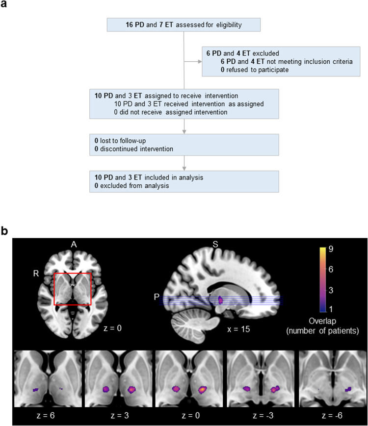
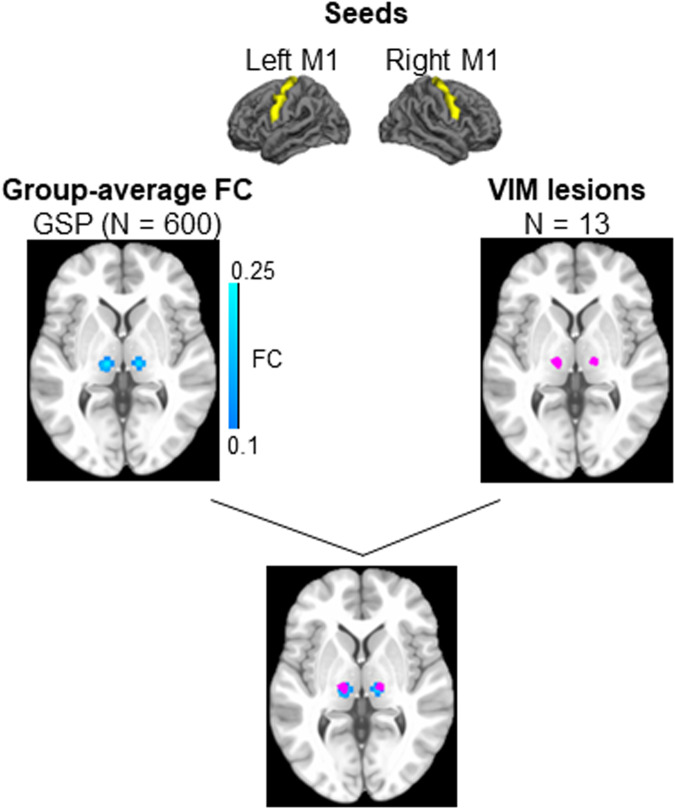
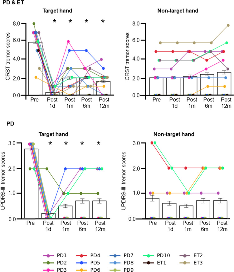
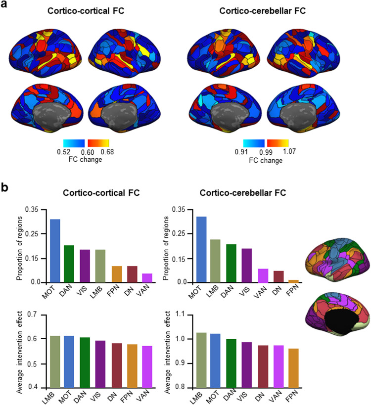
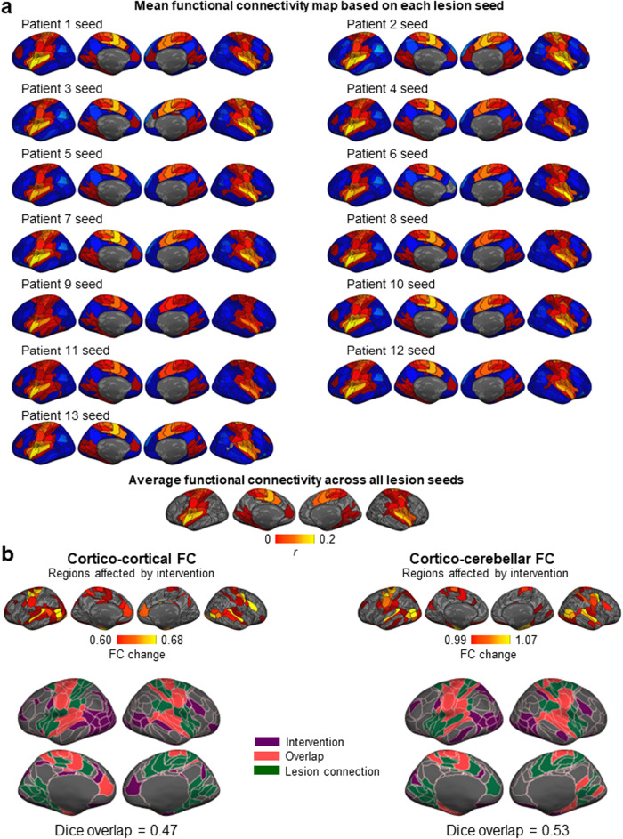
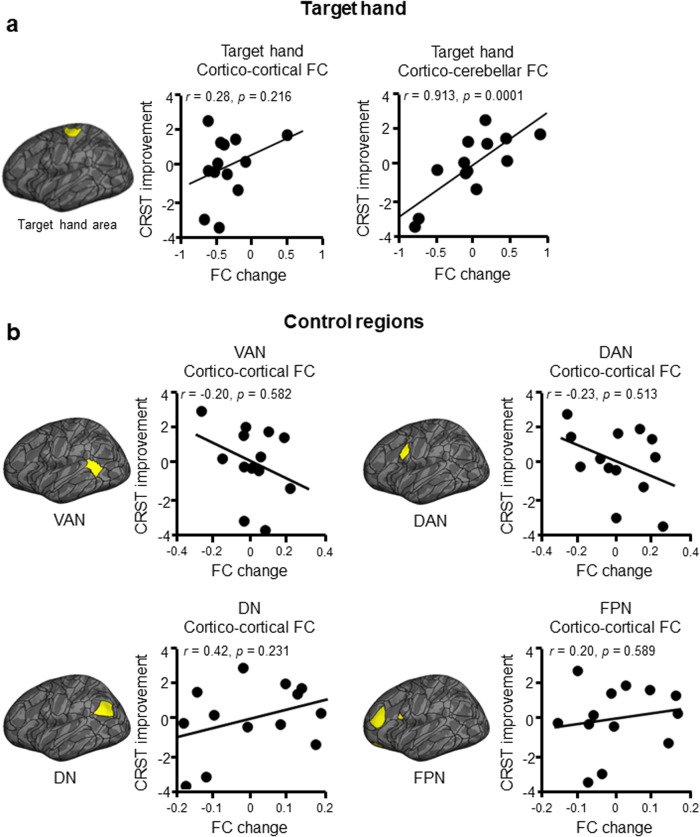
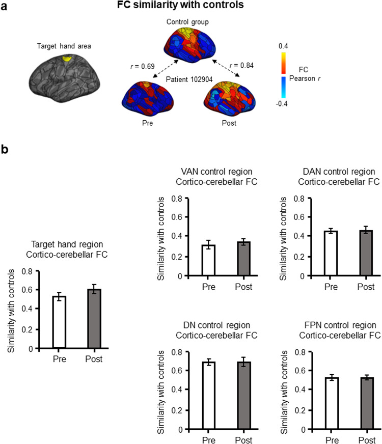
High-intensity Magnetic Resonance-guided Focused Ultrasound (MRgFUS) is a recent, non-invasive line of treatment for medication-resistant tremor. We used MRgFUS to produce small lesions in the thalamic ventral intermediate nucleus (VIM), an important node in the cerebello-thalamo-cortical tremor network, in 13 patients with tremor-dominant Parkinson's disease or essential tremor. Significant tremor alleviation in the target hand ensued (t(12) = 7.21, p < 0.001, two-tailed), which was strongly associated with the functional reorganization of the brain's hand region with the cerebellum (r = 0.91, p < 0.001, one-tailed). This reorganization potentially reflected a process of normalization, as there was a trend of increase in similarity between the hand cerebellar connectivity of the patients and that of a matched, healthy control group (n = 48) after treatment. Control regions in the ventral attention, dorsal attention, default, and frontoparietal networks, in comparison, exhibited no association with tremor alleviation and no normalization. More broadly, changes in functional connectivity were observed in regions belonging to the motor, limbic, visual, and dorsal attention networks, largely overlapping with regions connected to the lesion targets. Our results indicate that MRgFUS is a highly efficient treatment for tremor, and that lesioning the VIM may result in the reorganization of the cerebello-thalamo-cortical tremor network.


Conflict of interest statement

The authors declare no competing interests.

Figures

Fig. 1
Fig. 1. Study flow diagram and lesion sites in 13 patients with Parkinson’s disease and essential tremor.
a Flow diagram of the intervention study. In addition to the patients, 50 control participants were enrolled. Two were excluded because they did not meet study criteria, and 48 were retained. b The extent of lesion overlap across patients is color-coded according to the color bar, which depicts a range from one patient to the maximum overlap found—nine patients. Lesions were localized to the left hemisphere in nine patients, and to the right hemisphere in four patients. Axial slices are shown along with a midsagittal view of the ICBM52 MNI brain. The numbers below the axial slices indicate MNI z coordinates. The position of the axial slices is shown on the midsagittal view (purple lines). The lesion location is highly similar across patients, with 9/9 patients showing overlap in the left hemisphere and 4/4 patients showing overlap in the right hemisphere.
Fig. 2
Fig. 2. The VIM lesion sites correspond with the thalamus’s motor regions.
Using the GSP dataset (N = 600), we defined the thalamus’s motor regions using the primary motor cortices as seeds (top). We calculated the functional connectivity of these seeds within the thalamus (middle, left) and compared the resulting thalamic regions of interest with the lesion sites in the 13 patients (middle, right). There is substantial overlap between the two (bottom), indicating that the VIM lesion sites correspond with the thalamus’s motor regions.
Fig. 3
Fig. 3. MRgFUS successfully alleviated tremor in the target hand.
CRST tremor scores are shown for all 13 patients (TDPD and ET; top), for the target hand (left) and non-target hand (right), at each time point. There was a significant decrease in average target hand CRST tremor scores following the intervention (F(4,48) = 40.07, p < 0.001; top left). Scores decreased from baseline at all time points (all p’s < 0.001, Sidak correction; see Table 2). There was also a decrease in UPDRS-III target hand tremor scores after the procedure (F(4,36) = 26.70, p < 0.001; bottom left), which was significant at all time points (all p’s < 0.01, Sidak correction; see Table 2). Meanwhile, as expected, depending on the scale used, tremor in the non-target hand either increased following the intervention (CRST: F(4,48) = 3.01, p = 0.027, although post-hoc tests showed no significant different from baseline at any time point; top right) or did not change (UPDRS-III: F(4,36) = 1.35, p > 0.05; bottom right). Error bars represent standard errors of the mean. Each line connects datapoints from a given participant.
Fig. 4
Fig. 4. The motor, limbic, and visual large-scale functional networks are most affected by the VIM lesion.
a We investigated cortico-cortical (left) and cortico-cerebellar (right) functional connectivity to determine which regions exhibited an intervention effect, i.e., regions whose functional connectivity substantially changed from pre- to post-intervention. Regions in warm colors demonstrated an above-average intervention effect. The color bar indicates FC change, which was calculated as the dissimilarity between pre- and post-FC profiles while controlling for intra-individual variability (see Methods), with the middle value representing the mean. b Cortical regions were divided into the seven canonical large-scale functional networks (depicted on the right). The networks with the highest proportion of regions exhibiting an intervention effect at the level of cortico-cortical FC were the MOT, DAN, VIS, and LMB networks (top left panel). The average intervention effect of each network was calculated. The networks that demonstrated the largest average intervention effect in terms of their cortico-cortical FC were the LMB, MOT, DAN, and VIS networks (bottom left panel). In terms of cortico-cerebellar FC, the networks with the highest proportion of regions demonstrating an intervention effect were the MOT, LMB, DAN, and VIS networks (top right panel). The highest average intervention effect was found in the LMB, MOT, DAN, and VIS networks (bottom right panel). MOT sensorimotor network, DN default network, LMB limbic network, VIS visual network, VAN ventral attention network, DAN dorsal attention network, FPN frontoparietal network.
Fig. 5
Fig. 5. Regions functionally connected to the lesion sites exhibited a change in their functional connectivity following the intervention.
a Functional connectivity of the lesion site. The lesion site of each patient was used as a seed to generate a functional connectivity map, based on data from the control group (N = 48). The resulting FC maps are shown for each lesion site. The average FC map, across all 13 seeds, is shown at the bottom. The color bar represents FC strength (Pearson r). b The cortical maps depict the cortical regions whose functional connectivity substantially changed from pre- to post-intervention, according to their cortico-cortical (middle left panel) and cortico-cerebellar (middle right panel) functional connectivity. The color bar indicates FC change, and only values above the mean are shown. We then calculated the extent of overlap between the regions connected to the lesion sites and the regions that demonstrated an intervention effect. The overlap was Dice = 0.47 for cortico-cortical connections (bottom left panel), and Dice = 0.53 for cortico-cerebellar connections (bottom right panel). These substantial overlaps indicate that the functional reorganization that occurred after the intervention mainly affected the regions that are functionally connected to the lesion sites.
Fig. 6
Fig. 6. Association between FC change and tremor alleviation.
a Partial Spearman correlations, covaried with age and sex, between FC change and tremor alleviation demonstrated a non-significant, positive relationship for the cortical target hand region’s cortical FC (r = 0.28, p = 0.22, one-tailed; left), and a significant, positive relationship for its cerebellar FC (r = 0.91, p < 0.001, one-tailed; right). b The same analysis, performed in four control regions selected at random, showed non-significant relationships for the cortical FC of the VAN control region (r = −0.20, p = 0.58, two-tailed; right), DAN control region (r = −0.23, p = 0.51, two-tailed), DN control region (r = 0.42, p = 0.23, two-tailed), and FPN control region (r = 0.20, p = 0.59, two-tailed). DN default network, VAN ventral attention network, DAN dorsal attention network, FPN frontoparietal network.
Fig. 7
Fig. 7. Cortico-cerebellar FC similarity before and after the intervention.
a The left hemisphere hand region is highlighted on a cortical map (left). For each patient, we calculated the similarity in functional connectivity patterns between their cortical target hand region and the control group’s left hemisphere hand region, before and after the MRgFUS intervention. An example is shown for a randomly chosen patient, whereby the FC similarity is r = 0.69 before the intervention, and r = 0.84 after the intervention. The increased similarity from pre- to post-intervention indicates a normalization of the target hand region’s functional organization following VIM lesioning. b We averaged the FC similarity for the target hand region across patients at each time point. There was a non-significant trend of increase in cortico-cerebellar FC similarity from pre- to post-intervention (t(12) = −1.18, p = 0.13, one-tailed; mean difference = −0.07, one-sided 95% CI [−0.19, 0.04]). The four control regions, in comparison, showed either comparatively smaller or no change in FC similarity, and were all non-significant (VAN: t(12) = −0.71, p = 0.49, two-tailed; mean difference = −0.03, two-sided 95% CI [−0.11, 0.06]); DAN: t(12) = −0.33, p = 0.74, two-tailed; mean difference = −0.01, two-sided 95% CI [−0.08, 0.06]; DN: t(12) = 0.03, p = 0.96, two-tailed; mean difference = 0.00, two-sided 95% CI [−0.06, 0.06]; FPN: t(12) = 0.04, p = 0.97, two-tailed; mean difference = 0.00, two-sided 95% CI [−0.07, 0.07]). DN default network, VAN ventral attention network, DAN dorsal attention network, FPN frontoparietal network.

Similar articles

Cited by

References

    1. Deuschl G, Bain P, Brin M, Committee AHS. Consensus statement of the movement disorder society on tremor. Mov. Disord. 1998;13:2–23. doi: 10.1002/mds.870131303. - DOI - PubMed
    1. Louis ED, Rios E, Henchcliffe C. How are we doing with the treatment of essential tremor (ET)? Persistence of patients with ET on medication: data from 528 patients in three settings. Eur. J. Neurol. 2010;17:882–884. doi: 10.1111/j.1468-1331.2009.02926.x. - DOI - PMC - PubMed
    1. Meng Y, Hynynen K, Lipsman N. Applications of focused ultrasound in the brain: from thermoablation to drug delivery. Nat. Rev. Neurol. 2021;17:7–22. doi: 10.1038/s41582-020-00418-z. - DOI - PubMed
    1. Dormont D, et al. Chronic thalamic stimulation with three-dimensional MR stereotactic guidance. Am. J. Neuroradiol. 1997;18:1093–1107. - PMC - PubMed
    1. Hallett M. Tremor: pathophysiology. Parkinsonism Related Disord. 2014;20:S118–S122. doi: 10.1016/S1353-8020(13)70029-4. - DOI - PubMed