. 2023 Jun 15;9(1):91.
doi: 10.1038/s41531-023-00542-9.

AAV-mediated expression of a new conformational anti-aggregated α-synuclein antibody prolongs survival in a genetic model of α-synucleinopathies

Affiliations

AAV-mediated expression of a new conformational anti-aggregated α-synuclein antibody prolongs survival in a genetic model of α-synucleinopathies

Matthias Düchs et al. NPJ Parkinsons Dis. .

Abstract

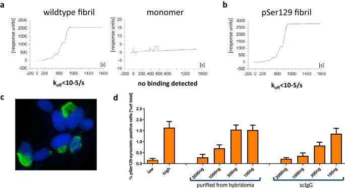
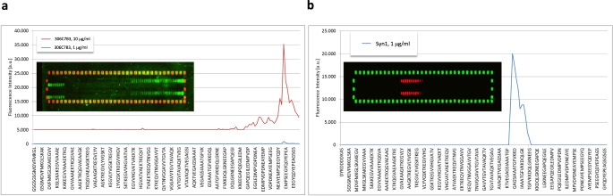
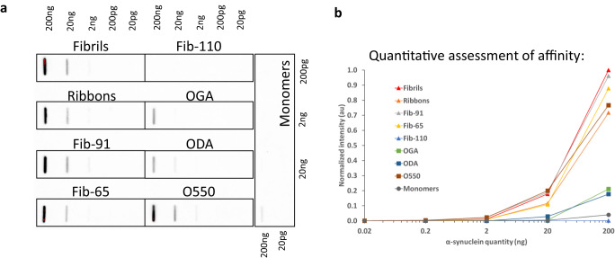
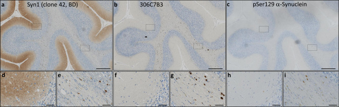
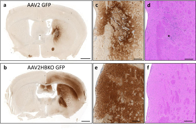
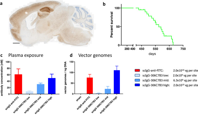
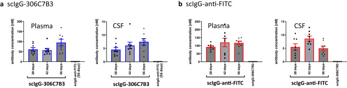
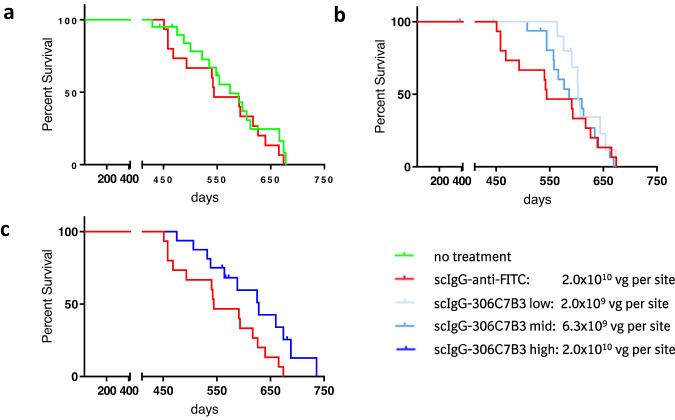
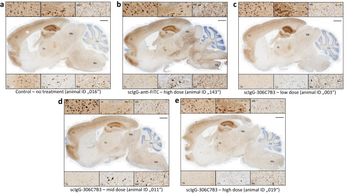
Prion-like transmission of pathology in α-synucleinopathies like Parkinson's disease or multiple system atrophy is increasingly recognized as one potential mechanism to address disease progression. Active and passive immunotherapies targeting insoluble, aggregated α-synuclein are already being actively explored in the clinic with mixed outcomes so far. Here, we report the identification of 306C7B3, a highly selective, aggregate-specific α-synuclein antibody with picomolar affinity devoid of binding to the monomeric, physiologic protein. 306C7B3 binding is Ser129-phosphorylation independent and shows high affinity to several different aggregated α-synuclein polymorphs, increasing the likelihood that it can also bind to the pathological seeds assumed to drive disease progression in patients. In support of this, highly selective binding to pathological aggregates in postmortem brains of MSA patients was demonstrated, with no staining in samples from other human neurodegenerative diseases. To achieve CNS exposure of 306C7B3, an adeno-associated virus (AAV) based approach driving expression of the secreted antibody within the brain of (Thy-1)-[A30P]-hα-synuclein mice was used. Widespread central transduction after intrastriatal inoculation was ensured by using the AAV2HBKO serotype, with transduction being spread to areas far away from the inoculation site. Treatment of (Thy-1)-[A30P]-hα-synuclein mice at the age of 12 months demonstrated significantly increased survival, with 306C7B3 concentration reaching 3.9 nM in the cerebrospinal fluid. These results suggest that AAV-mediated expression of 306C7B3, targeting extracellular, presumably disease-propagating aggregates of α-synuclein, has great potential as a disease-modifying therapy for α-synucleinopathies as it ensures CNS exposure of the antibody, thereby mitigating the selective permeability of the blood-brain barrier.


Conflict of interest statement

None of the authors declare a competing financial or non-financial interest. All authors except M.N., R.M. and J.S. were employed by Boehringer Ingelheim, a privately owned pharmaceutical company, at the time this work was executed. Nobody holds stock or stock options of Boehringer Ingelheim. No patent applications have been filed related to this work.

Figures

Fig. 1
Fig. 1. In vitro characterization of 306C7B3.
a Surface plasmon based affinity determination of 306C7B3 for fibrillar human α-synuclein (left) as well as for monomeric human α-synuclein (right). b Similar measurement as in (a), but using in vitro Ser129-phosphorylasted fibrillar human α-synuclein for the affinity determination. c Immunofluorescence staining of SH-SY5Y cells stably overexpressing A53T-mutated α-synuclein 2 days after electroporation with fibrillar α-synuclein. Intracellular mesh-like aggregates of pSer129 positive α-synuclein (green, Epitomics pSer129 antibody) are used for quantitative determination of functional activity of anti-α-synuclein antibodies (blue: DAPI staining). d Functional characterization of 306C7B3. Increasing amounts of purified antibody derived either from the original hybridoma clone or from a scIgG-based expression construct were preincubated with an excess of monomeric human α-synuclein, followed by addition of PFF α-synuclein and electroporation into SH-SY5Y cells stably overexpressing human A53T α-synuclein. High content-based analysis of cells stained for pSer129-positive intracellular aggregates demonstrates efficient inhibition of intracellular seeding by IgG-306C7B3 purified from the original hybridoma clone as well as single-chain IgG 306C7B3 (scIgG) derived from an expression construct. Error bars: standard deviation (s.d.).
Fig. 2
Fig. 2. PEPperMAP epitope mapping.
a Either 1 μg/ml (blue line) or 10 μg/ml (red line) of IgG 306C7B3 were incubated with membranes spotted with 140 different 15mer peptides (each peptide printed in duplicate to be present in the top and bottom part of the array), representing the full human α-synuclein sequence with each peptide having a 14 amino acid overlap with the preceding peptide with the human α-synuclein sequence being elongated by neutral GSGSGSG linkers to avoid truncated peptides. Inset shows microarray stained with 10 μg/ml 306C7B3 (green: 306C7B3 signal, red: HA signal). Outer spots correspond to control peptides used to orient the microarray. Some cross-reactivity against the control peptides is observed for 306C7B3 at this high antibody concentration. Figure shows signal intensity obtained for individual peptide spots, identifying the epitope of 306C7B3 as being “YQDYEPE” (amino acids 133–139 of full-length human α-synuclein) located in the C-terminus of α-synuclein with overall low binding intensity toward the microarray spotted α-synuclein peptides. b Similar approach to determine the epitope of Syn1 at a concentration of 1 μg/ml (blue line, red: Syn1 signal, green: HA signal), identifying the epitope of Syn1 as being “AATGFVKK” (amino acids 90–97 of full-length human α-synuclein). Note that the exemplified peptide sequences do not reflect all tested 15mer peptides for better visualization—identical peptide arrays were used for the analysis of the different antibodies.
Fig. 3
Fig. 3. Filter trap assessment of 306C7B3 binding toward different in vitro generated assemblies of human α-synuclein.
Fibrils, ribbons, fibrils 65, fibrils 91, fibrils 110, on fibrillar assembly pathway oligomers (O550), dopamine stabilized (ODA) and glutaraldehyde stabilized (OGA) oligomers were spotted in increasing amounts on nitrocellulose filters and tested with 306C7B3. a Images obtained after ECL based visualization. b Quantification of signals using a Chemidoc MP imaging system. Blots derive from the same experiment and were processed in parallel.
Fig. 4
Fig. 4. Immunohistochemical staining of adjacent sections of cerebellar tissue derived from a patient with multiple system atrophy (age at death 74 years, Braak&Braak stage I–II).
a Staining performed with the Syn1 antibody detecting monomeric α-synuclein in the molecular layer as well as aggregated forms of α-synuclein in the white matter fiber tracts. b 306C7B3 staining for aggregated α-synuclein in the white matter fiber tracts only. c Control staining against pSer129-positive α-synuclein aggregates demonstrating a similar staining pattern compared to 306C7B3. Scale bars: 500 mm. d, f, h Enlargement of the gray matter area indicated above (black box on the left, approximate location). e, g, i Enlargement of fiber tract staining in the white matter as indicated above (black box on the right, approximate location). Scale bars enlargements: 50 μm.
Fig. 5
Fig. 5. 306C7B3 does not label other pathological aggregates.
Immunohistochemical analysis of 306C7B3 revealed no labeling of pathological deposits characteristic for other neurodegenerative diseases, including Abeta-positive senile plaques in Alzheimer’s disease (AD, a, b), tau-positive tangles and tufted astrocytes in progressive supranuclear palsy (PSP, c, d), TDP-43-positive inclusions in frontotemporal lobar degeneration with TDP-43 pathology (FTLD-TDP, e, f), or FUS-positive inclusions in frontotemporal lobar degeneration with FET pathology (FTLD-FET, g, h). Immunohistochemistry on formalin fixed paraffin embedded adjacent tissue sections for 306C7B3 (b, d, f, h) and Abeta (a), pTau (c), pTDP-43 (e), or FUS (g). Scale bar 200 μm (ad), 40 μm (eh).
Fig. 6
Fig. 6. Expression of GFP in the murine striatum after AAV inoculation.
One μl of self-complementating (sc) AAV preparations (total of 2.8 × 108 vector genomes) coding for GFP under the control of the ubiquitous CMV-promoter were stereotactically inoculated into the murine striatum. Immunohistochemical analysis was done 21 days after virus administration. a AAV2 based transduction with limited spread of the viral preparation (anti-GFP IHC). b Significant spread of the viral transduction observed with AAV2HBKO based viral preparations (anti-GFP IHC). c, e Enlargement of the transduced striatum (anti-GFP IHC). d, f H&E staining of adjacent sections in the area of the enlargements (striatum), demonstrating cellular infiltrates in the inoculation area of the AAV2-GFP viral preparation (marked by the asterisk), no such infiltrations were observed in AAV2HBKO treated animals. Scale bars a, b: 1 mm, enlargements cf: 200 μm.
Fig. 7
Fig. 7. In vivo model to assess functional activity of the scIgG-306C7B3.
a Sagital section of the brain of a (Thy-1)-[A30P]-hα-synuclein mouse overexpressing mutant A30P α-synuclein under the control of the neuronal Thy-1 promoter euthanized due to a loss of righting reflex. Shown is a staining for pSer129-positive α-synuclein aggregates. Most aggregates are found in the hindbrain regions and areas of the Medulla oblongata. Scale bar 1 mm. b Kaplan–Meier plot of non-treated (Thy-1)-[A30P]-hα-synuclein mice demonstrating age-dependent survival based on loss of righting reflex. c Animals received bilateral stereotactic inoculation of each 1 μl of AAV2HBKO preparations carrying transgenes coding for either scIgG-306C7B3 or scIgG-antiFITC control antibody at the indicated viral titers (left). Animals were inoculated at the age of 12 months and analyzed for plasma exposure after loss of righting reflex (mean plus s.e.m.) analyzed with MSD-based ELISAs corresponding to the analyzed samples. No CSF samples had been taken from the actual in vivo study analyzing the functional activity of scIgG-306C7B3 for practical reasons. d Post-mortem analysis of brain tissue for transduction efficacy (vg = vector genomes per ng of genomic DNA), demonstrating dose-dependent transduction (mean plus s.e.m.). Sham = non-inoculated controls.
Fig. 8
Fig. 8. Pilot pharmacokinetic study to determine scIgG-306C7B3 and scIgG-anti-FITC exposure.
(Thy-1)-[A30P] hα-synuclein mice were unilaterally inoculated into the striatum at 2.0 × 1010 vector genomes with either AAV2HBKO-scIgG-anti-FITC or AAV2HBKO-scIgG-306C7B3. Plasma as well as CSF fluid was analyzed in transduced animals after 28, 42 and 56 days (separate cohorts for each individual time point). a Plasma and cerebrospinal fluid (CSF) exposure measuring 306C7B3 with a MSD based ELISA. b Plasma and CSF exposure of the control anti-FITC antibody. Analysis as in (a). Taking all data together, an approximate ratio of plasma to CSF exposure of 12.6:1 has been calculated for scIgG-306C7B3. Data presented as mean plus s.e.m. (n = 8 animals per group).
Fig. 9
Fig. 9. In vivo functional activity of scIgG-306C7B3 based on prolonged survival of AAV2-HBKO treated (Thy-1)-[A30P]-hα-synuclein mice.
a Comparison of non-treated with control antibody treated (scIgG-anti-FITC) animals. No detrimental effect of the AAV-treatment could be observed (p = 0.47). b Low and mid doses of AAV2HBKO coding for scIgG-306C7B3 demonstrate delayed initial mortality but no overall increase in survival (p = 0.99 and 0.71, respectively). c Significant increased survival observed in high dose AAV2HBKO-scIgG-306C7B3 compared to control antibody treated animals (p = 0.03; comparison toward non-treated animals p = 0.09).
Fig. 10
Fig. 10. IHC analysis of the animals involved in the in vivo study.
Formalin-fixed brain tissue from all animals included in the study were tested via pSer129-α-synuclein immunohistochemistry for strong pathology, corresponding to the symptom of loss of righting reflex at time of euthanasia. Examples of the obtained staining’s for representative animals from each cohort are shown (ae). See Supplementary Table 1 for details on the individual animals. High magnifications of the indicated areas are shown above and below the full brain images. CC cerebral cortex, Hi hippocampus, Mb midbrain, Th thalamus, Ce cerebellum, Me medulla. Scale bar full brain image: 1 mm, higher magnifications: 50 μm.

Similar articles

Cited by

References

    1. Shahmoradian SH, et al. Lewy pathology in Parkinson’s disease consists of crowded organelles and lipid membranes. Nat. Neurosci. 2019;22:1099–1109. doi: 10.1038/s41593-019-0423-2. - DOI - PubMed
    1. Oliveira LMA, et al. Alpha-synuclein research: defining strategic moves in the battle against Parkinson’s disease. NPJ Parkinsons Dis. 2021;7:65. doi: 10.1038/s41531-021-00203-9. - DOI - PMC - PubMed
    1. Spillantini MG, et al. Alpha-synuclein in Lewy bodies. Nature. 1997;388:341–346. doi: 10.1038/42166. - DOI - PubMed
    1. Goedert M, Spillantini MG, Del Tredici K, Braak H. 100 years of Lewy pathology. Nat. Rev. Neurol. 2013;9:13–24. doi: 10.1038/nrneurol.2012.242. - DOI - PubMed
    1. Schneider SA, Alcalay RN. Neuropathology of genetic synucleinopathies with parkinsonism: review of the literature. Mov. Disord. 2017;32:1504–1523. doi: 10.1002/mds.27193. - DOI - PMC - PubMed