. 2023 Jul;24(7):1161-1172.
doi: 10.1038/s41590-023-01513-1. Epub 2023 Jun 15.

ChAdOx1 nCoV-19 (AZD1222) vaccine-induced Fc receptor binding tracks with differential susceptibility to COVID-19

Affiliations

ChAdOx1 nCoV-19 (AZD1222) vaccine-induced Fc receptor binding tracks with differential susceptibility to COVID-19

Paulina Kaplonek et al. Nat Immunol. 2023 Jul.

Abstract

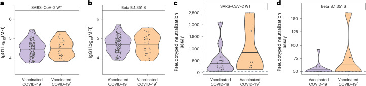
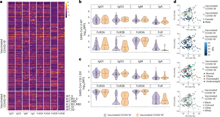
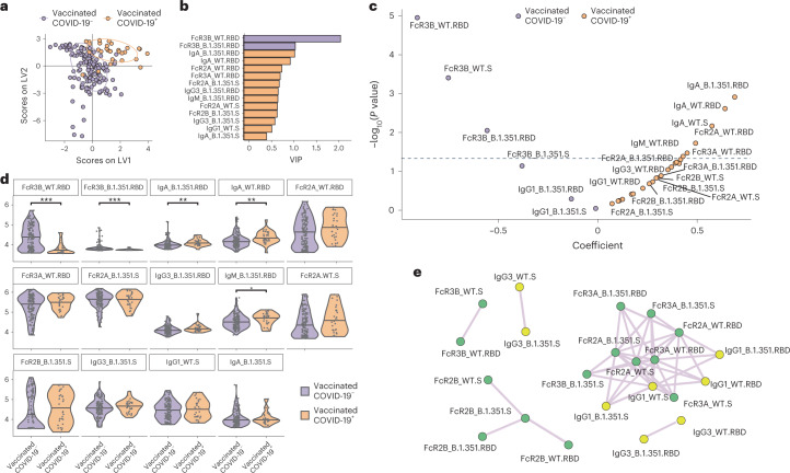
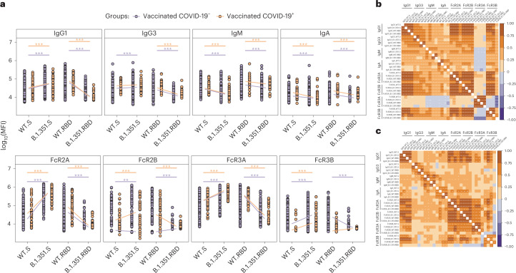
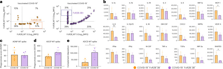
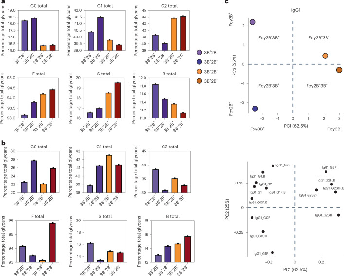
Despite the success of COVID-19 vaccines, severe acute respiratory syndrome coronavirus 2 (SARS-CoV-2) variants of concern have emerged that can cause breakthrough infections. Although protection against severe disease has been largely preserved, the immunological mediators of protection in humans remain undefined. We performed a substudy on the ChAdOx1 nCoV-19 (AZD1222) vaccinees enrolled in a South African clinical trial. At peak immunogenicity, before infection, no differences were observed in immunoglobulin (Ig)G1-binding antibody titers; however, the vaccine induced different Fc-receptor-binding antibodies across groups. Vaccinees who resisted COVID-19 exclusively mounted FcγR3B-binding antibodies. In contrast, enhanced IgA and IgG3, linked to enriched FcγR2B binding, was observed in individuals who experienced breakthrough. Antibodies unable to bind to FcγR3B led to immune complex clearance and resulted in inflammatory cascades. Differential antibody binding to FcγR3B was linked to Fc-glycosylation differences in SARS-CoV-2-specific antibodies. These data potentially point to specific FcγR3B-mediated antibody functional profiles as critical markers of immunity against COVID-19.


Conflict of interest statement

G.A. is a founder/equity holder in Seromyx Systems Inc. and Leyden Labs. She has served as a scientific advisor for Sanofi Vaccines and has collaborative agreements with GSK, Merck, Abbvie, Sanofi, Medicago, BioNtech, Moderna, BMS, Novavax, SK Bioscience, Gilead and Sanaria. The remaining authors declare no conflicting interests.

Figures

Fig. 1
Fig. 1. Equivalent WT and beta (B.1.351) IgG1 S-specific antibody levels and neutralization titers across vaccinees who either developed COVID-19 or were uninfected.
a,b, Violin plots showing the univariate comparison of WT (a) and beta B.1.351 (b) SARS-CoV-2 S-specific IgG levels between vaccinees who resisted COVID-19 (n = 140) and individuals who developed COVID-19 (n = 30) over the study period. MFI, mean fluorescence intensity. c,d, SARS-CoV-2 WT (c) and beta B.1.351 (d) neutralization titers measured for vaccinees who resisted COVID-19 (n = 28) or developed beta VOC COVID-19 (n = 12). A Mann–Whitney U-test was used to define differences and the Benjamini–Hochberg method was used to adjust for multiple comparisons, with an adjusted P (Padj): ***P < 0.001; **P< 0.01; *P < 0.05. Source data
Fig. 2
Fig. 2. Diverging antibody Fc profiles across ChAdOx1 nCoV-19 vaccinees who did or did not develop COVID-19.
a, Heatmap summarizing the SARS-COV-2 WT and beta (B.1.351)-specific IgG1, IgG3, IgA1 and IgM titers, as well as the ability of SARS-CoV-2-specific antibodies to bind to the low-affinity Fcγ-receptors (FcγR2A, Fcγ2B, Fcγ3a and Fcγ3b) across the vaccinees who did not (n = 140) or did develop COVID-19 (n = 30). Each column represents a distinct feature that was analyzed in the plasma and each row a different plasma sample. Titers and FcR data were first log(transformed) and z-scores are shown for comparison. b,c, Violin plot showing univariate comparisons of WT (b) and beta (c) SARS-CoV-2 S-specific Fc-antibody profiles between the groups. A Mann–Whitney U-test, with a correction for multiple comparison using the Benjamini–Hochberg method, was used to test for differences across the groups. d, A PCA applied to all samples and data, including vaccinees who did and did not develop COVID-19, to examine the impact of different demographic parameters on antibody profiles. In each panel, samples are colored based on sex, age, BMI and race, demonstrating limited effects of these demographic characteristics on shaping vaccine-induced humoral profiles. ***P < 0.001; **P < 0.01; *P < 0.05. Source data
Fig. 3
Fig. 3. FcγR3B-biased SARS-CoV-2-binding responses track with enhanced protection from beta VOC-induced disease.
a, A LASSO used to reduce the feature dimensionality and ultimately select antibody features that discriminated between vaccinees who resisted COVID-19 and those who developed the disease over the study period. The PLS-DA was then used to visualize the separation between the samples based on the LASSO-selected features, where each dot represents an individual vaccinee. Violet dots represent vaccinees who resisted COVID-19 disease over the study period and orange dots the vaccinees who developed COVID-19 over the study period. b, Bar graph showing the ranking of the LASSO-selected features based on a VIP score. c, The LME model depicting the overall differences in antibody features between individuals who resisted COVID-19 (left side) and individuals who developed COVID-19 (right side). The models were corrected for sex, age, BMI, race, alcohol and smoking status. The x axis depicts the effect size between the groups and the y axis shows the statistical significance. The hatched line depicts the significance cut-off after multiple comparisons. d, Violin plots showing the univariate comparisons of LASSO-selected features between the vaccinees who resisted COVID-19 and those who developed disease over the study period. Statistical differences were defined using a Mann–Whitney U-test and a correction for multiple comparisons, using the Benjamini–Hochberg method, and all P values were adjusted (***P < 0.001; **P < 0.01; *P < 0.05). e, Network analysis showing the additional antibody features, which were correlated with the LASSO-selected features and are likely to be important in driving the separation. The network was built using a threshold of absolute Spearman’s ρ < 0.7 and Benjamini–Hochberg-adjusted Padj < 0.01. Nodes were colored based on the type of measurement: antibody titers and FcR binding. The connecting lines denote all positive correlations (no negative correlations were observed). Source data
Fig. 4
Fig. 4. WT and beta RBD and S-specific Fcγ2B and Fcγ3B differ across vaccinees who developed COVID-19 or resisted disease.
a, Dot plots showing the univariate comparisons of S- and RBD-specific antibody levels to the WT or beta S (left) or RBD (left) across the vaccinees who resisted or developed COVID-19 over the study period. Differences were defined using a Wilcoxon’s rank-sum test, and all P values were corrected for multiple comparisons using the Benjamini–Hochberg method with: ***P < 0.001; **P < 0.01; *P < 0.05. b,c, Correlation matrices depicting the Spearman’s correlations between SARS-CoV-2 WT and beta B.1.351 S-specific antibody features in vaccinated individuals who resisted (b) or developed (c) COVID-19. Significance values were corrected for multiple comparisons using the Benjamini–Hochberg method and shown as: ***P < 0.001; **P < 0.01; *P < 0.05. The lower triangle shows P values, whereas the upper triangle shows Padj values. Source data
Fig. 5
Fig. 5. FcγR2B+3B- and FcγR2B3B+-binding IgG show different abilities to drive antibody-dependent effector functions as well as cytokine and chemokine production.
a, The selection strategy of COVID-19+FcR2B+3B and COVID-19FcR2B3B+ pools based on the ability of WT S-specific IgG to bind to FcγR2B and FcγR3B receptors. Subjects in the top quartile (marked with boxes) were selected and pooled (pool of n = 5). b, ADCD. c,d, ADCP (c) and ADNP (d) in COVID-19+FcR2B+3B and COVID-19FcR2B3B+ pools (pool of n = 5). Bars show the mean value with s.d. Dots represents replicates (n = 4 and n = 6). Samples were run in technical duplicates and two (ADCD) to three (ADCP and ADNP) biological replicates. Unpaired Student’s t-test and Padj (***P < 0.001; **P < 0.01; *P < 0.05) were used. e, Cytokine production by isolated human neutrophils stimulated with COVID-19+FcR2B+3B and COVID-19FcR2B3B+ pools (pool of n = 5). Bars show the mean with s.d. Dots represent replicates (n = 4, technical duplicates of two biological replicates with different blood donors). Unpaired Student’s t-test and Padj (***P < 0.001; **P < 0.01; *P < 0.05) were used. GM-CSF, granulocyte–macrophage colony-stimulating factor; IFNγ, interferon-γ; MCP, monocyte chemoattractant protein; MIP, macrophage inflammatory protein; TGFα; transforming growth factor α; TNF-α, tumor necrosis factor α; VEGF, vascular endothelial growth factor. Source data
Fig. 6
Fig. 6. The capacity of SARS-CoV-2 S-specific IgG1 to bind to Fcγ2B and Fcγ3B receptors is regulated by the Fc-fragment glycosylation pattern.
a,b, The overall representation (the percentage of total glycans) of major sugar classes, including galactose (agalactose: G0; single galactose: G1; digalactose: G2; fucose (F), sialic acid (S) and a bisecting GlcNAc (B)) across SARS-CoV-2 S-specific low and high FcγR2- and FcγR3B-binding IgG1 (FcγR2B+3B+, FcγR2B+3B, FcγR2B3B) (a) and total antigen unspecific+, FcγR2B3B groups (b) measured by LC–MS. Each bar shows the average of two replicates for FcγR2B+3B+, FcγR2B+3B, FcγR2B3B+ and FcγR2B3B samples (pool of n = 5 per sample). c, PCA applied to all samples and data to examine the impact of various glycoforms on FcγR2B and FcγR3B binding for IgG1. Source data

References

    1. COVID Data Tracker. Centers for Disease Control and Preventionhttps://covid.cdc.gov/covid-data-tracker/#datatracker-home (2021).
    1. Liu C, et al. Reduced neutralization of SARS-CoV-2 B.1.617 by vaccine and convalescent serum. Cell. 2021;184:4220–4236.e4213. doi: 10.1016/j.cell.2021.06.020. - DOI - PMC - PubMed
    1. Graham MS, et al. Changes in symptomatology, reinfection, and transmissibility associated with the SARS-CoV-2 variant B.1.1.7: an ecological study. Lancet Public Health. 2021;6:e335–e345. doi: 10.1016/S2468-2667(21)00055-4. - DOI - PMC - PubMed
    1. Kustin T, et al. Evidence for increased breakthrough rates of SARS-CoV-2 variants of concern in BNT162b2-mRNA-vaccinated individuals. Nat. Med. 2021;27:1379–1384. doi: 10.1038/s41591-021-01413-7. - DOI - PMC - PubMed
    1. Ramesh, S. et al. Emerging SARS-CoV-2 variants: a review of its mutations, its implications and vaccine efficacy. Vaccines10.3390/vaccines9101195 (2021). - PMC - PubMed

Publication types