

Arterial spin labeling MRI applied to migraine: current insights and future perspectives
- PMID: 37322466
- PMCID: PMC10273543
- DOI: 10.1186/s10194-023-01597-y
Arterial spin labeling MRI applied to migraine: current insights and future perspectives
Abstract
Introduction: Advanced neuroimaging techniques have extensively contributed to elucidate the complex mechanisms underpinning the pathophysiology of migraine, a neurovascular disorder characterized by episodes of headache associated with a constellation of non-pain symptoms. The present manuscript, summarizing the most recent progresses of the arterial spin labelling (ASL) MRI techniques and the most significant findings from ASL studies conducted in migraine, is aimed to clarify how ASL investigations are contributing to the evolving insight on migraine pathophysiology and their putative role in migraine clinical setting. ASL techniques, allowing to quantitatively demonstrate changes in cerebral blood flow (CBF) both during the attacks and in the course of interictal period, could represent the melting point between advanced neuroimaging investigations, conducted with pure scientific purposes, and conventional neuroimaging approaches, employed in the diagnostic decision-making processes.
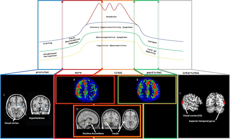
Main body: Converging ASL evidences have demonstrated that abnormal CBF, exceeding the boundaries of a single vascular territory, with biphasic trend dominated by an initial hypoperfusion (during the aura phenomenon but also in the first part of the headache phase) followed by hyperperfusion, characterizes migraine with aura attack and can represent a valuable clinical tool in the differential diagnosis from acute ischemic strokes and epileptic seizures. Studies conducted during migraine without aura attacks are converging to highlight the involvement of dorsolateral pons and hypothalamus in migraine pathophysiology, albeit not able to disentangle their role as "migraine generators" from mere attack epiphenomenon. Furthermore, ASL findings tend to support the presence of perfusion abnormalities in brain regions known to be involved in aura ignition and propagation as well as in areas involved in multisensory processing, in both patients with migraine with aura and migraine without aura.
Conclusion: Although ASL studies have dramatically clarified quality and timing of perfusion abnormalities during migraine with aura attacks, the same cannot be said for perfusion changes during migraine attacks without aura and interictal periods. Future studies with more rigorous methodological approaches in terms of study protocol, ASL technique and sample selection and size are mandatory to exploit the possibility of better understanding migraine pathophysiology and identifying neuroimaging biomarkers of each migraine phase in different migraine phenotypes.
Keywords: Arterial spin labelling; Cerebral blood flow; MRI; Migraine with aura; Migraine without aura.
© 2023. The Author(s).
Conflict of interest statement
The authors declare no competing interests.
Figures
Similar articles
-
Magnetic resonance imaging arterial-spin-labelling perfusion alterations in childhood migraine with atypical aura: a case-control study.Dev Med Child Neurol. 2016 Sep;58(9):965-9. doi: 10.1111/dmcn.13123. Epub 2016 Apr 6. Dev Med Child Neurol. 2016. PMID: 27060350
-
Magnetic resonance imaging in children presenting migraine with aura: Association of hypoperfusion detected by arterial spin labelling and vasospasm on MR angiography findings.Cephalalgia. 2018 Apr;38(5):949-958. doi: 10.1177/0333102417723570. Epub 2017 Jul 24. Cephalalgia. 2018. PMID: 28738690
-
Cerebral blood flow alterations in migraine patients with and without aura: An arterial spin labeling study.J Headache Pain. 2022 Oct 4;23(1):131. doi: 10.1186/s10194-022-01501-0. J Headache Pain. 2022. PMID: 36195842 Free PMC article.
-
Perfusion imaging by arterial spin labeling in migraine: A literature review.J Cereb Blood Flow Metab. 2024 Aug;44(8):1253-1270. doi: 10.1177/0271678X241237733. Epub 2024 Mar 14. J Cereb Blood Flow Metab. 2024. PMID: 38483125 Free PMC article. Review.
-
Shedding light on migraine with aura: the clarifying role of advanced neuroimaging investigations.Expert Rev Neurother. 2019 Aug;19(8):739-750. doi: 10.1080/14737175.2019.1638252. Epub 2019 Jul 3. Expert Rev Neurother. 2019. PMID: 31267785 Review.
Cited by
-
Connectivity of the insular subdivisions differentiates posttraumatic headache-associated from nonheadache-associated mild traumatic brain injury: an arterial spin labelling study.J Headache Pain. 2024 Jun 19;25(1):103. doi: 10.1186/s10194-024-01809-z. J Headache Pain. 2024. PMID: 38898386 Free PMC article.
-
Dynamics of cerebral blood flow following sertraline treatment in adolescent depression.Front Psychiatry. 2025 Jul 18;16:1521565. doi: 10.3389/fpsyt.2025.1521565. eCollection 2025. Front Psychiatry. 2025. PMID: 40756314 Free PMC article.
-
Cortical Hyperperfusion on MRI Arterial Spin-Labeling during the Interictal Period of Patients with Migraine Headache.AJNR Am J Neuroradiol. 2024 Jun 7;45(6):686-692. doi: 10.3174/ajnr.A8208. AJNR Am J Neuroradiol. 2024. PMID: 38663988 Free PMC article.
-
Reversible Perfusion Changes during Acute Attacks in Glucose Transporter Type 1 Deficiency Syndrome: A Pediatric Case Series.AJNR Am J Neuroradiol. 2025 Feb 3;46(2):395-400. doi: 10.3174/ajnr.A8506. AJNR Am J Neuroradiol. 2025. PMID: 39788627
-
Hyperperfusion of bilateral amygdala in patients with chronic migraine: an arterial spin-labeled magnetic resonance imaging study.J Headache Pain. 2023 Oct 18;24(1):138. doi: 10.1186/s10194-023-01668-0. J Headache Pain. 2023. PMID: 37848831 Free PMC article.
References
-
- Headache Classification Committee of the International Headache Society (IHS) The International Classification of Headache Disorders, 3rd edition (2018) Cephalalgia: an international journal of headache, 38(1), 1–211 - PubMed
-
- Alsop DC, Detre JA, Golay X, Günther M, Hendrikse J, Hernandez-Garcia L, Lu H, MacIntosh BJ, Parkes LM, Smits M, van Osch MJ, Wang DJ, Wong EC, Zaharchuk G. Recommended implementation of arterial spin-labeled perfusion MRI for clinical applications: a consensus of the ISMRM perfusion study group and the european consortium for ASL in dementia. Magn Reson Med. 2015;73(1):102–116. doi: 10.1002/mrm.25197. - DOI - PMC - PubMed
Publication types
MeSH terms
LinkOut - more resources
Full Text Sources
Miscellaneous
