. 2023 Jun 15;18(1):72.
doi: 10.1186/s13020-023-00758-0.

A novel dammarane triterpenoid alleviates atherosclerosis by activating the LXRα pathway

Affiliations

A novel dammarane triterpenoid alleviates atherosclerosis by activating the LXRα pathway

Yan Huang et al. Chin Med. .

Abstract

Background: We have previously demonstrated that ginsenoside compound K can attenuate the formation of atherosclerotic lesions. Therefore, ginsenoside compound K has potential for atherosclerosis therapy. How to improve the druggability and enhance the antiatherosclerotic activity of ginsenoside compound K are the core problems in the prevention and treatment of atherosclerosis. CKN is a ginsenoside compound K derivative that was previously reported to have excellent antiatherosclerotic activity in vitro, and we have applied for international patents for it.

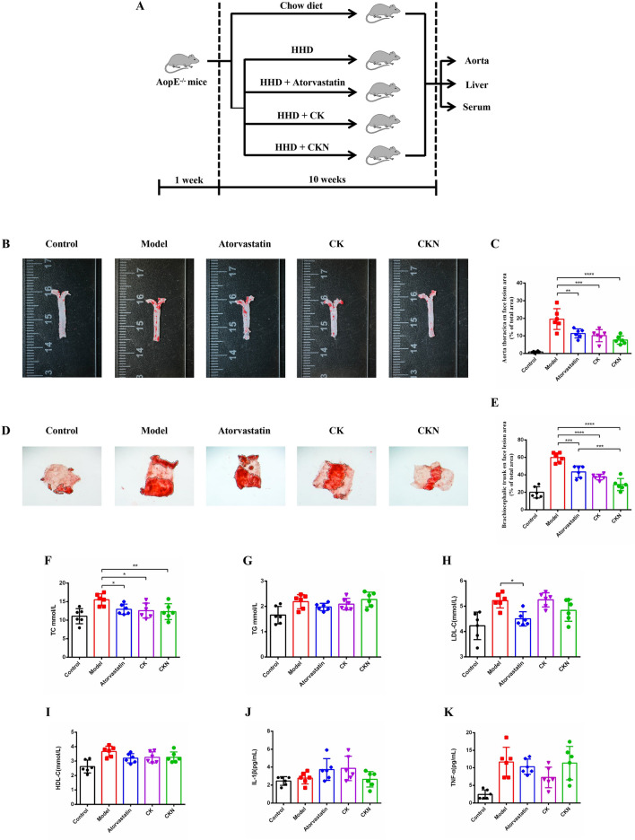
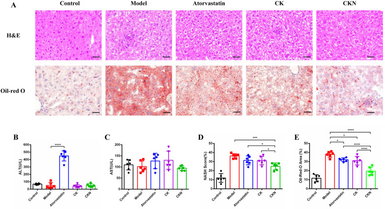
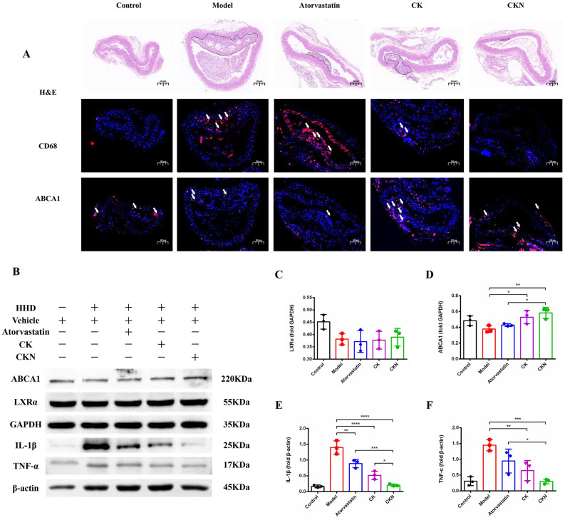
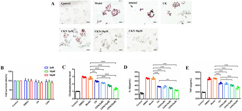
Methods: Male C57BL/6 ApoE-/- mice were fed a high-fat and high-choline diet to induce atherosclerosis and were subjected to in vivo studies. In vitro, the CCK-8 method was applied to evaluate cytotoxicity in macrophages. Foam cells were utilized, and cellular lipid determination was performed for in vitro studies. The area of atherosclerotic plaque and fatty infiltration of the liver were measured by image analysis. Serum lipid and liver function were determined by a seralyzer. Immunofluorescence and western blot analysis were conducted to explore the alterations in the expression levels of lipid efflux-related proteins. Molecular docking, reporter gene experiments and cellular thermal shift assays were used to verify the interaction between CKN and LXRα.

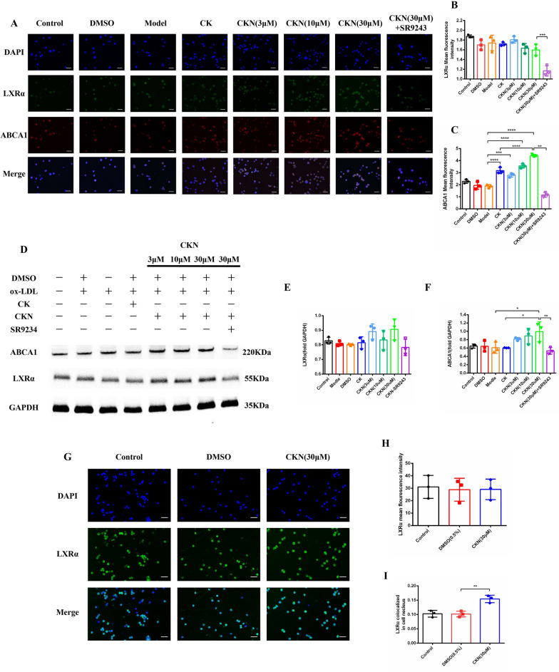
Results: After confirming the therapeutic effects of CKN, molecular docking, reporter gene experiments and cellular thermal shift assays were used to predict and investigate the antiatherosclerotic mechanisms of CKN. CKN exhibited the greatest potency, with a 60.9% and 48.1% reduction in en face atherosclerotic lesions on the thoracic aorta and brachiocephalic trunk, reduced plasma lipid levels and decreased foam cell levels in the vascular plaque content in HHD-fed ApoE-/- mice. Moreover, CKN in the present study may exert its antiatherosclerotic effects through activated ABCA1 by promoting LXRα nuclear translocation and reducing the adverse effects of LXRα activation.

Conclusions: Our results revealed that CKN prevented the formation of atherosclerosis in ApoE-/- mice by activating the LXRα pathway.

Keywords: ATP-binding cassette A1; Antiatherosclerosis; Derivative; Ginsenoside compound K; LXRα; Nuclear translocation.


Conflict of interest statement

The authors declare that they have no competing interests.

Figures

Fig. 1
Fig. 1
CKN attenuated the formation of atherosclerotic lesions in ApoE−/− mice. The design of the experiments (A). Representative images of aortic lesions in the thoracic aorta (B) and quantification of atherosclerotic lesions shown as the percentage of thoracic aorta (C). Representative images of aortic lesions in the brachiocephalic trunk (D) and quantification of atherosclerotic lesions shown as a percentage of the brachiocephalic trunk (E). The serum lipid levels of total cholesterol (TC) (F), total triglyceride (TG) (G), low-density lipoprotein-cholesterol (LDL-C) (H) and high-density lipoprotein-cholesterol (HDL-C) (I). Serum and local levels of IL-1β (J) and TNF-α (K) were modulated by CKN. Data are presented as mean ± SD (n = 6) and analyzed by ANOVA with Dunnett’s post-hoc analysis. *P < 0.05, **P < 0.01; ***P < 0.001, ****P < 0.0001
Fig. 2
Fig. 2
CKN did not cause hepatotoxicity. Representative images of H&E- and Oil Red O-stained (A) liver sections. Scale bars: 500 µm. Detection of ALT (B) and AST (C) in ApoE−/− mice after different treatments. Changes in the NASH SCORE in each group (D). Quantification of the lipid content in the liver with Oil Red O staining (n = 6) (E). Data are presented as mean ± SD (n = 6) and analyzed by ANOVA with Dunnett’s post-hoc analysis. *P < 0.05, ***P < 0.001, ****P < 0.0001
Fig. 3
Fig. 3
CKN reduced the inflammatory reaction in the aorta. Representative histochemistry images of aortic root cross-sections stained with CD68 antibody and ABCA1 antibody showed that CKN treatment decreased macrophage infiltration in the aorta. Scale bars: 100 µm (A). The levels of LXR signaling pathway and inflammatory cytokine proteins in atherosclerotic mice were determined using Western blotting (B). Western blot analysis of LXRα (C) and ABCA1 (D) in aortic tissue treated with CKN. The western blotting results showed that CKN treatment significantly decreased the levels of IL-1β (E) and TNF-α (F) in aortic tissue. Data are presented as mean ± SD (n = 3) and analyzed by ANOVA with Dunnett’s post-hoc analysis. *P < 0.05, **P < 0.01; ***P < 0.001, ****P < 0.0001
Fig. 4
Fig. 4
CKN inhibited the formation of foam cells. The concentrations of cholesteryl ester in foam cells decreased after RAW264.7 macrophages were treated with CK (30 μM) or CKN (3, 10, and 30 μM). Scale bars: 50 µm (A and C). The cellular toxicity of CKN was analysed using the CCK-8 method (B). Cellular IL-1β (D) and TNF-α (E) levels decreased after treatment with CKN (3, 10, and 30 μM). Data are presented as mean ± SD (n = 3), and significance was determined by ANOVA with Dunnett’s post-hoc analysis. *P < 0.05, **P < 0.01; ***P < 0.001, ****P < 0.0001
Fig. 5
Fig. 5
The pharmacological effects of CKN may be associated with the LXRα pathway. RAW264.7 macrophages were stimulated with 50 µg/mL ox-LDL, 50 µg/mL ox-LDL + CKN (30 μM) and 50 µg/mL ox-LDL + CKN (3, 10, and 30 μM) for 24 h. The cells were fixed with 4% paraformaldehyde and stained with DIPY (blue fluorescence), anti-LXRα antibody (green fluorescence) and anti-ABCA1 antibody (red fluorescence). Scale bars: 50 µm (A). Quantitative analysis of LXRα and ABCA1 fluorescence intensity (B and C). Western blotting was used to examine LXRα and ABCA1 protein expression in CKN-treated or untreated macrophages (D). Western blot analysis of LXRα (E) and ABCA1 (F) in macrophages treated with CKN. RAW264.7 macrophages were treated with CKN (100 μM) for 24 h. LXRα (green fluorescence) was found in the cytoplasm of RAW264.7 cells. Scale bars: 50 µm (G). CKN treatment did not change the total expression of LXRα (H). After CKN treatment, the LXRα (green fluorescence) signal was significantly decreased in the cytoplasm but increased in the nucleus (blue fluorescence) (I). Data are presented as mean ± SD (n = 3) and were analyzed by ANOVA with Dunnett’s post-hoc analysis. *P < 0.05, **P < 0.01; ***P < 0.001
Fig. 6
Fig. 6
CKN is a novel LXRα agonist. Docking of CKN to mouse LXRα was simulated by SYBYL-X 2.0 (A). HEK293T cells were treated with different concentrations of CKN, and GW3965 (10 μM) was used as a positive control (B). The RAW264.7 cell proteins were mixed with compound CKN (30 μM), and the control group was treated with 0.5% DMSO. The proteins were heated at 45 to 69 °C under the same conditions using a heat block for 2 min. The supernatant was collected for western blotting (C). Diagram of the molecular mechanisms of CKN (D). Data are presented as mean ± SD (n = 3) and were analyzed by ANOVA with Dunnett’s post-hoc analysis

Similar articles

References

    1. GBD 2019 Risk Factors Collaborators Global burden of 87 risk factors in 204 countries and territories, 1990–2019: a systematic analysis for the global burden of disease study 2019. Lancet. 2020;396:1223–1249. doi: 10.1016/S0140-6736(20)30752-2. - DOI - PMC - PubMed
    1. Tsao CW, Aday CW, Almarzooq ZI, et al. Heart disease and stroke statistics-2022 update: a report from the American heart association. Circulation. 2022;145:e153–e639. doi: 10.1161/CIR.0000000000001052. - DOI - PubMed
    1. Yang S, Yuan HQ, Hao YM, et al. Macrophage polarization in atherosclerosis. Clin Chim Acta. 2020;501:142–146. doi: 10.1016/j.cca.2019.10.034. - DOI - PubMed
    1. Koelwyn GJ, Corr EM, Erbay E, et al. Regulation of macrophage immunometabolism in atherosclerosis. Nat Immunol. 2018;19:526–537. doi: 10.1038/s41590-018-0113-3. - DOI - PMC - PubMed
    1. Chistiakov DA, Melnichenko AA, Myasoedova VA, et al. Mechanisms of foam cell formation in atherosclerosis. J Mol Med. 2017;95:1153–1165. doi: 10.1007/s00109-017-1575-8. - DOI - PubMed