. 2023 Jun 15;67(2):3663.
doi: 10.4081/ejh.2023.3663.

Sodium hyaluronate promotes proliferation, autophagy, and migration of corneal epithelial cells by downregulating miR-18a in the course of corneal epithelial injury

Affiliations

Sodium hyaluronate promotes proliferation, autophagy, and migration of corneal epithelial cells by downregulating miR-18a in the course of corneal epithelial injury

Yingzhuo Guo et al. Eur J Histochem. .

Abstract

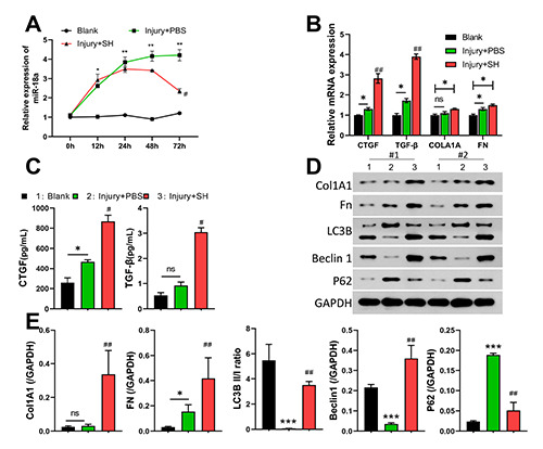
Corneal epithelium can resist the invasion of external pathogenic factors to protect the eye from external pathogens. Sodium hyaluronate (SH) has been confirmed to promote corneal epithelial wound healing. However, the mechanism by which SH protects against corneal epithelial injury (CEI) is not fully understood. CEI model mice were made by scratching the mouse corneal epithelium, and in vitro model of CEI were constructed via curettage of corneal epithelium or ultraviolet radiation. The pathologic structure and level of connective tissue growth factor (CTGF) expression were confirmed by Hematoxylin and Eosin staining and immunohistochemistry. CTGF expression was detected by an IHC assay. The levels of CTGF, TGF-β, COLA1A, FN, LC3B, Beclin1, and P62 expression were monitored by RT-qPCR, ELISA, Western blotting or immunofluorescence staining. Cell proliferation was detected by the CCK-8 assay and EdU staining. Our results showed that SH could markedly upregulate CTGF expression and downregulate miR-18a expression in the CEI model mice. Additionally, SH could attenuate corneal epithelial tissue injury, and enhance the cell proliferation and autophagy pathways in the CEI model mice. Meanwhile, overexpression of miR-18a reversed the effect of SHs on cell proliferation and autophagy in CEI model mice. Moreover, our data showed that SH could induce the proliferation, autophagy, and migration of CEI model cells by downregulating miR-18a. Down-regulation of miR-18a plays a significant role in the ability of SH to promote corneal epithelial wound healing. Our results provide a theoretical basis for targeting miR-18a to promote corneal wound healing.


Figures

Figure 1.
Figure 1.
SH markedly attenuated corneal epithelial tissue injury and upregulated CTGF in CEI model mice. A) Hematoxylin and eosin staining was used to observe pathological changes in the corneal epithelium of CEI model mice after treatment with SH. B,b) HIC was used to confirm the effect of SH on CTGF expression in mice with CEI. Magnification: B), x100; b) x200.
Figure 2.
Figure 2.
SH markedly downregulated miR-18a expression and mediated autophagy-related protein expression in mice with CEI. CEI model mice were established and then treated with SH. A) Changes in miR-18a expression in mice were identified by using qPCR at 0, 12, 24, 48, and 72 h. B) RT-qPCR analyses of CTGF, TGF-β, Col1A1, and FN expression. C) The concentrations of CTGF and TGF-β were detected with ELISA kits at 72 h post injury. D,E) Western blotting was used to monitor changes in Col1A1, FN, LC3B, Beclin 1, and P62 expression in corneal tissues at 72 h post injury.
Figure 3.
Figure 3.
Overexpression of miR-18a attenuated the effects of SH on miR-18a, and autophagy-related pathways, CTGF and TGF-β level in CEI model mice. CEI model mice were treated with SH and injected with miR-18a mimics or an inhibitor, respectively. A) MiR-18a levels were determined by RT-qPCR. The levels of CTGF and TGF-β in each group were detected using RT-qPCR (B) and ELISA kits. C,D) Changes in LC3B, Beclin 1, and P62 expression were detected by Western blotting.
Figure 4.
Figure 4.
Treatment with miR-18a dramatically reversed the induction effect of SH on the cell viability, CTGF and TGF-β of CEI model cells. CEI cell model was constructed using HCECs that were exposed to UV radiation. Next, the HCECs were treated with SH and miR-18a mimics. A) RT-qPCR analysis of miR-18a. B) CCK8 assays were performed to assess changes in cell viability. C) ELISA kits were used to detect the concentrations of CTGF and TGF-β. D) Edu assays revealed changes in cell proliferation. Magnification: x100.
Figure 5.
Figure 5.
SH induced autophagy and cell migration by downregulating miR-18a in CEI model cells. CEI model cells were treated with SH and miR-18a mimics, respectively. A) Cell migration was analyzed by the Transwell assay, and the numbers of migrated cells in each group were counted. B) Western blotting showed changes in LC3B, Beclin1, and P62 expression. C) LC3B expression was examined by IF staining. Magnification: x200.

Similar articles

Cited by

References

    1. Ljubimov AV, Saghizadeh M. Progress in corneal wound healing. Prog Retin Eye Res 2015; 49:17-45. - PMC - PubMed
    1. Wilson SE, Torricelli AAM, Marino GK. Corneal epithelial basement membrane: Structure, function and regeneration. Exp Eye Res 2020;194:108002. - PMC - PubMed
    1. Ma X, Wang Q, Song F, Li Y, Li J, Dou S, et al. . Corneal epithelial injury-induced norepinephrine promotes Pseudomonas aeruginosa keratitis. Exp Eye Res 2020;195:108048. - PubMed
    1. Raghunathan VK, Thomasy SM, Strom P, Yanez-Soto B, Garland SP, Sermeno J, et al. . Tissue and cellular biomechanics during corneal wound injury and repair. Acta Biomater 2017; 58:291-301. - PMC - PubMed
    1. Chen W, Shi XL, He XH, Mao YH, Li C, Dong N. Loteprednol combined with sodium hyaluronate in the treatment of dry eye disease and its effect on TNF-α and CXCL10 in tears. J Biol Regul Homeost Agents 2020;34:1825-9. - PubMed