. 2021 Oct 30;1(2):20210011.
doi: 10.1002/EXP.20210011. eCollection 2021 Oct.

Biomaterials and nanomedicine for bone regeneration: Progress and future prospects

Affiliations

Biomaterials and nanomedicine for bone regeneration: Progress and future prospects

Jun Zhou et al. Exploration (Beijing). .

Abstract

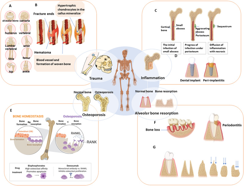
Bone defects pose a heavy burden on patients, orthopedic surgeons, and public health resources. Various pathological conditions cause bone defects including trauma, tumors, inflammation, osteoporosis, and so forth. Auto- and allograft transplantation have been developed as the most commonly used clinic treatment methods, among which autologous bone grafts are the golden standard. Yet the repair of bone defects, especially large-volume defects in the geriatric population or those complicated with systemic disease, is still a challenge for regenerative medicine from the clinical perspective. The fast development of biomaterials and nanomedicine favors the emergence and promotion of efficient bone regeneration therapies. In this review, we briefly summarize the progress of novel biomaterial and nanomedical approaches to bone regeneration and then discuss the current challenges that still hinder their clinical applications in treating bone defects.

Keywords: biomaterials; bone defects; nanomedicine; scaffolds; tissue engineering.


Conflict of interest statement

Wei Tao and Omid C. Farokhzad are members of the Exploration editorial board. Omid C. Farokhzad has financial interests in Selecta Biosciences, Tarveda Therapeutics, XLINK Therapeutics, PrognomIQ, and Seer.

Figures

FIGURE 1
FIGURE 1
Main causes of bone loss. (A) Common fracture locations include the craniofacial (alveolar bone, calvaria), long bones (femur, tibia, humerus), wrist (radius/ ulna), ankle (above the joint, distal tibia/fibula), and vertebral sites. (B) The process of normal fracture healing through the formation of a cartilaginous callus. (C) Progress of osteomyelitis (OM). A local abscess creates diffuse infection and produces a region of necrotic bone tissue. (D) Unlike non‐inflammatory dental implants (left), peri‐implantitis (right) may lead to loss of supporting tissue and implant failure. (E) Bone homeostasis and common dynamic cycle (left) of bone formation and bone resorption. At right is bone remodeling in osteoporosis, in which bone resorption by osteoclasts exceeds bone formation mediated by osteoblasts. (F) Bone loss as a cause of high‐morbidity periodontitis. (G) Alveolar ridge resorption post‐extraction of the tooth, from left to right, pre‐extraction, post‐extraction, high and well rounded, knife ridge, low and well rounded, and depressed
FIGURE 2
FIGURE 2
The key mechanisms of cellular and molecular biology in bone. (A) Differentiation process of osteoblasts and osteoclasts. Bone marrow mesenchymal stem cells (MSCs) are multipotent cells with the capacity to differentiate toward osteoblast (OB), chondrocyte, and adipocyte lineages. The largest cellular population in bone, osteocytes are derived from OBs. Osteoporosis represents an increase in bone marrow fat tissue due to a shift in the differentiation of MSCs to adipocytes rather than to osteoblasts. Therefore, the fate of MSCs affects the proportion of various types of bone cells and bone mass overall. On the other hand, osteoclasts (OCs), which facilitate bone resorption, originate from the monocyte‐macrophage lineage. (B) Effects of the Wnt β‐catenin signaling pathway. The Wnt/β‐catenin signaling pathway is transduced by the stabilization of β‐catenin following the interaction between a specific Wnt ligand and its designated receptors. Wnt has been considered the main regulator of osteogenesis. The activation of the Wnt signaling pathway can promote the formation of bone. (C) Cross‐talk and effects of the two key pathways in bone metabolism (PTH and Wnt signaling) on MSC, pre‐OB, OB, and indirect function on osteoclasts. Both PTH and Wnt promote the proliferation of MSCs and the commitment of these cells to the OB lineage, whereas PTH can also stimulate OB to produce RANKL, which facilitates pre‐OC differentiation to OC. The Wnt pathway produces osteoprotegerin (OPG), hindering OC differentiation and function by suppressing RANKL. Sclerostin (Sost) and Dkk1, activated by Wnt, can suppress Wnt activity, creating a negative feedback loop
FIGURE 3
FIGURE 3
Nanomedicine and biomaterials for bone repair. (A) Utility of bisphosphonates for targeted bone mineral delivery in its unconjugated/ conjugated form to various pharmaceuticals, nanoparticles, fluorophores, chelation complexes, and macromolecules. Bisphosphonates can also be used for drug delivery, radiotherapy, and diagnosis. Reproduced with permission.[ 39 ] Copyright 2015, Elsevier. (B) The three main scaffold fabrication techniques for bone regeneration. The first uses synthetic bone graft substitutes for medical device(MD) class II, the second a synergy of bioactive molecules in a ceramic scaffold for MD class III, and the third a tissue engineering‐based approach with stem cells in a scaffold, with or without bioactive molecules called advanced therapeutic medicinal products (ATMP). Reproduced with permission.[ 13 ] Copyright 2018, Elsevier. (C) The development of nanomaterials with biomimicry considering various factors such as interconnected macropores, microporous topography, composition, mineralization, and use of bioactive molecules. These nanomaterials function as in vivo bioreactors for in situ bone regeneration. Reproduced with permission.[ 8 ] Copyright 2017, RSC Pub
FIGURE 4
FIGURE 4
Multifunctional materials and combination therapies by Black phosphorus (BP). (A) Application of BP in bone regeneration using the photothermal conversion of BP under near‐infrared (NIR), with PLGA and BPs@PLGA. (i) 3D reconstruction of bone by Micro‐CT. (ii) Sequential fluorescence labeling of newly formed bone. (iii) Representative histologic graphs with toluidine blue staining. (iv) MSCs cultured on BPs@PLGA membrane with NIR irradiation. (v) Cell viability analysis for biocompatibility of MSCs on PLGA and BPs@PLGA with or without NIR irradiation. ** denotes p  <  0.01 compared with the PLGA group. Reproduced with permission.[ 13 ] Copyright 2019, Elsevier. (B) In vivo osteogenesis performance of the bifunctional 3D‐printed bioglass (BG) scaffold designed using 2D BP nanosheets. (i–v) Micro‐CT 3D imaging from rat calvaria 8 weeks after implantation. Bone defects were restored with BP‐BG scaffold (left) and BG scaffold (right) as control. (i) 3D reconstruction image of micro‐CT in calvaria. The two defects in calvaria represent newborn tissues in the middle of holes that were too thin to be identified by the 3D reconstruction software. BP‐BG group (ii,iii) and BG group (iv,v) were acquired using black (ii,iv) and white (iii,v) substrates. When the color of substrates reverses from black to white, the newborn osseous tissue can be visualized. (vi) Schematic illustration of the fabrication process for BP‐BG scaffold and the stepwise therapeutic strategy for the ablation of osteosarcoma and in situ osteogenesis. Reproduced with permission.[ 65 ] Copyright 2018, John Wiley and Sons
FIGURE 5
FIGURE 5
Targeting skeletal endothelium to ameliorate bone loss. (A) (i) The vasculature is widely distributed in the skeleton. (ii) Type H vessels are the primary factor regulating and maintaining perivascular osteoprogenitors and promoting angiogenesis. They are rich in young people (left) and traumatic sites in bone (right), but much less frequent among the elderly (middle). (ii) Reprinted with permission.[ 67 ] (B) Administration of recombinant SLIT3 has a salutary influence on bone fracture healing, as well as OVX‐induced bone loss in mouse femur. (i) Representative micro‐CT (ii) Representative H&E staining and endomucin (EMCN) immunohistochemistry (IHC) images 3 weeks after open fracture in the midshaft. The boxes represent the fracture site. Arrowheads highlight EMCN‐positive vessels. (iii–v) Non‐union frequency, micro‐CT analysis of BV/TV in callus region. (iv) Shn3+/+ Slit3+/+, Shn3+/+ Slit3−/−, Shn3−/− Slit3+/+, and Shn3−/− Slit3−/−, EMCN‐positive vessel numbers (v, left) and maximum compressive loading (v, right) of the fractured femora 28 days after open midshaft fracture in femur. (vi,vii) Representative micro‐CT (vi) H&E staining (vii) and EMCN IHC images of mouse femurs 3 weeks after fracturing with i.v. injection of SLIT3 or PBS. (viii–x) Micro‐CT measurement of BV/TV in callus area, EMCN‐positive vessel number and volume (ix), and maximum compressive load and stiffness (x) of femurs 3 weeks post‐fracture with i.v. injection of SLIT3 or PBS. (xi) Test of fracture callus BV/TV (left) and maximum loading (right) of mouse femurs excised 3 weeks after fracturing with insertion of a gelatin sponge soaked with SLIT3 or vehicle. (xii) Representative confocal images of CD31 (green) and EMCN (red) dual‐immunostained callus sections of mouse femurs 3 weeks after fracturing with insertion of a gelatin sponge soaked with SLIT3 or vehicle (high power, insert). Arrowheads highlight CD31hiEMCNhi vessels. (xiii,xiv) Representative μCT images of the trabecular bone in the distal femur (xiii) and relative BV/TV. Values represent mean ± s.e.m. Reproduced with permission.[ 68 ] Copyright 2018, Springer Nature

Similar articles

Cited by

References

    1. Rammelt S., EFORT Open Rev. 2016, 1, 239. - PMC - PubMed
    1. Liu Z., Yuan X., Liu M., Fernandes G., Zhang Y., Yang S., Ionita C. N., Yang S., Mol. Ther. 2018, 26, 199. - PMC - PubMed
    1. Li M., Liu J., Cui X., Sun G., Hu J., Xu S., Yang F., Zhang L., Wang X., Tang P., Regen. Biomater. 2019, 6, 373. - PMC - PubMed
    1. Liao Y., Zhang X. L., Li L., Shen F. M., Zhong M. K., Br. J. Clin. Pharmacol. 2014, 78, 718. - PMC - PubMed
    1. Zhang X., Koo S., Kim J. H., Huang X., Kong N., Zhang L., Zhou J., Xue J., Harris M. B., Tao W., Kim J. S., Matter 2021, 4, 2727.

LinkOut - more resources