. 2023 Jul:231:112087.
doi: 10.1016/j.matdes.2023.112087. Epub 2023 Jun 11.

Osteoporosis and Covid-19: Detected similarities in bone lacunar-level alterations via combined AI and advanced synchrotron testing

Affiliations

Osteoporosis and Covid-19: Detected similarities in bone lacunar-level alterations via combined AI and advanced synchrotron testing

Federica Buccino et al. Mater Des. 2023 Jul.

Abstract

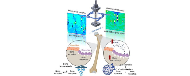
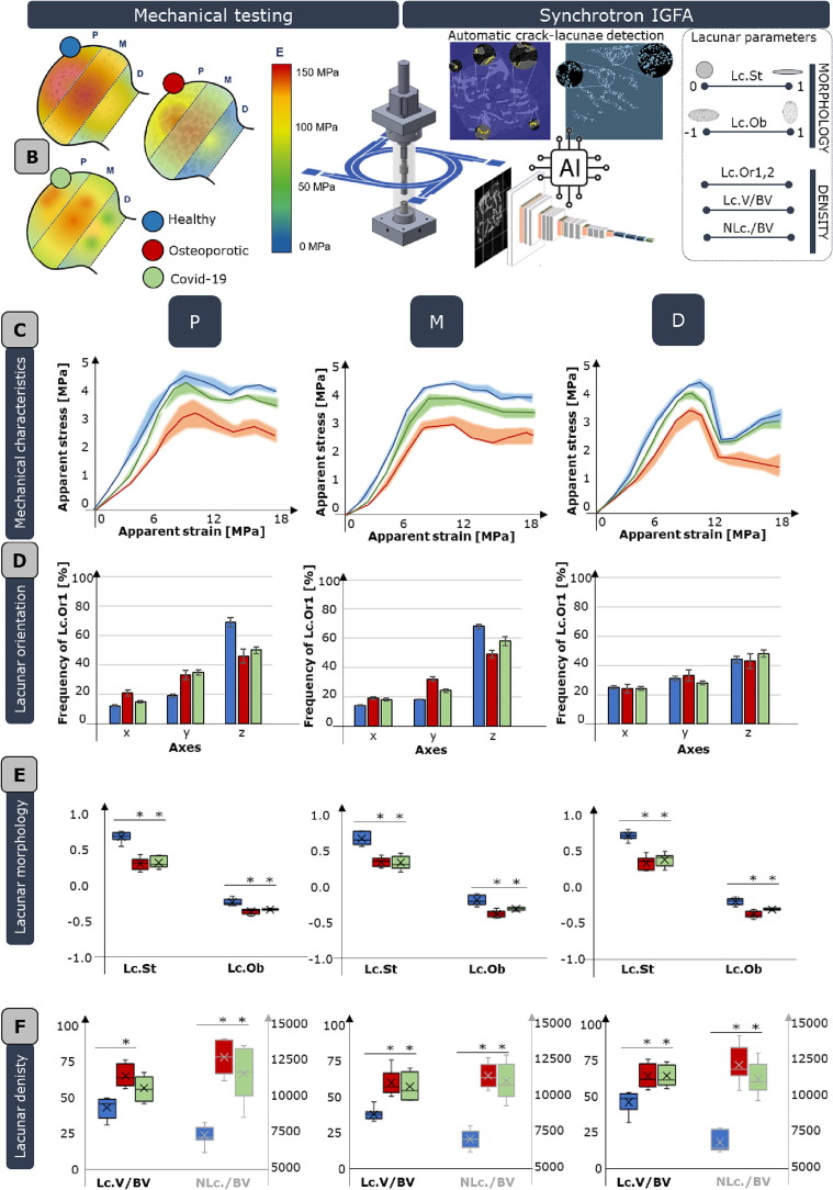
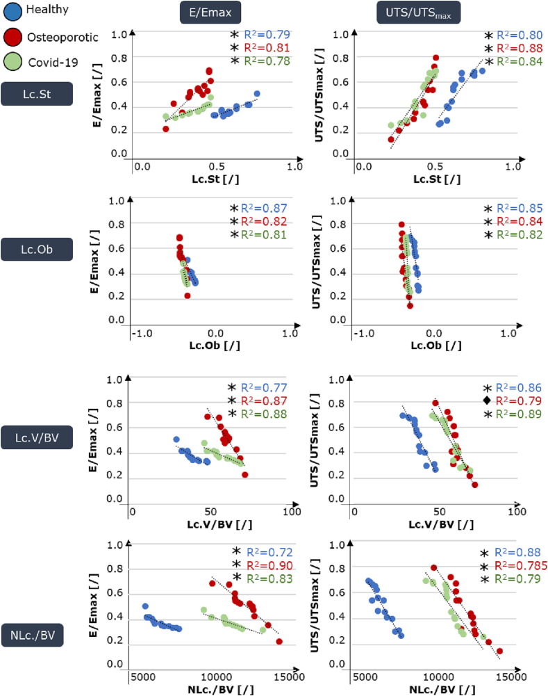
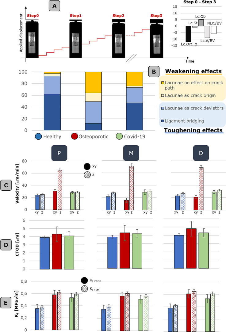
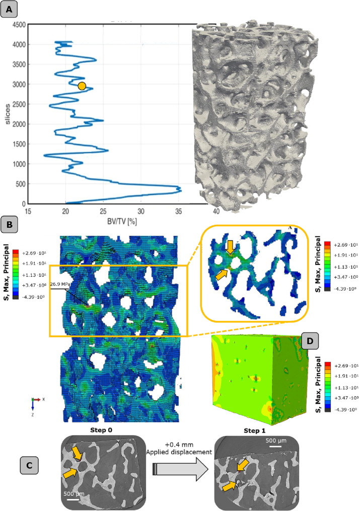
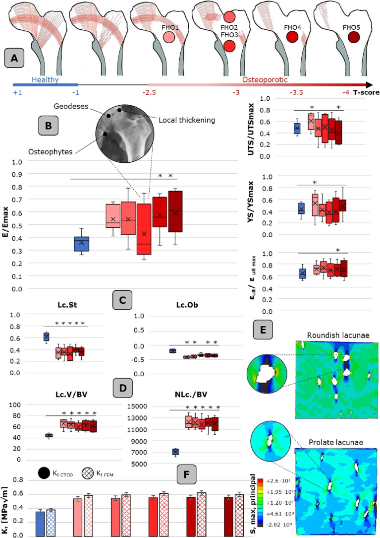
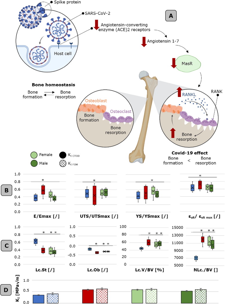
While advanced imaging strategies have improved the diagnosis of bone-related pathologies, early signs of bone alterations remain difficult to detect. The Covid-19 pandemic has brought attention to the need for a better understanding of bone micro-scale toughening and weakening phenomena. This study used an artificial intelligence-based tool to automatically investigate and validate four clinical hypotheses by examining osteocyte lacunae on a large scale with synchrotron image-guided failure assessment. The findings indicate that trabecular bone features exhibit intrinsic variability related to external loading, micro-scale bone characteristics affect fracture initiation and propagation, osteoporosis signs can be detected at the micro-scale through changes in osteocyte lacunar features, and Covid-19 worsens micro-scale porosities in a statistically significant manner similar to the osteoporotic condition. Incorporating these findings with existing clinical and diagnostic tools could prevent micro-scale damages from progressing into critical fractures.

Keywords: Bone lacunae; Covid-19; Micro-scale; Osteoporosis; Synchrotron.


Conflict of interest statement

The authors declare that they have no known competing financial interests or personal relationships that could have appeared to influence the work reported in this paper.

Figures

None
Graphical abstract
Fig. 1
Fig. 1
Meso- and micro-scale bone characteristics in dependence of external loading scenario. A. harvesting site, i.e. femoral heads, and the complexity of femur multi-scale architecture. At the macro-scale (on the left), the loading distribution is reported (JRF: joint reaction force, Fabd: force due to the abductor muscles, Fip: force due to the iliopsoas muscle, Fvl: force due to vastus lateralis muscle). At the meso-scale, trabeculae are arranged along principal tensile and compressive groups of fibers. At the micro-scale, lacunae are the sites where osteocyte reside. B. testing equipment, comprising an ad hoc micro-compression device and synchrotron in situ imaging. This allows to map the average Young modulus inside healthy (blue dot), osteoporotic (red dot) and Covid-19 (green dot) femoral heads (on the left). On the right, an artificial intelligence-based tool, specifically a convolutional neural network is exploited for the automatic segmentation of micro-cracks and lacunae, which morpho-densitometric parameters are visually schematized (including: Lacunar Stretch, Lc.St., Lacunar Oblateness, Lc.Ob., Lacunar Orientation 1 and 2, Lc.Or1,2, lacunar volume density, Lc.V/BV, and lacunar number density, NLc./BV). C. Meso-scale mechanical curves of healthy, osteoporotic and Covid-19 femoral heads are reported in terms of apparent stress-apparent strain and clustered with respect to the harvesting region (proximal, P, medial, M and distal, D). Standard deviations of data are reported in a lighter shade. D. lacunar orientation of the three physio-pathological conditions with respect to the ×, y and z (loading direction) axis of the sample and the harvesting region. E. lacunar morphological parameters including stretch and oblateness. F. lacunar densitometric parameters, including lacunar volume and lacunar number with respect to the bone volume. Boxplots are shown with mean, median, lower and upper quantile and whiskers. * p-value < 0.05. (For interpretation of the references to colour in this figure legend, the reader is referred to the web version of this article.)
Fig. 2
Fig. 2
Correlation between osteocyte lacunar morpho-densitometric features and meso-scale mechanical characteristics of the samples tested inside the synchrotron, including normalized Young modulus and normalized ultimate tensile stress. The scatter plots include best fitted lines and related R-square for each physio-pathological condition. *p-values < 0.001, ♦ p-value < 0.05.
Fig. 3
Fig. 3
Lacunar morpho-densitometric feature effects on micro-damage occurrence A. Synchrotron image-guided failure assessment conducted at four different stages: i.e., uncompressed state (named step 0), after four applied displacement levels of 0.1 mm (step 1), after additional four applied displacement levels of 1 mm (step 2) and at the complete failure of the sample (step 3). The radiographs of an exemplificative sample are reported for each step. At step 3, the femoral head bone sample is fully collapsed. The differences in lacunar morpho-densitometric parameters between Step0 and Step 3 are reported on the right. B. Quantification of toughening and weakening effects occurring at the micro-scale in all three physio-pathological conditions. In > 60% of the healthy cases is detected the presence of ligament bridging. C. Micro-crack velocity along xy plane and z direction, corresponding to the applied displacement. D. Crack-tip opening displacement (CTOD) values. E. Stress intensity factor, KI, values extrapolated from the quantification of the CTOD and from the finite element analysis. All values indicated in C, D and E are reported for all the three physio-pathological conditions, considering P, M, D zone. Histograms are reported with data standard deviation bars.
Fig. 4
Fig. 4
Multi-scale numerical modeling of trabecular bone. A. full bone (global level) and indication of the Bone volume ratio (BV/TV). The yellow dot indicates the critical zone where average low values of BV/TV are detected. B. localization of the failure band, in the proximity of the maximum principal stresses region. C. localization of the most stressed/strained trabecula and validation of the models with synchrotron images at increasing applied displacement levels. D. evaluation of the stress state at the lacunar level by means of a finite element model in the most stressed region of the failure band. (For interpretation of the references to colour in this figure legend, the reader is referred to the web version of this article.)
Fig. 5
Fig. 5
Osteoporosis effect of bone meso- and micro-scale morpho-mechanical characteristics. A. progressive deterioration of the principal and secondary tensile and compression group. The osteoporotic FH are reported with respect to their T-score. B. Mechanical characteristics (normalized with respect to the maximum value detected in each FH) of the osteoporotic FHs with respect to the healthy control group. Reported values are: E/Emax, UTS/UTSmax, YS/YSmax and εult/ εult_max. C. Lacunar morphological parameters and D. lacunar densitometric parameters of the osteoporotic FHs with respect to the healthy condition. E. computational models evidencing the stress state in presence of osteoporotic micro-porosities, i.e. roundish lacunae (upper model) and healthy, i.e. flattened prolate lacunae (bottom model). For these lacunar level models, boundary conditions are extrapolated from the global full bone model, as detailed in section 2.7. F. stress intensity factors calculated based on the crack-tip opening displacement (CTOD) and the finite element models for each osteoporotic FH and the healthy case. Boxplots are shown with mean, median, lower and upper quantile and whiskers. * p-value < 0.05. Histograms are reported with data standard deviation bars.
Fig. 6
Fig. 6
Covid-19 effects on bone meso-and micro-scale morpho-mechanical characteristics. A. Schematic of Covid-19 hypothesized effects on bone remodeling mechanisms. Covi-19 targets angiotensin-converting enzyme (ACE2) receptor, producing a downregulation of Angiotensin 1–7/MasR cascade. ACE2 and MasR are expressed by osteoblasts and osteoclasts, meanwhile, the activation of ACE2/Ang-(1–7)/MasR axis reduce the bone resorptive milieu by inhibiting the expression of receptor activator of nuclear factor-kappaB ligand (RANKL). B. Meso-scale mechanical characteristics (normalized with respect to the maximum value detected in each FH) of both male and female Covid-19 FH, with respect to the healthy and osteoporotic cases. Reported values are: E/Emax, UTS/UTSmax, YS/YSmax and εult/ εult_max. C. Lacunar morphological parameters and lacunar densitometric parameters of the Covid-19 FHs with respect to the healthy/osteoporotic condition. E. stress intensity factor calculated based on the crack-tip opening displacement (CTOD) and the finite element models for Covid-19 FH and the healthy/osteoporotic cases. Boxplots are shown with mean, median, lower and upper quantile and whiskers. * p-value < 0.05. Histograms are reported with data standard deviation bars.

Similar articles

Cited by

References

    1. Qing H., Ardeshirpour L., Divieti Pajevic P., Dusevich V., Jähn K., Kato S., Wysolmerski J., Bonewald L.F. Demonstration of osteocytic perilacunar/canalicular remodeling in mice during lactation. J. Bone Miner. Res. 2012;27(5):1018–1029. - PMC - PubMed
    1. Nicolella D.P., Moravits D.E., Gale A.M., Bonewald L.F., Lankford J. Osteocyte lacunae tissue strain in cortical bone. J. Biomech. 2006;39(9):1735–1743. - PMC - PubMed
    1. Buccino F., Colombo C., Vergani L.M. A Review on Multiscale Bone Damage: From the Clinical to the Research Perspective. Materials (Basel) 2021;14(5):1240. - PMC - PubMed
    1. Gauthier R., Langer M., Follet H., Olivier C., Gouttenoire P.-J., Helfen L., Rongiéras F., Mitton D., Peyrin F. 3D micro structural analysis of human cortical bone in paired femoral diaphysis, femoral neck and radial diaphysis. J. Struct. Biol. 2018;204(2):182–190. - PubMed
    1. Varga P., Hesse B., Langer M., Schrof S., Männicke N., Suhonen H., Pacureanu A., Pahr D., Peyrin F., Raum K. Synchrotron X-ray phase nano-tomography-based analysis of the lacunar–canalicular network morphology and its relation to the strains experienced by osteocytes in situ as predicted by case-specific finite element analysis. Biomech. Model. Mechanobiol. 2015;14(2):267–282. - PubMed

LinkOut - more resources