Case Reports
. 2023 Jun 13;11(6):e7514.
doi: 10.1002/ccr3.7514. eCollection 2023 Jun.

A case of intradural lumbar disc herniation

Affiliations
Case Reports

A case of intradural lumbar disc herniation

Utsav Bhattarai et al. Clin Case Rep. .

Abstract

Key clinical message: MRI remains the best tool in the diagnosis of this disease entity however preoperative diagnosis remains a difficult task. A high degree of suspicion is raised when intraoperative findings and preoperative image description become incompatible.

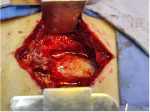
Abstract: Lumbar disc herniation into the dural space is a rare phenomenon of lumbar disc degeneration with an unclear remaining pathogenesis. Intraoperative ultrasonography and histopathological examination of resected specimen help in the diagnosis of intradural disc herniation. Prompt surgery is recommended due to the high incidence of cauda equina syndrome.

Keywords: disc; intradural; lumbar; spine.


Conflict of interest statement

None of the authors have potential conflicts of interest to be disclosed.

Figures

FIGURE 1
FIGURE 1
MRI lumbar T1 & T2 axial and T2 sagittal revealed focal posterocentral disc bulge with central and B/l paracentral extrusion of the disc elements in the superior and inferior spinal canal at L5‐S1 level causing severe narrowing of the central spinal canal and compression of the descending nerve roots as well as traversing S1 left nerve root.
FIGURE 2
FIGURE 2
Intraoperative image showing tense and bulging mass inside the thecal sac at L5‐S1 level.
FIGURE 3
FIGURE 3
After removal a hard fungating mass similar to tissue texture of nucleus pulposus was revealed.
FIGURE 4
FIGURE 4
Histopathological examination showing disc tissue.

References

    1. Schisano G, Franco A, Nina P. Intraradicular and intradural lumbar disc herniation: experiences with nine cases. Surg Neurol. 1995;44(6):536‐543. doi:10.1016/0090-3019(95)00248-0 - DOI - PubMed
    1. Fairbank JCT, Pynsent PB. The oswestry disability index. Spine (Phila Pa 1976). 2000;25:2940‐2953. doi:10.1097/00007632-200011150-00017 - DOI - PubMed
    1. Kim HS, Eun JP, Park JS. Intradural migration of a sequestrated lumbar disc fragment masquerading as a spinal intradural tumor. J Korean Neurosurg Soc. 2012;52:156‐158. doi:10.3340/jkns.2012.52.2.156 - DOI - PMC - PubMed
    1. Öztürk A, Avci E, Yazgan P, Torun F, Yücetaş Ş, Karabaé H. Intraradural herniation of intervertebral disc at the level of lumbar 1‐lumbar 2. Turk Neurosurg. 2007;17:134‐137. - PubMed
    1. Liu CC, Huang CT, Lin CM, Liu KN. Intradural disc herniation at L5 level mimicking an intradural spinal tumor. Eur Spine J. 2011;20:326‐329. doi:10.1007/s00586-011-1772-z - DOI - PMC - PubMed

Publication types

LinkOut - more resources