Case Reports
. 2023 May 15;15(5):e39031.
doi: 10.7759/cureus.39031. eCollection 2023 May.

Candida dubliniensis Fungemia Leading to Infective Endocarditis and Septic Pulmonary Emboli

Affiliations
Case Reports

Candida dubliniensis Fungemia Leading to Infective Endocarditis and Septic Pulmonary Emboli

Abtin Jafroodifar et al. Cureus. .

Abstract

Illicit drugs, especially those injected intravenously, are becoming increasingly more common worldwide. Individuals who use intravenous drugs often reuse or share needles which predisposes them to life-threatening infections. We present the case of a patient who was injecting intravenous drugs into her internal jugular vein, which eventually led to acutely worsening sepsis secondary to fungal infective endocarditis and bilateral septic pulmonary emboli. Transthoracic echocardiogram demonstrated multilobulated and spherical vegetations on the tricuspid and mitral valves, respectively. On computed tomography of the thorax, numerous cavitary lesions and ground-glass opacities were present in both lungs. Multiple hyperdense, linear structures consistent with broken needles were seen on chest radiography. It is important for radiologists to recognize the possibility of broken needles in patients with a history of intravenous drug use as astute recognition of broken needles may lead to better source control and improved outcomes.

Keywords: broken needle fragments; candida dubliniensis; computed tomography (ct) imaging; fungemia; infective endocarditis.


Conflict of interest statement

The authors have declared that no competing interests exist.

Figures
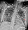
Figure 1
Figure 1. Portable 30 degrees upright anteroposterior chest radiograph demonstrates numerous bilateral lung opacities with central areas of lucency (red arrows). There is no obvious pleural effusion. The cardiac silhouette appears enlarged.
Figure 2
Figure 2. Axial CT images of the thorax with lung algorithm demonstrate numerous cavitary lesions throughout all lobes of the lungs. The largest lesion measures 3.5 cm in the greatest dimension in the anterior left upper lobe (red arrow in A). There are scattered areas of bronchiectasis (yellow arrow in C).
Figure 3
Figure 3. Axial (A) and coronal (B) CT images of the chest in the lung window demonstrate dependent ground-glass opacities involving bilateral lower lobes (red arrows in A). Cavitary lesions are present throughout the lungs (yellow arrows in B).
Figure 4
Figure 4. Axial (A), coronal (B), and sagittal (C) CT images of the abdomen with intravenous contrast demonstrate a 6.9 cm linear hypodensity in the splenic parenchyma (red arrow in A, B, and C) with a small amount of perisplenic fluid (yellow arrow in B), consistent with a grade 3 laceration.
Figure 5
Figure 5. Portable semi-erect anterioposterior radiograph of the chest demonstrates a left pneumothorax (delineated by red arrows in A) without mediastinal shift. A subsequent radiograph of the chest demonstrates a large right pneumothorax (delineated by red arrows in B) with a possible right-to-left mediastinal shift. Thin, linear, metallic foreign bodies are noted involving the base of the neck (yellow arrows in B). A left-sided chest tube is present (dark gray arrow in B).
Figure 6
Figure 6. Portable semi-erect anterioposterior radiographs of the chest (A, B) demonstrate bilateral large pleural effusions with areas of loculations. Focal areas of aeration are present (red arrows in A). The patient is status post-sternotomy with sternal wires and closure devices in place. Numerous thoracic drains are in place (yellow arrows in B).
Figure 7
Figure 7. Coronal contrast-enhanced CT images of the chest in the lung window (A, B) demonstrate near-complete opacification of all lobes of the lungs bilaterally, with a small focal area of aeration involving the left lower lobe (green arrow in A). Multiple cystic collections are also present throughout the lungs (red arrows in B).

References

    1. United Nations on Drugs and Crime. Prevalence-general. Published. [ May; 2023 ]. 2021. https://dataunodc.un.org/data/drugs/Prevalence-general https://dataunodc.un.org/data/drugs/Prevalence-general
    1. United Nations on Drugs and Crime. People Who Inject Drugs. Published. [ May; 2023 ]. 2021. https://dataunodc.un.org/data/drugs/People Injucting drugs https://dataunodc.un.org/data/drugs/People Injucting drugs
    1. Injection drug use and HIV/AIDS transmission in China. Chu TX, Levy JA. Cell Res. 2005;15:865–869. - PubMed
    1. Needle embolism in intravenous drug abuse. Monroe EJ, Tailor TD, McNeeley MF, Lehnert BE. Radiol Case Rep. 2012;7:714. - PMC - PubMed
    1. Intravenous drug users and broken needles--a hidden risk? Norfolk GA, Gray SF. Addiction. 2003;98:1163–1166. - PubMed

Publication types

LinkOut - more resources