

Comparison of bilateral differential characteristics of corneal biomechanics between keratoconus and normal eyes
- PMID: 37324412
- PMCID: PMC10267412
- DOI: 10.3389/fbioe.2023.1163223
Comparison of bilateral differential characteristics of corneal biomechanics between keratoconus and normal eyes
Abstract
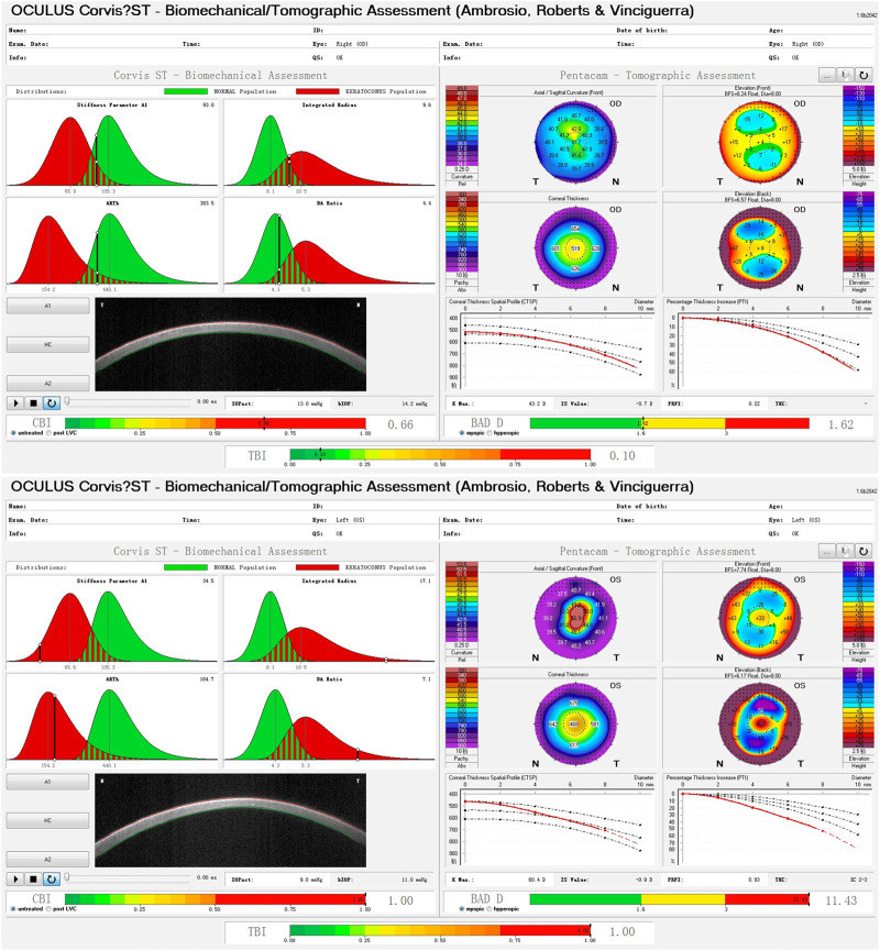
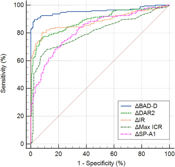
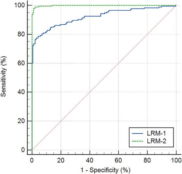
Purpose: To compare bilateral differences in corneal biomechanics between keratoconus and normal eyes. Methods: In this case-control study, 346 eyes of 173 patients (aged 22.1 ± 6.1 years) with keratoconus (KC group) and 378 eyes of 189 patients (aged 26.7 ± 5.6 years) with ametropia (control group) were enrolled. Corneal tomography and biomechanical properties were examined using Pentacam HR and Corvis ST, respectively. The corneal biomechanical parameters were compared between eyes with forme fruste keratoconus (FFKC) and normal eyes. Bilateral differences in corneal biomechanical parameters were compared between the KC and control groups. Receiver operating characteristic (ROC) analysis was used to assess discriminative efficacies. Results: The areas under the ROC curves (AUROCs) of stiffness parameter at the first applanation (SP-A1) and Tomographic and Biomechanical Index (TBI) for identifying FFKC were 0.641 and 0.694, respectively. The bilateral differential values of major corneal biomechanical parameters were significantly increased in the KC group (all p < 0.05), except for the Corvis Biomechanical Index (CBI). The AUROCs of the bilateral differential values of the deformation amplitude ratio at 2 mm (ΔDAR2), Integrated Radius (ΔIR), SP-A1 (ΔSP-A1), and the maximum inverse concave radius (ΔMax ICR) for discriminating keratoconus were 0.889, 0.884, 0.826, and 0.805, respectively. The Logistic Regression Model-1 (comprising of ΔDAR2, ΔIR, and age) and the Logistic Regression Model-2 (comprising of ΔIR, ΔARTh, ΔBAD-D, and age) had AUROCs of 0.922 and 0.998, respectively, for discriminating keratoconus. Conclusion: The bilateral asymmetry of corneal biomechanics was significantly increased in keratoconus compared with normal eyes, which may be helpful for the early detection of keratoconus.
Keywords: biomechanics; corneal ectasia; forme fruste keratoconus; keratoconus; tomography; topography.
Copyright © 2023 Xian, Zhao, Sun, Zhang, Ding, Liu, Li, Ding, Jiang, Zhou and Shen.
Conflict of interest statement
The authors declare that the research was conducted in the absence of any commercial or financial relationships that could be construed as a potential conflict of interest.
Figures
Similar articles
-
Application of a scheimpflug-based biomechanical analyser and tomography in the early detection of subclinical keratoconus in chinese patients.BMC Ophthalmol. 2021 Sep 20;21(1):339. doi: 10.1186/s12886-021-02102-2. BMC Ophthalmol. 2021. PMID: 34544392 Free PMC article.
-
Comparison of the morphological and biomechanical characteristics of keratoconus, forme fruste keratoconus, and normal corneas.Semin Ophthalmol. 2021 Nov 17;36(8):671-678. doi: 10.1080/08820538.2021.1896752. Epub 2021 Mar 18. Semin Ophthalmol. 2021. PMID: 33734947
-
Biomechanical properties analysis of forme fruste keratoconus and subclinical keratoconus.Graefes Arch Clin Exp Ophthalmol. 2023 May;261(5):1311-1320. doi: 10.1007/s00417-022-05916-y. Epub 2022 Nov 28. Graefes Arch Clin Exp Ophthalmol. 2023. PMID: 36441226
-
Early diagnosis of keratoconus using corneal biomechanics and OCT derived technologies.Eye Vis (Lond). 2025 May 12;12(1):18. doi: 10.1186/s40662-025-00435-3. Eye Vis (Lond). 2025. PMID: 40350508 Free PMC article. Review.
-
Progress of corneal morphological examination combined with biomechanical examination in preoperative screening for keratorefractive surgery.Indian J Ophthalmol. 2023 Jun;71(6):2369-2378. doi: 10.4103/ijo.IJO_1377_22. Indian J Ophthalmol. 2023. PMID: 37322646 Free PMC article. Review.
Cited by
-
Corneal Stress Distribution Evolves from Thickness-Driven in Normal Corneas to Curvature-Driven with Progression in Keratoconus.Ophthalmol Sci. 2023 Jul 20;4(2):100373. doi: 10.1016/j.xops.2023.100373. eCollection 2024 Mar-Apr. Ophthalmol Sci. 2023. PMID: 37868791 Free PMC article.
-
Keratoconus cone location influences ocular biomechanical parameters measured by the Ocular Response Analyzer.Eye Vis (Lond). 2024 Jan 3;11(1):2. doi: 10.1186/s40662-023-00371-0. Eye Vis (Lond). 2024. PMID: 38167119 Free PMC article.
-
Intraocular pressure and ocular biomechanical parameters vary between generations of the ocular response analyzer in healthy and ectatic eyes.Front Med (Lausanne). 2025 Jul 24;12:1605641. doi: 10.3389/fmed.2025.1605641. eCollection 2025. Front Med (Lausanne). 2025. PMID: 40776931 Free PMC article.
-
The application of corneal biomechanical interocular asymmetry for the diagnosis of keratoconus and subclinical keratoconus.Front Bioeng Biotechnol. 2023 Oct 6;11:1266940. doi: 10.3389/fbioe.2023.1266940. eCollection 2023. Front Bioeng Biotechnol. 2023. PMID: 37869711 Free PMC article.
-
Squishy matters - Corneal mechanobiology in health and disease.Prog Retin Eye Res. 2024 Mar;99:101234. doi: 10.1016/j.preteyeres.2023.101234. Epub 2024 Jan 2. Prog Retin Eye Res. 2024. PMID: 38176611 Free PMC article. Review.
References
LinkOut - more resources
Full Text Sources