

Assessment and diagnosis of right ventricular failure-retrospection and future directions
- PMID: 37324632
- PMCID: PMC10268009
- DOI: 10.3389/fcvm.2023.1030864
Assessment and diagnosis of right ventricular failure-retrospection and future directions
Abstract
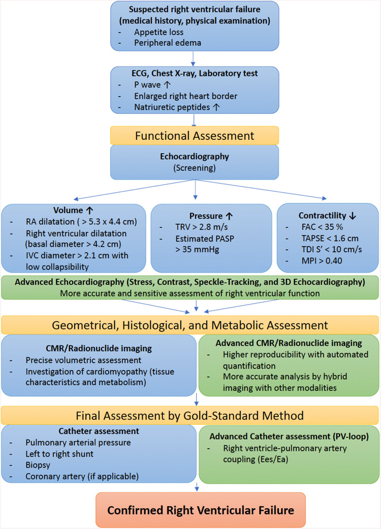
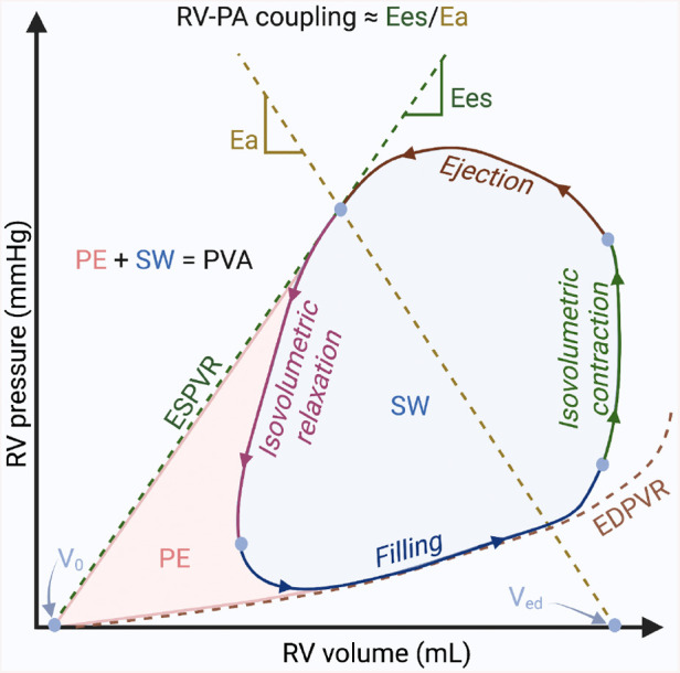
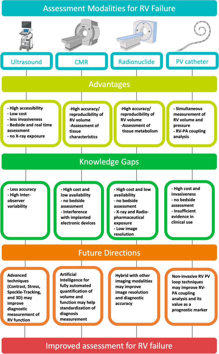
The right ventricle (RV) has a critical role in hemodynamics and right ventricular failure (RVF) often leads to poor clinical outcome. Despite the clinical importance of RVF, its definition and recognition currently rely on patients' symptoms and signs, rather than on objective parameters from quantifying RV dimensions and function. A key challenge is the geometrical complexity of the RV, which often makes it difficult to assess RV function accurately. There are several assessment modalities currently utilized in the clinical settings. Each diagnostic investigation has both advantages and limitations according to its characteristics. The purpose of this review is to reflect on the current diagnostic tools, consider the potential technological advancements and propose how to improve the assessment of right ventricular failure. Advanced technique such as automatic evaluation with artificial intelligence and 3-dimensional assessment for the complex RV structure has a potential to improve RV assessment by increasing accuracy and reproducibility of the measurements. Further, noninvasive assessments for RV-pulmonary artery coupling and right and left ventricular interaction are also warranted to overcome the load-related limitations for the accurate evaluation of RV contractile function. Future studies to cross-validate the advanced technologies in various populations are required.
Keywords: assessment; future development.; knowledge gap; parameters; right ventricular failure.
© 2023 Ro, Sato, Ijuin, Sela, Fior, Heinsar, Kim, Chan, Nonaka, Lin, Bassi, Platts, Obonyo, Suen and Fraser.
Conflict of interest statement
The authors declare that the research was conducted in the absence of any commercial or financial relationships that could be construed as a potential conflict of interest.
Figures
References
-
- Voelkel NF, Quaife RA, Leinwand LA, Barst RJ, McGoon MD, Meldrum DR, et al. Right ventricular function and failure: report of a national heart, lung, and blood institute working group on cellular and molecular mechanisms of right heart failure. Circulation. (2006) 114(17):1883–91. 10.1161/CIRCULATIONAHA.106.632208 - DOI - PubMed
-
- Heidenreich PA, Bozkurt B, Aguilar D, Allen LA, Byun JJ, Colvin MM, et al. 2022 Aha/Acc/Hfsa guideline for the management of heart failure: a report of the American college of cardiology/American heart association joint committee on clinical practice guidelines. Circulation. (2022) 145(18):e895–1032. 10.1161/CIR.0000000000001063 - DOI - PubMed
-
- McDonagh TA, Metra M, Adamo M, Gardner RS, Baumbach A, Böhm M, et al. 2021 ESC guidelines for the diagnosis and treatment of acute and chronic heart failure: developed by the task force for the diagnosis and treatment of acute and chronic heart failure of the European society of cardiology (ESC). with the special contribution of the heart failure association (HFA) of the ESC. Eur J Heart Fail. (2022) 24(1):4–131. 10.1002/ejhf.2333 - DOI - PubMed
Publication types
LinkOut - more resources
Full Text Sources
Miscellaneous