Review
. 2022 May 23;2(6):20210157.
doi: 10.1002/EXP.20210157. eCollection 2022 Dec.

Recent applications of immunomodulatory biomaterials for disease immunotherapy

Affiliations
Review

Recent applications of immunomodulatory biomaterials for disease immunotherapy

Huaxing Dai et al. Exploration (Beijing). .

Abstract

Immunotherapy is used to regulate systemic hyperactivation or hypoactivation to treat various diseases. Biomaterial-based immunotherapy systems can improve therapeutic effects through targeted drug delivery, immunoengineering, etc. However, the immunomodulatory effects of biomaterials themselves cannot be neglected. In this review, we outline biomaterials with immunomodulatory functions discovered in recent years and their applications in disease treatment. These biomaterials can treat inflammation, tumors, or autoimmune diseases by regulating immune cell function, exerting enzyme-like activity, neutralizing cytokines, etc. The prospects and challenges of biomaterial-based modulation of immunotherapy are also discussed.

Keywords: biomaterials; disease treatment; immunomodulatory effect; immunotherapy.


Conflict of interest statement

The authors declare no conflict of interest.

Figures

FIGURE 1
FIGURE 1
Diagram of representative inorganic and organic biomaterials with immunomodulatory functions
FIGURE 2
FIGURE 2
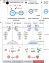
(A) Schematic illustration depicting that artificially reprogrammed HION@Macs target tumors through active chemotaxis and magnet guidance, produce inflammatory factors (such as TNF‐α, NO, and ROS) to suppress tumors, and re‐educate in situ M2 macrophages into a proinflammatory M1 phenotype for synergistic cancer‐specific therapy. Reproduced with permission.[ 43 ] Copyright 2019, Wiley‐VCH. (B) Mn2+ ions enhance the sensitivity of the cGAS‐STING signaling pathway to DNA viruses. Reproduced with permission.[ 55 ] Copyright 2020, Elsevier. (C) Schematic diagram of the synthesis and mechanism of Cu5.4O ultrasmall nanoparticles. Reproduced with permission.[ 69 ] Copyright 2018, Springer Nature
FIGURE 3
FIGURE 3
Silicon nanoparticles can regulate T cell activation and improve immune checkpoint therapy. (A) Nonfunctionalized ultrasmall silica nanoparticles directly activate T cells selectively. Reproduced with permission.[ 79 ] Copyright 2018, American Chemical Society. (B) A single intraperitoneal injection of mesoporous silica nanoparticles (MSNs) can improve the efficacy of PD‐1 in tumor immunotherapy. Reproduced under the terms of the Creative Commons Attribution‐NonCommercial license.[ 90 ] Copyright 2021, Society for Immunotherapy of Cancer
FIGURE 4
FIGURE 4
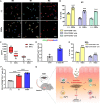
Graphene quantum dots (GQDs) promote M2 polarization of macrophages. (A,B) Primary CD14+ macrophage‐like cells are polarized into M0‐, M1‐, and M2‐type cells in the presence of GQDs. (C) Cytokine concentration in the supernatant of M1 macrophages. (D) M1‐induced cells in the presence of GQDs were cocultured with naïve CD4+ T cells supplemented with IL‐2 and TGF‐β1, and the proportions of Tregs were investigated by flow cytometry. (E) Flow cytometry analysis of M2b polarization in vivo. (F) Flow cytometry to evaluate the proportion of M2b macrophages in the peritoneum of mice with chronic colitis. (G) Schematic diagram of the mechanism of action of GQDs. Reproduced under the terms of the Creative Commons Attribution‐NonCommercial license.[ 102 ] Copyright 2020, American Association for the Advancement of Science
FIGURE 5
FIGURE 5
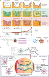
(A) The use of L‐ or D‐MAP in wound healing models. In the case of d‐MAP, the hydrogel activates the adaptive immune system, leading to tissue remodeling and skin regeneration as the adaptive immune system degrades the D‐MAP scaffold. Reproduced with permission.[ 117 ] Copyright 2021, Springer Nature. (B) Schematic of the minimalist design of the PC7A nanovaccine. Reproduced with permission.[ 121 ] Copyright 2017, Springer Nature. (C) Cationic nanoparticles have high free DNA binding capacity, which can effectively inhibit the activation of TLR9 and inhibit the inflammatory response of rheumatoid arthritis. Reproduced with permission.[ 125 ] Copyright 2018, Springer Nature
FIGURE 6
FIGURE 6
(A) Schematic diagram and TEM image of HABN obtained by self‐assembly of hyaluronic acid (HA)‐bilirubin (BR). HABN accumulates in the inflamed colon and has a therapeutic effect on acute colitis. Reproduced with permission.[ 140 ] Copyright 2020, Springer Nature. (B) Schematic representation of the utilization of DNA nanodevices for efficient cancer immunotherapy. Reproduced with permission.[ 146 ] Copyright 2020, Springer Nature
FIGURE 7
FIGURE 7
(A) Preparation of anti‐COVID‐19 nanodecoys by fusing cellular membrane nanovesicles derived from genetically edited 293T/ACE2 and THP‐1 cells. Nanodecoys fight COVID‐19 infection by neutralizing SARS‐CoV‐2 and inflammatory cytokines. Reproduced under the terms of the Creative Commons Attribution‐NonCommercial license.[ 164 ] Copyright 2020, United States National Academy of Sciences. (B) Schematic illustration of the inhaled ACE2‐engineered microfluidic microsphere for neutralization of COVID‐19 and calming of the cytokine storm. Reproduced with permission.[ 167 ] Copyright 2020, Elsevier
FIGURE 8
FIGURE 8
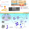
(A) A schematic diagram of neutrophil membrane‐coated nanoparticles inhibiting synovial inflammation and improving joint destruction in inflammatory arthritis. Reproduced with permission.[ 173 ] Copyright 2018, Springer Nature. (B) Mesenchymal stem cell exosomes promote cartilage repair and regeneration through various mechanisms, such as promoting cartilage proliferation, migration, and matrix synthesis, reducing cell apoptosis, and regulating the immune response. Reproduced with permission.[ 195 ] Copyright 2018, Elsevier. (C) Schematic diagram of the preparation and mechanism of implantable blood clot vaccine. Reproduced under the terms of the Creative Commons Attribution‐NonCommercial license.[ 215 ] Copyright 2020, American Association for the Advancement of Science

References

    1. Oyler‐Yaniv A., Oyler‐Yaniv J., Whitlock B. M., Liu Z., Germain R. N., Huse M., Altan‐Bonnet G., Krichevsky O., Immunity 2017, 46, 609. - PMC - PubMed
    1. Rankin L., Artis D., Cell 2018, 173, 554. - PubMed
    1. Disis M., J. Clin. Oncol. 2010, 28, 4531. - PMC - PubMed
    1. Borgoni S., Kudryashova K. S., Burka K., de Magalhaes J. P., Ageing Res. Rev. 2021, 70, 101410. - PubMed
    1. Wykes M., Lewin S., Nat. Rev. Immunol. 2018, 18, 91. - PMC - PubMed

LinkOut - more resources