. 2023 May 11;19(9):2663-2677.
doi: 10.7150/ijbs.81809. eCollection 2023.

Heme-binding protein 1 delivered via pericyte-derived extracellular vesicles improves neurovascular regeneration in a mouse model of cavernous nerve injury

Affiliations

Heme-binding protein 1 delivered via pericyte-derived extracellular vesicles improves neurovascular regeneration in a mouse model of cavernous nerve injury

Jiyeon Ock et al. Int J Biol Sci. .

Abstract

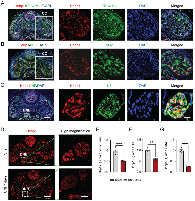
As a peripheral nerve injury disease, cavernous nerve injury (CNI) caused by prostate cancer surgery and other pelvic surgery causes organic damage to cavernous blood vessels and nerves, thereby significantly attenuating the response to phosphodiesterase-5 inhibitors. Here, we investigated the role of heme-binding protein 1 (Hebp1) in erectile function using a mouse model of bilateral CNI, which is known to promote angiogenesis and improve erection in diabetic mice. We found a potent neurovascular regenerative effect of Hebp1 in CNI mice, demonstrating that exogenously delivered Hebp1 improved erectile function by promoting the survival of cavernous endothelial-mural cells and neurons. We further found that endogenous Hebp1 delivered by mouse cavernous pericyte (MCP)-derived extracellular vesicles promoted neurovascular regeneration in CNI mice. Moreover, Hebp1 achieved these effects by reducing vascular permeability through regulation of claudin family proteins. Our findings provide new insights into Hebp1 as a neurovascular regeneration factor and demonstrate its potential therapeutic application to various peripheral nerve injuries.

Keywords: Cavernous nerve injury; Extracellular vesicles; Heme-binding protein 1; Neurovascular regeneration; Peripheral nerve injury.


Conflict of interest statement

Competing Interests: The authors have declared that no competing interest exists.

Figures

Figure 1
Figure 1
Hebp1 expression is decreased in the penis of CNI-induced ED mice. (A-C) Representative (left) and high-magnification (right) images of double-immunofluorescence staining of normal mouse CC and DNB tissue using antibodies against Hebp1 (red), PECAM-1 (A; green), NG2 (B; green), and NF (C; green). Nuclei were labeled with the DNA dye DAPI (blue). Scale bars, 400 µm (left), 100 µm (right top and middle), and 25 µm (right bottom). (D) Representative (left) and high-magnification (right) images of Hebp1 (red) immunofluorescence staining in penis tissues from sham-operated and CNI-induced ED mice (7 days after CNI). (E-G) Quantitative analyses of Hebp1 expression in total penis tissue (E), the CC (F), and DNBs (G) were performed using an image analyzer. Results are presented as means ± SEM (n = 4; **P < 0.01, ***P < 0.001 vs. sham operation groups). The relative ratio of the sham operation group was defined as 1. CC, corpus cavernosum; DNBs, dorsal nerve bundles; ED, erectile dysfunction; CNI, cavernous nerve injury; NF, neurofilament; DAPI, 4,6-diamidino-2-phenylindole.
Figure 2
Figure 2
Hebp1 protein improves erectile function in CNI-induced ED mice. (A) Representative ICP responses for sham-operated and CNI-induced ED mice stimulated 1 week after two intracavernous injections (administered on days -3 and 0) of Hebp1 protein (1.0 and 5 µg per mouse). The stimulus interval is indicated by a solid bar. (B, C) Ratios of mean maximal ICP and total ICP (area under the curve) to MSBP were calculated for each group. Results are presented as means ± SEM (n = 5). (D, E) Immunofluorescence staining of CC and DNB tissues for PECAM-1 (D; green), NG2 (D; red), and NF (E; green) after ICP studies. Nuclei were labeled with the DNA dye DAPI (blue). Scale bars, 100 µm (D) and 25 µm (E). (F) NF (green) immunofluorescence staining in mouse MPGs exposed to LPS (2.5 μg/ml) and then treated with Hebp1 (5 µg/ml) or PBS for 5 days. Scale bar, 100 μm. (G-I) Quantitative analysis of CC endothelial cell (G, PECAM-1), pericyte (H, NG2) and neuronal cell (I, NF) contents using an image analyzer. Results are presented as means ± SEM (n = 5). (J) Neurite sprouting, quantified using an image analyzer. Results are presented as means ± SEM (n = 4; *P < 0.05, **P < 0.01, ***P < 0.001). The relative ratio of the sham operation or control group was defined as 1. ED, erectile dysfunction; CNI, cavernous nerve injury; NF, neurofilament; DAPI, 4,6-diamidino-2-phenylindole; MPG, major pelvic ganglion; PBS, phosphate-buffered saline; LPS, lipopolysaccharide; MSBP, mean systolic blood pressure; DNB, dorsal nerve bundles; ns, not significant.
Figure 3
Figure 3
Hebp1 protein reduces apoptosis of pericytes and induces their proliferation in the penis of CNI-induced ED mice. (A) TUNEL assay (green) and NG-2 (red) immunofluorescence staining of mouse cavernous tissues in sham-operated and CNI-induced ED mice 1 week after two intracavernous injections (administered on days -3 and 0) of Hebp1 protein (5 µg per mouse in 20 µl). Nuclei were labeled with the DNA dye DAPI. Scale bar, 100 μm. (B) Immunofluorescence staining of CC tissue for phospho-histone H3 (PH3; red) and PDGFRβ (green) using the same samples as above. Scale bar, 100 μm. (C, D) Number of TUNEL-positive (C) or PH3-positive (D) pericytes, quantified using an image analyzer. Results are presented as means ± SEM (n = 4). TUNEL, terminal deoxynucleotidyl transferase-mediated deoxyuridine triphosphate nick end labeling; DAPI, 4,6-diamidino-2-phenylindole; ED, erectile dysfunction; CNI, cavernous nerve injury; PBS, phosphate-buffered saline; ns, not significant.
Figure 4
Figure 4
Hebp1 is delivered by MCP-EVs and contributes to nerve regeneration. (A) Representative transmission electron microscopy (TEM) phase images for detecting isolated MCP-EVs. Scale bar = 100 nm. (B) Representative Western blot for Hebp1, EV positive markers (CD63, CD9 and CD81) or EV negative marker (GM130) in MCPs and MCP-EVs. (C, D) Expression of Hebp1 in MCP-EVs infected with lentivirus expressing shHebp1 or shCon at three doses (1 × 10³, 5 × 10⁴, and 1 × 10⁴ TU/ml culture medium) for at least 3 days. (E, F) Densitometric quantification of Hebp1 protein bands using an image analyzer. Results are presented as means ± SEM (n = 4). (G, H) Immunofluorescence staining for NF in mouse MPG tissues exposed to LPS (2.5 μg/ml) and other indicated conditions for 5 days (G). Lengths of NF-positive neurites in MPG tissues, quantified using an image analyzer (H). Results are presented as means ± SEM (n = 4). Scale bar, 100 µm. (I, J) Migration assays of mouse Schwann cells exposed to LPS (2.5 μg/ml) and other indicated conditions for 24 hours (I). Number of migrated cells in the red dotted rectangle, quantified using an image analyzer (J). Results are presented as means ± SEM (n = 4; **P < 0.01, ***P < 0.001). The relative ratio of the shCon (control) group was defined as 1. MCPs, mouse cavernous pericytes; EVs, extracellular vesicles; LPS, lipopolysaccharide; MPG, major pelvic ganglion; PBS, phosphate-buffered saline; ns, not significant.
Figure 5
Figure 5
Hebp1 is delivered by MCP-EVs and contributes to nerve regeneration. (A) Representative ICP responses for sham-operated or CNI-induced ED mice stimulated 1 week after two intracavernous injections (administered on days -3 and 0) of shCon MCP-EVs (10 µg per mouse in 20 µl PBS), shHebp1 MCP-EVs (10 µg per mouse in 20 µl PBS), or PBS only. The stimulus interval is indicated by a solid bar. (B, C) Ratios of mean maximal ICP and total ICP (area under the curve) to MSBP, calculated for each group. Results are presented as means ± SEM (n = 5). (D, E) Immunofluorescence staining for PECAM-1 (D; green), NG2 (D; red), and NF (E; green) in CC and DNB tissues after ICP studies. Nuclei were labeled with the DNA dye DAPI (blue). Scale bars, 100 µm (D) and 25 µm (E). (F-H) Quantitative analysis of CC endothelial cell (F, PECAM-1), pericyte (G, NG2) and neuronal cell (H, NF) contents using an image analyzer. Results are presented as means ± SEM (n = 4; *P < 0.05, **P < 0.01, ***P < 0.001). The relative ratio of the sham operation group was defined as 1. ED, erectile dysfunction; MCPs, mouse cavernous pericytes; EVs, extracellular vesicles; PBS, phosphate-buffered saline; MSBP, mean systolic blood pressure; DAPI, 4,6-diamidino-2-phenylindole; DNB, dorsal nerve bundles; ns, not significant.
Figure 6
Figure 6
Identification of the signaling pathway mediating the effects of MCP-EV-delivered Hebp1 in CNI-induced ED mice. (A) Heatmap of log2-normalized intensities of significantly differentially expressed genes in Signaling Explorer Antibody Arrays from sham-operated and CNI-induced ED mice that received two intracavernous injections (administered on days -3 and 0) of shHebp1 MCP-EVs (10 µg per mouse in 20 µl PBS), shCon MCP-EVs (10 µg per mouse in 20 µl PBS), or PBS only. Color density shows fold-changes, with different multiples indicated by different colors. Red, up-regulated; blue, down-regulated. (B) Proteins significantly regulated by MCP-EV-delivered Hebp1 in CNI-induced ED mice, identified by a PPI network of differentially expressed genes. The thickness of lines corresponds to the strength of the interaction between the proteins. Red box highlights claudin family genes. (C) Representative Western blots for claudin -1, -2, -3, and -11 in CC tissue from the indicated groups. (D) Ox-LDL (green) immunostaining in CC tissues from the indicated groups. Nuclei were labeled with the DNA dye DAPI (blue). Scale bar, 100 µm. (E-I) Expression of the indicated proteins, quantified by densitometric analysis of the corresponding protein bands, and Ox-LDL immunopositive areas, determined using an image analyzer. Results are presented as means ± SEM (n = 4; *P < 0.05, ***P < 0.001). The relative ratio of the sham operation group was defined as 1. ED, erectile dysfunction; MCPs, mouse cavernous pericytes; EVs, extracellular vesicles; PBS, phosphate-buffered saline; Ox-LDL, oxidized low-density lipoprotein; DAPI, 4,6-diamidino-2-phenylindole; ns, not significant.
Figure 7
Figure 7
Schematic depiction of the proposed mechanism for MCP-EV-delivered Hebp1 in CNI-induced ED mice. EVs, extracellular vesicles; ECs, endothelial cells; Ox-LDL, oxidized low-density lipoprotein; CNI, cavernous nerve injury; ED, erectile dysfunction.

References

    1. Taketani S, Adachi Y, Kohno H, Ikehara S, Tokunaga R, Ishii T. Molecular characterization of a newly identified heme-binding protein induced during differentiation of urine erythroleukemia cells. J Biol Chem. 1998;273:31388–94. - PubMed
    1. Chua JJE. HEBP1 - An early trigger for neuronal cell death and circuit dysfunction in Alzheimer's disease. Semin Cell Dev Biol. 2022. - PubMed
    1. Yagensky O, Kohansal-Nodehi M, Gunaseelan S, Rabe T, Zafar S, Zerr I, Increased expression of heme-binding protein 1 early in Alzheimer's disease is linked to neurotoxicity. Elife. 2019. 8. - PMC - PubMed
    1. Devosse T, Dutoit R, Migeotte I, De Nadai P, Imbault V, Communi D. et al. Processing of HEBP1 by cathepsin D gives rise to F2L, the agonist of formyl peptide receptor 3. J Immunol. 2011;187:1475–85. - PubMed
    1. Yin GN. Pericyte-derived heme-binding protein 1 promotes angiogenesis and improves erectile function in diabetic mice. Investig Clin Urol. 2022;63:464–74. - PMC - PubMed

Publication types