

Case Reports
doi: 10.1016/j.hrcr.2023.02.003.
eCollection 2023 May.
Nonsustained premature atrial contraction ablation guided by single-beat mapping using charge density mapping
Affiliations
- PMID: 37324964
- PMCID: PMC10265183
- DOI: 10.1016/j.hrcr.2023.02.003
Item in Clipboard
Case Reports
Nonsustained premature atrial contraction ablation guided by single-beat mapping using charge density mapping
HeartRhythm Case Rep.
.
No abstract available
Keywords: Atrial tachycardia; Catheter ablation; Left atrial appendage; Premature atrial contraction; Pulmonary veins; Sequential mapping; Supraventricular tachycardia; Trigger mapping.
Figures
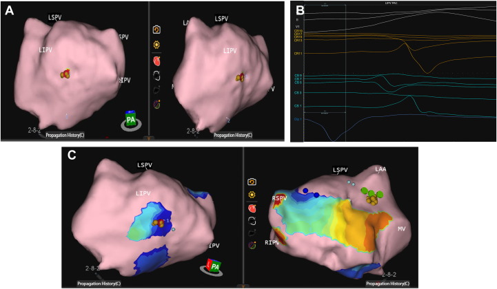
A: Premature atrial contraction (PAC) location: posterior base of the left inferior pulmonary vein (LIPV; red disc). Dipole 1 at the earliest source of charge (ie, origin of PAC). Gold dots indicate where ablation occurred. B: Dipole 1 (blue) signal at origin of the LIPV PAC map∼25 ms pre–P wave. CR unipolar signals in orange; CS unipolar signals in light blue; body surface leads II, V1, and V6 in white. C: Map visualization of the activation at the location of abrupt change in dipole activation around LIPV from -QS to R wave on the unipolar leads. Gold dots indicate where ablation occurred. LAA = left atrial appendage; LSPV = left superior pulmonary vein; RIPV = right inferior pulmonary vein; RSPV = right superior pulmonary vein; MV = mitral valve; PA = posterior anterior.

A: Premature atrial contraction (PAC) location: anterior aspect of left atrial appendage (LAA; green discs). Tachycardia mapped to the location of the LAA PAC. Dipole 1 at the earliest source of charge on LAA PAC map. Gold dots indicate where ablation occurred. B: Dipole 1 signal at origin of charge activation on the LAA PAC map ∼25 ms pre–P wave. CR unipolar signals in orange; CS unipolar signals in light blue; body surface leads II, V1, V6 in white. LSPV = left superior pulmonary vein; RIPV = right inferior pulmonary vein; RSPV = right superior pulmonary vein; MV = mitral valve; R.PA = right posterior anterior.
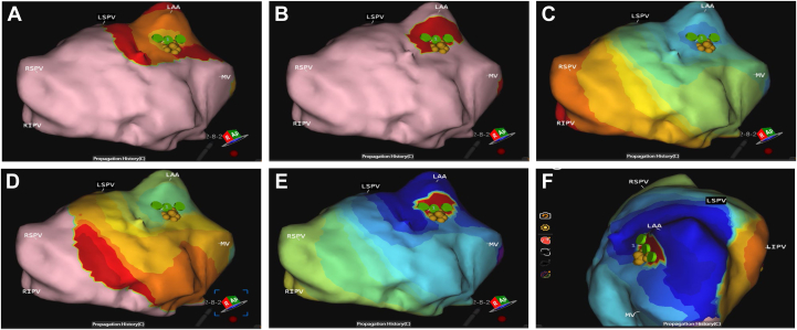
A–F: Series of images visualizing the focal activation of our tachycardia source via single-position mapping. Origin at the anterior aspect of left atrial appendage (LAA), same area of interest localized while mapping premature atrial contractions (PACs). Secondary firing at source of PAC within the single P-wave morphology used to map. LSPV = left superior pulmonary vein; RIPV = right inferior pulmonary vein; RSPV = right superior pulmonary vein; MV = mitral valve; R.PA = right posterior anterior.
Similar articles
-
[Electrophysiological findings and ablation strategies in patients with atrial tachyarrhythmias after left atrial circumferential ablation in the treatment of atrial fibrillation].Zhonghua Xin Xue Guan Bing Za Zhi. 2007 Feb;35(2):119-22. Zhonghua Xin Xue Guan Bing Za Zhi. 2007. PMID: 17445402 Chinese.
-
Advanced electrophysiologic mapping systems: an evidence-based analysis.Ont Health Technol Assess Ser. 2006;6(8):1-101. Epub 2006 Mar 1. Ont Health Technol Assess Ser. 2006. PMID: 23074499 Free PMC article.
-
Single-beat global atrial mapping facilitates the treatment of short-lived atrial tachycardias and infrequent premature atrial contractions.J Interv Card Electrophysiol. 2023 Jun;66(4):951-959. doi: 10.1007/s10840-022-01405-8. Epub 2022 Oct 25. J Interv Card Electrophysiol. 2023. PMID: 36282368 Free PMC article.
-
Emergent Zero-Fluoroscopy Mapping and Thoracoscopic Ectomy of Appendage in Pregnant Women with Life-Threatening Atrial Tachycardia: A Case Report and Literature Review.Medicina (Kaunas). 2023 Mar 8;59(3):528. doi: 10.3390/medicina59030528. Medicina (Kaunas). 2023. PMID: 36984528 Free PMC article. Review.
-
[Catheter ablation of supraventricular tachycardia].Herzschrittmacherther Elektrophysiol. 2019 Dec;30(4):336-342. doi: 10.1007/s00399-019-00654-x. Epub 2019 Nov 11. Herzschrittmacherther Elektrophysiol. 2019. PMID: 31713026 Review. German.
References
-
- Chieng D., Lahiri A., Sugumar H., et al. Multipolar mapping with the high-density grid catheter compared with conventional point-by-point mapping to guide catheter ablation for focal arrhythmias. J Cardiovasc Electrophysiol. 2020;31:2288–2297. - PubMed
-
- Gunawardene M.A., Hartmann J., Kottmaier M., et al. [Focal atrial tachycardias: diagnostics and therapy] Herzschrittmacherther Elektrophysiol. 2022;33:467–475. - PubMed
-
- Bala G., De Asmundis C., Chierchia G.B. A novel noncontact high-resolution charge density mapping system to guide ablation of complex atrial arrhythmias: overview of device technology and application. Expert Rev Med Devices. 2021;18:343–350. - PubMed
Publication types
LinkOut - more resources
Full Text Sources
