

Organoids
- PMID: 37325195
- PMCID: PMC10270325
- DOI: 10.1038/s43586-022-00174-y
Organoids
Abstract
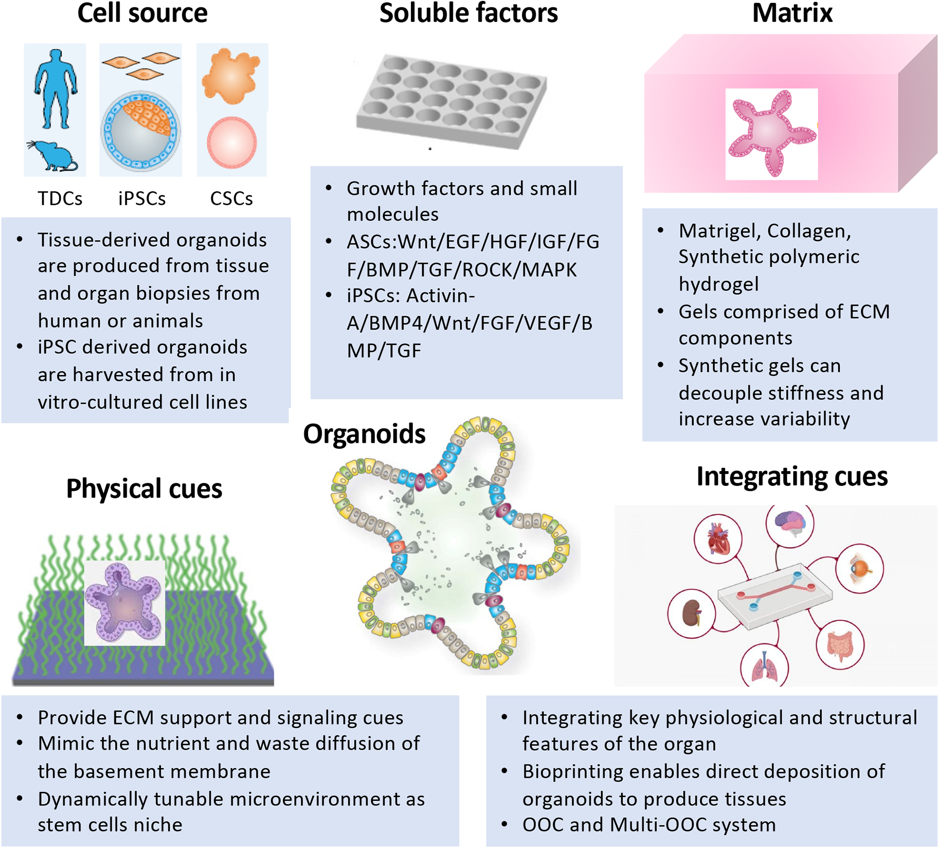
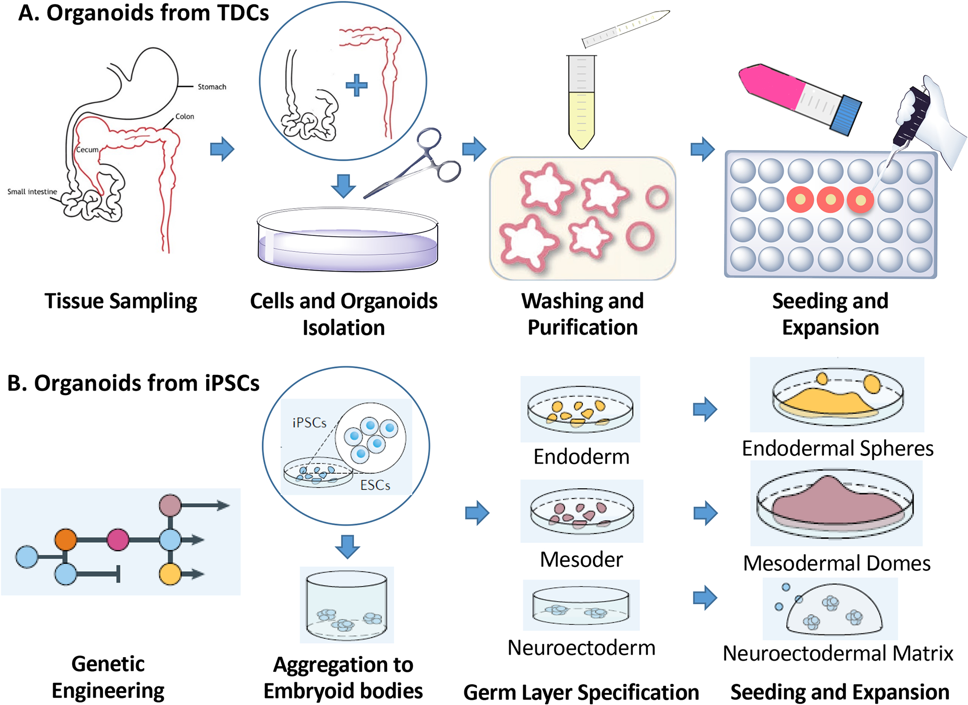
Organoids have attracted increasing attention because they are simple tissue-engineered cell-based in vitro models that recapitulate many aspects of the complex structure and function of the corresponding in vivo tissue. They can be dissected and interrogated for fundamental mechanistic studies on development, regeneration, and repair in human tissues. Organoids can also be used in diagnostics, disease modeling, drug discovery, and personalized medicine. Organoids are derived from either pluripotent or tissue-resident stem (embryonic or adult) or progenitor or differentiated cells from healthy or diseased tissues, such as tumors. To date, numerous organoid engineering strategies that support organoid culture and growth, proliferation, differentiation and maturation have been reported. This Primer serves to highlight the rationale underlying the selection and development of these materials and methods to control the cellular/tissue niche; and therefore, structure and function of the engineered organoid. We also discuss key considerations for generating robust organoids, such as those related to cell isolation and seeding, matrix and soluble factor selection, physical cues and integration. The general standards for data quality, reproducibility and deposition within the organoid community is also outlined. Lastly, we conclude by elaborating on the limitations of organoids in different applications, and key priorities in organoid engineering for the coming years.
Conflict of interest statement
Declaration of competing interests: MH is inventor in several patents on organoid technology. A.So and LL are inventors on a patent on organoid technology. A.So is a founder and owner of Icona BioDx. HY is inventor in several patents on cell-based models. The remaining authors declare no competing interests.
Figures
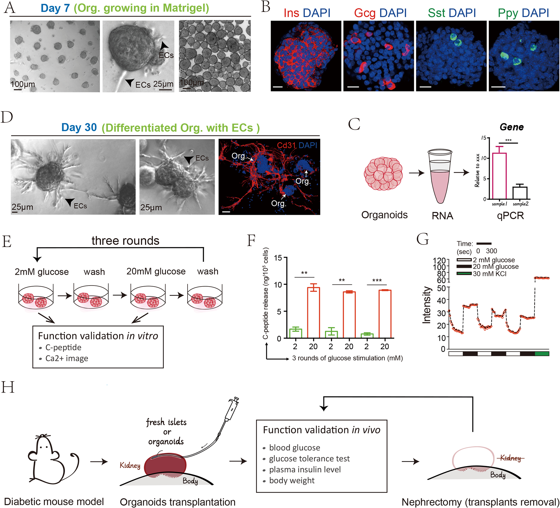
Representative view of pancreatic islet organoids.
Cell types and hormones secretion level validation by immunofluorescence or immunohistochemical staining.
Real-time qPCR analysis for some key transcription factors and differentiation markers.
The maturation of the organoids can be induced through prolonged culture for a total of 30 days at any passage.
Schematic of the pancreatic islet organoids function validation in vitro
Measurement of the secreted C-peptide by ELISA
Intensity of calcium signalling traces imaging indicating the capability of responding acutely to glucose
Schematic of the islet organoids function validation in vivo
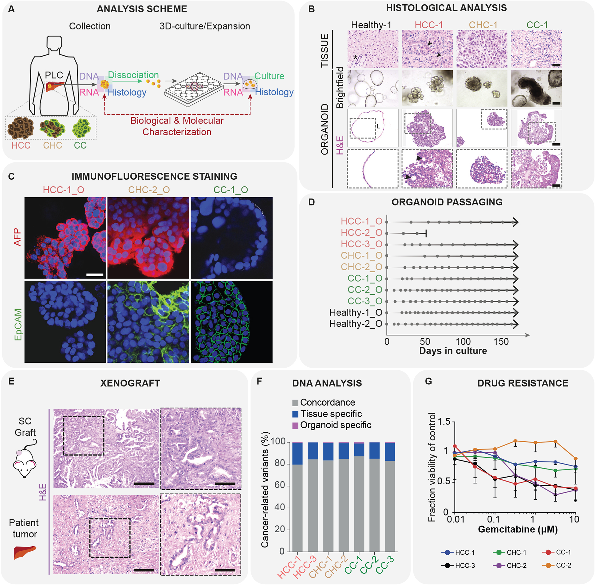
Isolation of cells from patient samples and organoid culture; schematic of tissue isolation and processing;
HCC, hepatocellular carcinoma; CC, cholangiocarcinoma; CHC, combined HCC/CC tumors.
Histological analysis of liver cancer samples: top, tissue; middle, organoid brightfield images; bottom, histological H&E staining of organoids; scale bar, middle row, 100 μm; top and bottom rows, 50 μm.
Analysis of specific marker gene expression: immunofluorescene staining for AFP (hepatocyte/HCC marker; red) and EpCAM (ductal/CC marker; green); blue - DAPI, scale bar, 30 μm.
Organoid formation efficiency: growth and splitting curves; dot, splitting time point, arrow, continuous expansion.
Transplantation into immunodeficient mice: xenograft and histopathology analysis, matching to the patient origianal tissue sample; scale bars, left, 125 μm; right, 62.5 μm
Analysis of genetic changes in the cancer organoids and their concordance to the mutations in the original tumor sample.
Organoid sensitivity to drugs: IC50 curves for gemcitabine treatment.
References
Grants and funding
LinkOut - more resources
Full Text Sources
Other Literature Sources