. 2023 Feb 1;8(5):501-514.
doi: 10.1016/j.jacbts.2022.11.005. eCollection 2023 May.

Obesity-Induced Coronary Microvascular Disease Is Prevented by iNOS Deletion and Reversed by iNOS Inhibition

Affiliations

Obesity-Induced Coronary Microvascular Disease Is Prevented by iNOS Deletion and Reversed by iNOS Inhibition

Soham A Shah et al. JACC Basic Transl Sci. .

Abstract

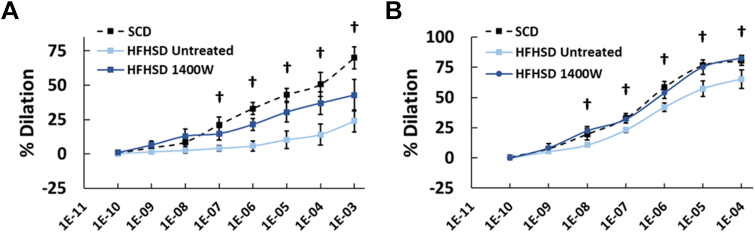
Coronary microvascular disease (CMD) caused by obesity and diabetes is major contributor to heart failure with preserved ejection fraction; however, the mechanisms underlying CMD are not well understood. Using cardiac magnetic resonance applied to mice fed a high-fat, high-sucrose diet as a model of CMD, we elucidated the role of inducible nitric oxide synthase (iNOS) and 1400W, an iNOS antagonist, in CMD. Global iNOS deletion prevented CMD along with the associated oxidative stress and diastolic and subclinical systolic dysfunction. The 1400W treatment reversed established CMD and oxidative stress and preserved systolic/diastolic function in mice fed a high-fat, high-sucrose diet. Thus, iNOS may represent a therapeutic target for CMD.

Keywords: HFpEF; cardiac MRI; coronary microvascular disease; iNOS.


Conflict of interest statement

This study was supported by the National Institutes of Health National Institutes of Biomedical Imaging and Biomedical Engineering (R01 EB001763), Bethesda, Maryland; National Institutes of Health National Heart, Lung, and Blood Institute (R01 HL162872), Bethesda, Maryland; U.S.-Israel Binational Science Foundation grant BSF2017200, Jerusalem, Israel; and National Institute of General Medical Sciences Medical Scientist Training Program T32 grant T32GM007267, Bethesda, Maryland. The authors have reported that they have no relationships relevant to the contents of this paper to disclose.

Figures

None
Graphical abstract
Figure 1
Figure 1
Experimental Protocols (A) Eight-week-old iNOS-/- (B6.129P2-Nos2tm1Lau/J) and WT (C57Bl/6J) mice underwent cardiac magnetic resonance, GTT, and start of radiotelemetry BP monitoring 2 weeks before starting an HFHSD or standard chow diet. Eighteen weeks after each diet, at 28 weeks of age, mice again underwent cardiac magnetic resonance and GTT, and ended radiotelemetry BP monitoring. Coronary arteriole vasoreactivity measurements were performed at 30 weeks of age. (B) Eight-week-old WT (C57Bl/6J) mice underwent cardiac magnetic resonance and GTT 2 weeks before starting an HFHSD. The mice also underwent cardiac magnetic resonance after 10 weeks of HFHSD and thereafter were randomized to 1400W treatment or no treatment and continued HFHSD for 8 weeks. Cardiac magnetic resonance and GTTs were repeated after 8 weeks of 1400W treatment or no treatment. Coronary arteriole vasoreactivity measurements were performed at 30 weeks of age. BP = blood pressure; CMR = cardiac magnetic resonance; GTT = glucose tolerance testing; HFHSD = high-fat high-sucrose diet; iNOS-/- = inducible nitric oxide synthase knockout; SCD = standard chow diet; WT = wild type.
Figure 2
Figure 2
Body Weight and GTT (A) Body weight measurements in grams for WT C57Bl/6J and iNOS-/- mice at baseline and 18 weeks post-SCD or post-HFHSD. (B, C) Average glucose tolerance curves for WT and iNOS-/- mice at baseline and post-SCD or post-HFHSD. (D) GTT AUC measurements for WT and iNOS-/- mice at baseline and 18 weeks post-SD or post-HFHSD. Data are shown as mean ± SD and compared either using 2-way ANOVA with a Tukey’s HSD test over mouse groups and diets or a paired Student’s t-test over timepoints. ∗P < 0.05 for indicated groups. ∗∗P < 0.01 for indicated groups. P < 0.01 HFHSD postdiet vs HFHSD baseline and SCD postdiet. ANOVA = analysis of variance; AUC = area under the curve; HSD = honestly significant difference; other abbreviations as in Figure 1.
Figure 3
Figure 3
Myocardial Perfusion and MPR (A) Example arterial spin labeling myocardial perfusion maps acquired at rest and during adenosine-induced stress in a midventricular slice of a WT mouse heart at baseline. (B) Rest and (C) stress perfusion measurements for WT and iNOS-/- mice at baseline and 18-weeks post-SCD or post-HFHSD. (D) MPR measurements for WT and iNOS-/- mice at baseline and post-SCD or post-HFHSD. Data are shown as mean ± SD and compared either using 2-way ANOVA with a Tukey’s HSD test over mouse groups and diets or a paired Student’s t-test over timepoints. ∗P < 0.05 for indicated groups. ∗∗P < 0.01 for indicated groups. MPR = myocardial perfusion reserve; other abbreviations as in Figures 1 and 2.
Figure 4
Figure 4
Coronary Arteriole Vasoreactivity Cumulative arteriolar dose-response curves to (A) adenosine and (B) sodium nitroprusside are shown for WT and iNOS-/- mice at 20 weeks post-SCD and post-HFHSD. Vasodilator concentrations are reported in mol/L. Data are shown as mean ± SD and compared using 2-way ANOVA with a Tukey’s HSD test over mouse groups and diets. P < 0.01 WT-HFHSD vs WT-SD and iNOS-/--HFHSD. P < 0.01 WT-HFHSD vs WT-SD and P < 0.05 WT-HFHSD vs iNOS-/--HFHSD. Abbreviations as in Figures 1 and 2.
Figure 5
Figure 5
Nitroxide-Enhanced MRI of Cardiovascular Oxidative Stress (A) Example dynamic nitroxide-enhanced magnetic resonance images before and after injection of 3CP demonstrate signal enhancement kinetics in a WT mouse at baseline. Also shown is a T1-weighted image (T1w) used to visualize myocardial borders. (B) Example contours of the left ventricle blood pool and myocardium used to estimate vascular and tissue 3CP concentrations, CV and CT, respectively. (C) Example CV(t) fit and 2CXRM fitting of CT(t) of a WT mouse at baseline and 18 weeks post-HFHSD. (D) The 3CP reduction rate, Kred, a 2CXRM metric of oxidative stress, in WT and iNOS-/- mice at baseline and post-SCD or post-HFHSD. Data are shown as mean ± SD and compared either using 2-way ANOVA with a Tukey’s HSD test over mouse groups and diets or a paired Student’s t-test over timepoints. ∗P < 0.05 for indicated groups. ∗∗P < 0.01 for indicated groups. 2CXRM = 2-compartment exchange and reduction model; [3CP] = concentration of 3-carbamoyl-proxyl; Kred = 3-carbamoyl-proxyl reduction rate; CT = vascular compartment; CV = vascular compartment; other abbreviations as in Figures 1 and 2
Figure 6
Figure 6
Systolic Strain and Diastolic Strain Rate (A) Example DENSE long-axis longitudinal strain maps in a WT mouse at baseline and 18 weeks post-HFHSD showing an impairment in contractile function. (B) Longitudinal strain measurements in WT and iNOS-/- mice at baseline and post-SD or post-HFHSD. (C) Diastolic strain rate measurements (seconds-1) in WT and iNOS-/- mice at baseline and post-SCD or post-HFHSD. Data are shown as mean ± SD and compared either using 2-way ANOVA with a Tukey’s HSD test over mouse groups and diets or a paired Student’s t-test over timepoints. ∗P < 0.05 for indicated groups. ∗∗P < 0.01 for indicated groups. DENSE = displacement encoding with stimulated echoes; other abbreviations as in Figures 1 and 2.
Figure 7
Figure 7
Untreated vs 1400W-Treated Mice (A) Body weight, (B) GTT AUC, (C) MPR, (D) oxidative stress as measured by Kred, (E) longitudinal strain, and (F) diastolic strain rate measurements in untreated and 1400W-treated WT mice at baseline, 10 weeks post-HFHSD, and 18 weeks post-HFHSD. Data are shown as mean ± SD and compared either using 2-way ANOVA with a Tukey’s HSD test over mouse groups and diets or a repeated-measures ANOVA over timepoints. ∗P < 0.05 for indicated groups. ∗∗P < 0.01 for indicated groups. Abbreviations as in Figures 1, 2, 3, and 5.
Figure 8
Figure 8
Coronary Arteriole Vasoreactivity in Untreated vs 1400W-Treated Mice Cumulative arteriolar dose-response curves to (A) adenosine and (B) sodium nitroprusside are shown for untreated and 1400W-treated WT mice at 20 weeks post-HFHSD. Results from age-matched WT mice fed an SCD are also shown for reference. Vasodilator concentrations are reported in mol/L. Data are shown as mean ± SD and compared using a Student’s t-test. P < 0.01, untreated vs 1400W.

References

    1. Kaski J.-C., Crea F., Gersh B.J., Camici P.G. Reappraisal of ischemic heart disease: fundamental role of coronary microvascular dysfunction in the pathogenesis of angina pectoris. Circulation. 2018;138:1463–1480. - PubMed
    1. Sinha A., Rahman H., Webb A., Shah A.M., Perera D. Untangling the pathophysiologic link between coronary microvascular dysfunction and heart failure with preserved ejection fraction. Eur Heart J. 2021;42:4431–4441. - PMC - PubMed
    1. Gdowski M.A., Murthy V.L., Doering M., Monroy-Gonzalez A.G., Slart R., Brown D.L. Association of isolated coronary microvascular dysfunction with mortality and major adverse cardiac events: a systematic review and meta-analysis of aggregate data. J Am Heart Assoc. 2020;9 - PMC - PubMed
    1. Yang J.H., Obokata M., Reddy Y.N.V., Redfield M.M., Lerman A., Borlaug B.A. Endothelium-dependent and independent coronary microvascular dysfunction in patients with heart failure with preserved ejection fraction. Eur J Heart Fail. 2020;22:432–441. - PubMed
    1. Murthy V.L., Naya M., Foster C.R., et al. Association between coronary vascular dysfunction and cardiac mortality in patients with and without diabetes mellitus. Circulation. 2012;126:1858–1868. - PMC - PubMed

LinkOut - more resources