. 2023 Jan 18;8(5):479-496.
doi: 10.1016/j.jacbts.2022.10.001. eCollection 2023 May.

Macrophage-Specific NLRC5 Protects From Cardiac Remodeling Through Interaction With HSPA8

Affiliations

Macrophage-Specific NLRC5 Protects From Cardiac Remodeling Through Interaction With HSPA8

Qing Yu et al. JACC Basic Transl Sci. .

Abstract

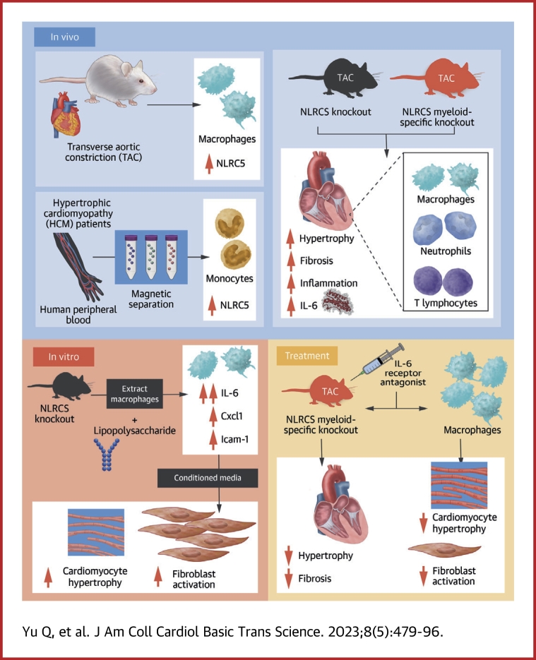
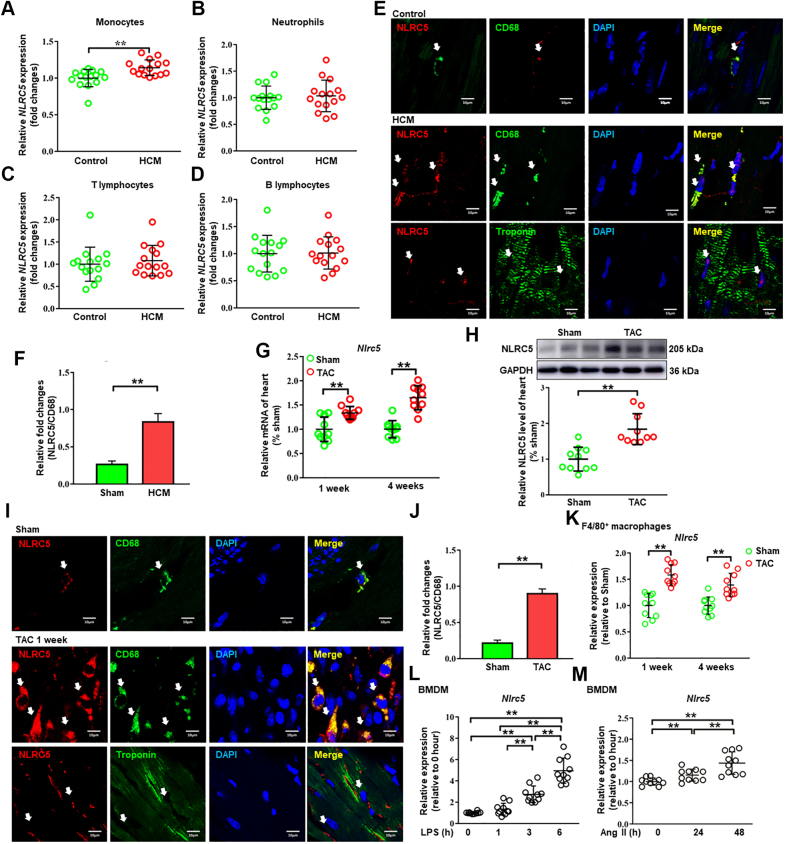
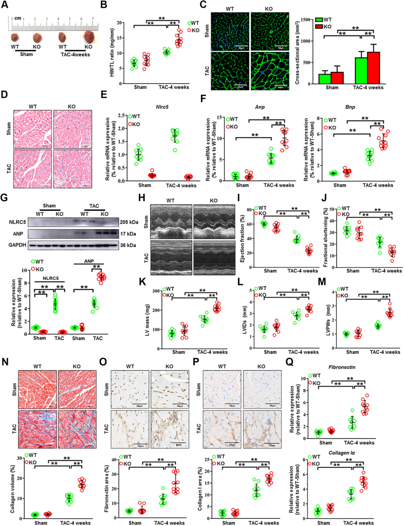
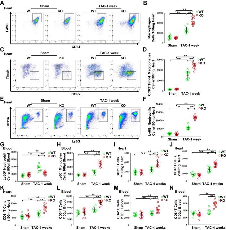
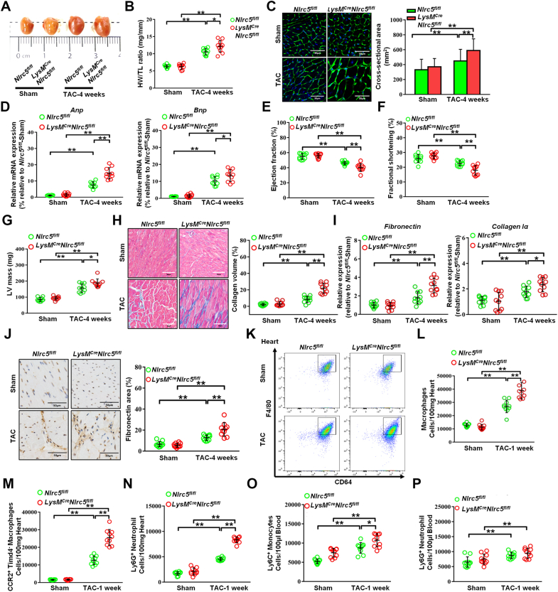
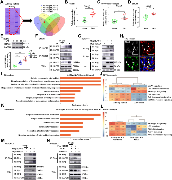
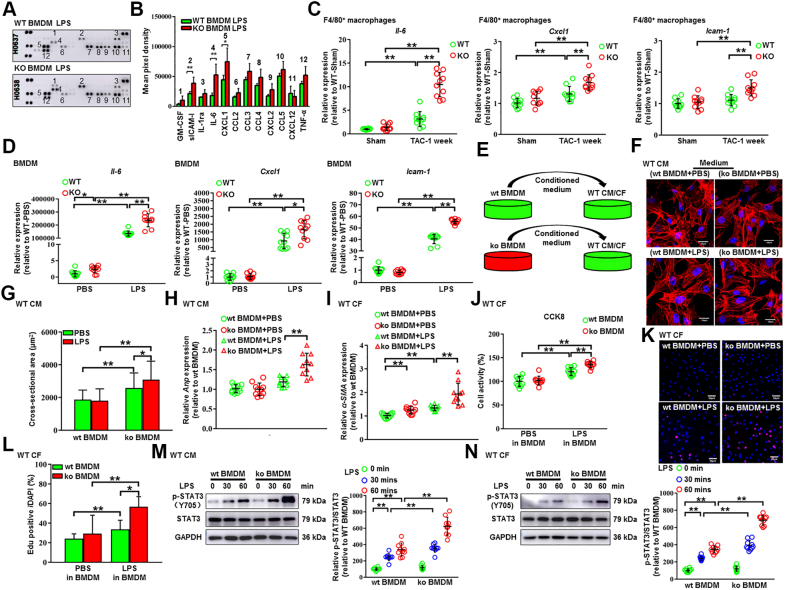
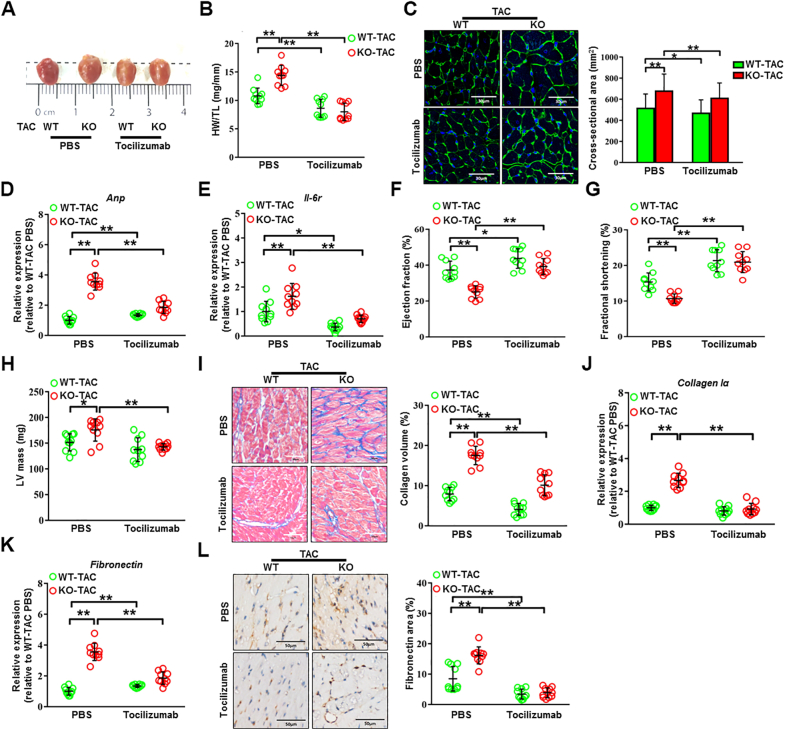
Macrophages regulate inflammation and the process of tissue repair. Therefore, a better understanding of macrophages in the pathogenesis of heart failure is needed. In patients with hypertrophic cardiomyopathy, NLRC5 was significantly increased in circulating monocytes and cardiac macrophages. Myeloid-specific deletion of NLRC5 aggravated pressure overload-induced pathological cardiac remodeling and inflammation. Mechanistically, NLRC5 interacted with HSPA8 and suppressed NF-κB pathway in macrophages. The absence of NLRC5 in macrophages promoted the secretion of cytokines such as interleukin-6 (IL-6), which affected cardiomyocyte hypertrophy and cardiac fibroblast activation. Tocilizumab, an anti-IL-6 receptor antagonist, may be a novel therapeutic strategy for cardiac remodeling and chronic heart failure.

Keywords: NOD-like receptor; cardiac remodeling; heart failure; immunomodulatory therapy; macrophages.


Conflict of interest statement

Funded by National Natural Science Foundation of China grants 81800424, 81900239, 81670746, 91939101, and 82070230, Clinical Research Plan of SHDC No. SHDC2020CR4019, and the Natural Science Foundation of Shanghai No. 20ZR1435300. The authors have reported that they have no relationships relevant to the contents of this paper to disclose.

Figures

None
Graphical abstract
Figure 1
Figure 1
NLRC5 Expression Was Increased in Monocytes and Cardiac Macrophages From HCM Patients and TAC-Operated Mice (A to D) The mRNA expression of NLRC5 was analyzed in monocytes (A), neutrophils (B), T lymphocytes (C), and B lymphocytes (D) isolated from the peripheral blood of healthy control subjects and hypertrophic cardiomyopathy (HCM) patients (n = 15 per group). (E) Representative immunofluorescence staining showed that NLRC5 (red) was up-regulated in CD68+ macrophages (green) in HCM patients as compared with control subjects, but NLRC5 was rarely located in cardiomyocytes (labeled with troponin T in green). Scale bars, 10 μm. (F) Quantitative analysis of the percentages of NLRC5 in CD68-stained macrophages in HCM patients compared with control subjects (n = 3 per group). Five fields per section from each sample were analyzed. (G) The mRNA levels of Nlrc5 in hearts were detected after 1 and 4 weeks of transverse aortic constriction (TAC) surgery (n = 10 per group). (H) Western blots and quantitative analyses were performed to determine the protein expression of NLRC5 in hearts of sham and TAC-treated mice for 4 weeks (n = 10 per group). (I) Representative immunofluorescence staining showed that NLRC5 (red) was constitutively colocalized with macrophages (labeled with CD68 in green) in hearts after 1 week of TAC operation and was rarely expressed in cardiomyocytes. Scale bars, 10 μm. (J) Quantitative analysis of the percentages of NLRC5 in CD68-stained macrophages in sham and TAC heart from C57BL/6 mice after 1-week TAC operation (n = 5 per group). Five fields per section from each sample were analyzed. (K) The mRNA expression of Nlrc5 was analyzed in F4/80+ macrophages isolated from hearts after 1 or 4 weeks of TAC (n = 10 per group). The control was heart tissue taken from sham mice at either 1 or 4 weeks after surgery. (L and M) The mRNA expression of Nlrc5 was analyzed in bone marrow–derived macrophages (BMDMs) with liposaccharide (LPS) treatment at 0, 1, 3, or 6 hours (L) and with angiotensin II (Ang II) treatment at 0, 24, or 48 hours (M) (n = 10 per group). Data are expressed as mean ± SD. ∗P < 0.05, ∗∗P < 0.01. Data in L and M were analyzed using repeated measures analysis of variance. Other data were analyzed using Student’s unpaired t-test.
Figure 2
Figure 2
NLRC5 Deletion Promoted Cardiac Remodeling Induced by Pressure Overload (A) Gross morphology of hearts from wild-type (WT) and knockout (KO) mice after 4 weeks of transverse aortic constriction (TAC). (B) The ratios of heart weight and tibia length (HW/TL) in WT and KO mice (n = 10 per group). (C) Histological analysis of cardiomyocyte area was measured by wheat germ agglutinin staining. Scale bars, 30 μm. (D) Representative images of transverse area of cardiomyocytes detected by hematoxylin and eosin staining. The cross-sectional areas of cardiomyocytes were analyzed by ImageJ (NIH) (n = 10 per group). Ten fields per section from each sample are analyzed. (E) The mRNA expression of Nlrc5 was assessed in heart tissues, normalized to Gapdh (n = 10 per group). (F) Relative mRNA expression of cardiac hypertrophy–related markers Anp and Bnp was assessed (n = 10 per group). (G) Representative Western blots determined the protein expression of ANP and NLRC5 in WT and KO mice after 4 weeks of TAC operation (n = 10 per group). (H to M) Echocardiographic analyses of ejection fraction, fractional shorting, left ventricular internal diameter at end-systole (LVIDs), left ventricular posterior wall diameter at end-systole (LVPWs), and left ventricular (LV) mass after 4 weeks of TAC operation (n = 10 per group). (N) Representative Masson’s trichrome staining and quantitative analysis of collagen volume of hearts from WT and KO mice after sham or TAC operation (n = 10 per group). Scale bars, 50 μm. (O and P) Immunohistochemistry staining and quantitative analysis of cardiac fibrosis reflected by fibronectin and collagen I in WT and KO mice with sham or TAC operation (n = 10 per group). Scale bars, 50 μm. (Q) Relative mRNA expression of cardiac fibrosis markers Fibronectin and Collagen Iα was measured in heart tissues from WT and KO mice with sham or TAC operation (n = 10 per group). Data are expressed as mean ± SD. ∗P < 0.05, ∗∗P < 0.01. Data in E and F were analyzed using Kruskal-Wallis followed by Dunn’s multiple comparison test. Other data were analyzed using 1-way analysis of variance followed by Tukey’s post hoc analysis.
Figure 3
Figure 3
NLRC5 Deficiency Promoted the Infiltration of Immune Cells After TAC Operation (A and B) Representative cytograms and quantitative analysis of F4/80+ CD64+MerTK+ macrophages in heart tissues (n = 10 per group). (C and D) Representative cytograms and quantitative analysis of CCR2+Timd4 macrophages in heart tissues (n = 10 per group). (E and F) Representative cytograms and quantitative analysis of Ly6G+ neutrophils in heart tissues (n = 10 per group). (G and H) Quantitative analysis of Ly6G+ neutrophils (G) and Ly6C+ monocytes (H) in peripheral blood after 1 week of TAC (n = 10 per group). (I to K) Quantitative analysis of CD3+ T cells (I), CD4+ T cells (J), and CD8+ T cells (K) in heart tissues after 4 weeks of TAC. (L to N) Quantitative analysis of CD3+ T cells (L), CD4+ T cells (M), and CD8+ T cells (N) in peripheral blood after 4 weeks of TAC (n = 10 per group). Data are expressed as mean ± SD. ∗P < 0.05, ∗∗P < 0.01. Data were analyzed using 1-way analysis of variance followed by Tukey’s post hoc analysis. Abbreviations as in Figure 2.
Figure 4
Figure 4
Myeloid-Specific NLRC5 Deficiency Exacerbated Cardiac Remodeling Induced by Pressure Overload (A) Gross morphologies of hearts from Nlrc5flox/flox (Nlrc5fl/fl) and LysM-Cre/Nlrc5flox/flox mice after 4 weeks of TAC. (B) The ratios of HW/TL in Nlrc5fl/fl and LysM-Cre/Nlrc5flox/flox mice after TAC (n = 10 per group). (C) Representative images of transverse area of cardiomyocytes detected by wheat germ agglutinin staining. Scale bars, 30 μm. The cross-sectional areas of cardiomyocytes were analyzed by ImageJ (NIH) (n = 10 per group). Ten fields per section from each sample were analyzed. (D) Relative mRNA expression of cardiac hypertrophy-related markers Anp and Bnp were assessed (n = 10 per group). (E to G) Echocardiography analysis of fractional shorting (E), ejection fraction (F), and LV mass (G) after 4 weeks of TAC operation (n = 10 per group). (H) Representative Masson’s trichrome staining and quantitative analysis of collagen volume of hearts from Nlrc5fl/fl and LysM-Cre/Nlrc5flox/flox mice after 4 weeks of TAC operation (n = 10 per group). Scale bars, 50 μm. (I) Relative mRNA expression of cardiac fibrosis markers Fibronectin and Collagen Iα were measured in heart tissues from Nlrc5fl/fl and LysM-Cre/Nlrc5flox/flox mice after 4 weeks of TAC operation (n = 10 per group). (J) Immunohistochemistry staining and quantitative analysis of cardiac fibrosis marker fibronectin in Nlrc5fl/fl and LysM-Cre/Nlrc5flox/flox mice with TAC operation (n = 10 per group). Scale bars, 50 μm. (K and L) Representative cytograms of F4/80+ CD64+MerTK+ macrophages in heart tissues (K) and quantitative analysis (L) (n = 10 per group). (M and N) Quantitative analysis of CCR2+Timd4 macrophages (M) and Ly6G+ neutrophils (N) in hearts from Nlrc5fl/fl and LysM-Cre/Nlrc5flox/flox mice after 1 week of TAC operation (n = 10 per group). (O and P) Quantitative analysis of Ly6C+ monocytes (O) and Ly6G+ neutrophils (P) in peripheral blood from Nlrc5fl/fl and LysM-Cre/Nlrc5flox/flox mice after 1 week of TAC operation (n = 10 per group). Data are expressed as mean ± SD. ∗P < 0.05, ∗∗P < 0.01. Data were analyzed using 1-way analysis of variance followed by Tukey’s post hoc analysis. Abbreviations as in Figure 2.
Figure 5
Figure 5
NLRC5 Directly Interacted With HSPA8 (A) The most potential functional binding targets of NLRC5 were evaluated by mass spectrometry of BMDMs with adenovirus (Ad) overexpressing NLRC5. Six potential target proteins were interacting with NLRC5 were screened through 3 batches of overexpressed NLRC5 compared with the result of a control group. (B) Relative mRNA expression of Hspa8 in hearts after TAC. (C) Relative mRNA expression of Hspa8 in F4/80+ macrophages isolated from hearts. (D) Relative mRNA expression of Hspa8 in BMDMs with or without LPS treatment. (E) Western blotting and quantitative analysis of HSPA8 and GAPDH in BMDMs from mice with or without LPS treatment. (F) Coimmunoprecipitation of NLRC5 and HSPA8 in BMDMs with and without LPS treatment. The lysates immunoprecipitated with anti-IgG serve as a negative control condition. (G) HEK293T cells were cotransfected with Flag-tagged NLRC5 and Myc-tagged HSPA8 plasmid. (H) Immunofluorescence staining of NLRC5 (red), Mac2 (grey), HSPA8 (green), and nuclei (blue) in hearts. Scale bars, 30 μm. (I) Gene ontology (GO) enrichment analysis and gene set variation analysis (GSVAs) of the expression of several genes with roles in different biological responses of Ad-Flag-NLRC5 compared with the Ad-negative control (Ad-NC) condition from RNA sequencing. (J) GSVAs of the expression of several genes were performed with role in different signaling pathways from RNA sequencing. (K and L) GO enrichment analysis and GSVAs analysis of the expression of several genes with role in different biological response and signaling pathways of Ad-Flag-NLRC5 plus siHSPA8 treatment compared with Ad-Flag-NLRC5 from RNA sequencing. (M) BMDMs were cotransfected with Flag-tagged NLRC5 and Myc-tagged HSPA8 plasmid. The lysates immunoprecipitated with anti-IgG serve as a negative control condition. (N) Coimmunoprecipitation of NLRC5 and IKKβ in BMDMs with or without siHSPA8 treatment. Data are expressed as mean ± SD. ∗P < 0.05, ∗∗P < 0.01. Data in B to D were analyzed using Student’s unpaired t-test. Data in panel E were analyzed using 1-way analysis of variance followed by Tukey’s post hoc analysis. IB = immunoblotting; IP = immunoprecipitation; WCL = whole cell lysis; other abbreviations as in Figures 1 and 2.
Figure 6
Figure 6
Deficiency of NLRC5 in Macrophages Facilitated Dysfunction of CMs and CFs Through Promoting the Secretion of IL-6 (A and B) The cell culture supernatants of BMDMs from WT and KO mice were detected by mouse cytokine array. BMDMs were treated with 100 ng/mL of LPS for 6 hours, and the relative mean pixel density was calculated (n = 3 per group). (C) Relative mRNA expression of proinflammatory cytokines Il-6, Cxcl1, and Icam-1 in F4/80+ macrophages isolated from WT and KO hearts was quantified by real-time polymerase chain reaction (n = 10 per group). (D) Relative mRNA expression of proinflammatory cytokines Il-6, Cxcl1, and Icam-1 in BMDMs from WT and KO mice were quantified by real-time polymerase chain reaction (n = 10 per group). (E) Experimental design. Conditional medium was collected from BMDMs of WT (wt BMDM) and KO (ko BMDM) mice in the presence or absence of LPS. The conditional medium was then used to culture WT cardiomyocytes (WT CM). (F and G) Representative immunofluorescent images and quantitative analysis of cross-sectional areas of CMs labeled by α-actinin after culturing with conditional medium. More than 100 CMs from each group were randomly selected. Scale bars, 50 μm. (H) Relative mRNA expression of Anp was quantified by real-time polymerase chain reaction (n = 10 per group). (I) Relative mRNA expression of α-SMA in CFs cultured with conditional medium from WT and KO BMDMs was quantified by real-time polymerase chain reaction (n = 10 per group). (J) The cell activity of cardiac fibroblasts (CFs) was measured by CCK8 kit (n = 10 per group). (K and L) The proliferation capacity of CFs was shown in EdU staining (n = 10 per group). Scale bars, 50 μm. (M) Representative Western blot and quantitative analysis of p-STAT3 and STAT3 in CMs with conditional medium from WT and KO BMDMs (n = 10 per group). (N) Representative Western blot and quantitative analysis of p-STAT3 and STAT3 in CFs with conditional medium from WT and KO BMDMs (n = 10 per group). Data are expressed as mean ± SD. ∗P < 0.05, ∗∗P < 0.01. Data were analyzed using 1-way analysis of variance followed by Tukey’s post hoc analysis. PBS = phosphate-buffered saline; other abbreviations as in Figures 1 and 2.
Figure 7
Figure 7
IL-6 Receptor Antagonist Tocilizumab Prevented Pathological Cardiac Remodeling Caused by NLRC5 Deficiency (A) Gross morphologies of hearts from WT and KO mice with and without tocilizumab after TAC surgery. (B) HW/TL in WT and KO mice with and without tocilizumab after TAC surgery (n = 10 per group). (C) Representative images and quantitative analysis of transverse area of CMs detected by wheat germ agglutinin staining (n = 10 per group). Scale bars, 30 μm. (D and E) Relative mRNA expression of Anp and Il-6r was assessed by real-time polymerase chain reaction (n = 10 per group). (F to H) Echocardiography analysis of fractional shorting, ejection fraction, and LV mass after 4 weeks of TAC were shown (n = 10 per group). (I) Representative Masson’s trichrome staining and quantitative analysis of heart sections from WT and KO mice with and without tocilizumab after TAC surgery (n = 10 per group). Scale bars, 50 μm. (J and K) Relative mRNA expression of cardiac fibrosis markers Fibronectin and Collagen Iα were measured by real-time polymerase chain reaction in hearts from WT and KO mice with and without tocilizumab (n = 10 per group). (L) Representative immunohistochemistry staining and quantification analysis of fibronectin in hearts from WT and KO mice with and without tocilizumab (n = 10 per group). Scale bars, 50 μm. Data are expressed as mean ± SD. ∗P < 0.05, ∗∗P < 0.01. Data were analyzed using 1-way analysis of variance followed by Tukey’s post hoc analysis. Abbreviations as in Figures 1, 2, and 6.

References

    1. Heidenreich P.A., Bozkurt B., Aguilar D., et al. 2022 AHA/ACC/HFSA guideline for the management of heart failure: a report of the American College of Cardiology/American Heart Association Joint Committee on Clinical Practice Guidelines. J Am Coll Cardiol. 2022;79:e263–e421. - PubMed
    1. Nagase T., So Y.G., Yasui H., et al. Observation of domain wall bimerons in chiral magnets. Nat Commun. 2021;12:3490. - PMC - PubMed
    1. Ridker P.M., Devalaraja M., Baeres F.M.M., et al. IL-6 inhibition with ziltivekimab in patients at high atherosclerotic risk (RESCUE): a double-blind, randomised, placebo-controlled, phase 2 trial. Lancet. 2021;397:2060–2069. - PubMed
    1. Ridker P.M., Everett B.M., Thuren T., et al. Antiinflammatory therapy with canakinumab for atherosclerotic disease. N Engl J Med. 2017;377:1119–1131. - PubMed
    1. Mouton A.J., Li X., Hall M.E., Hall J.E. Obesity, hypertension, and cardiac dysfunction: novel roles of immunometabolism in macrophage activation and inflammation. Circ Res. 2020;126:789–806. - PMC - PubMed

LinkOut - more resources