

Case Reports
doi: 10.1016/j.case.2022.12.014.
eCollection 2023 May.
Degenerative Mitral Stenosis: A Case-Based Review
Affiliations
- PMID: 37325463
- PMCID: PMC10264206
- DOI: 10.1016/j.case.2022.12.014
Item in Clipboard
Case Reports
Degenerative Mitral Stenosis: A Case-Based Review
CASE (Phila).
.
No abstract available
Keywords: Degenerative mitral disease; Echocardiography; Mitral annular calcification; Mitral stenosis; Multimodality imaging.
Figures


Two-dimensional TTE zoomed apical 4-chamber view, without (left) and with (right) color flow Doppler of the mitral inflow, demonstrates severe MAC (∗) and leaflet calcification (#) with associated proximal diastolic flow acceleration (A). Continuous-wave Doppler spectrum demonstrates an increased mean TMG of 10 mm Hg (B).

Two-dimensional TEE midesophageal apical 4-chamber view (0°), diastolic phase, demonstrates severe MAC (∗) with calcification of the mitral leaflets (#) and restricted leaflet opening (A). Continuous-wave Doppler spectrum across the mitral inflow demonstrates a normal TMG of 3 mm Hg (B). Spectral ghosting (mirroring artifact) is seen and should be avoided when tracing. The LVOT was measured in the apical 3-chamber, midsystolic view (120°) inner edge to inner edge at the hinge points (blue double arrow) while avoiding the calcification (∗) on the anterior mitral leaflet (C).

Three-dimensional TEE acquisition and multiplanar, diastolic reconstruction demonstrates the MV leaflets in 2 orthogonal planes (A, B), short-axis leaflet tip display (C), and volume-rendered reconstruction in the surgeon's view orientation from the perspective of the LA (D).

Two-dimensional TTE parasternal long-axis view (A) and apical 4-chamber (B) diastolic images demonstrate moderate posterior MAC (∗). The continuous-wave Doppler spectral display of the mitral inflow obtained from the apical 4-chamber view demonstrates an increased TMG of 9 mm Hg (C).
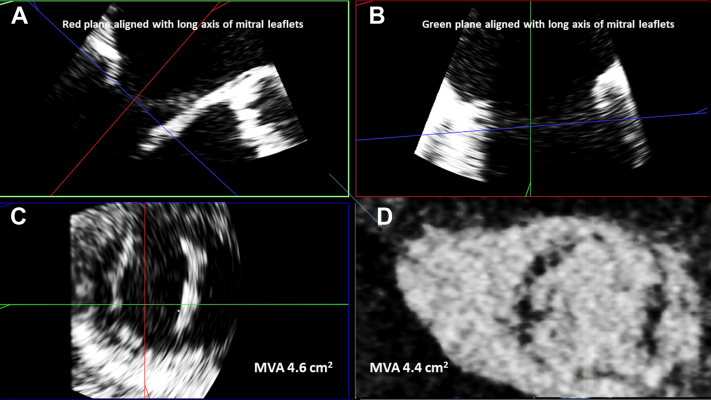
Three-dimensional TEE acquisition and multiplanar, diastolic reconstruction demonstrates the MV leaflets in 2 orthogonal planes (A, B) and short-axis leaflet tip display (C), which allows for direct planimetry (MVA = 4.4 cm2). A corresponding CCT-derived short-axis slice of the MV leaflet tips (D) during diastole demonstrates a similar MVA by planimetry (MVA = 4.4 cm2).

Etiology, outcomes, and associations of DMS. ESRD, End-stage renal disease; LV, left ventricle; HOCM, hypertrophic cardiomyopathy; MR, mitral regurgitation; TR, tricuspid regurgitation.

Proposed algorithm featuring summary of steps for echocardiographic evaluation of DMS. CW, Continuous wave.
References
-
- Otto C.M., Nishimura R.A., Bonow R.O., Carabello B.A., Erwin J.P., III, Gentile F., et al. 2020 ACC/AHA guideline for the management of patients with valvular heart disease: executive summary: a report of the American College of Cardiology/American Heart Association Joint Committee on clinical practice guidelines. J Am Coll Cardiol. 2021;77:450–500. - PubMed
-
- Kato N., Guerrero M., Padang R., Amadio J.M., Eleid M.F., Scott C.G., et al. Prevalence and natural history of mitral annulus calcification and related valve dysfunction. Mayo Clin Proc. 2022;97:1094–1107. - PubMed
-
- Nishimura R.A., Vahanian A., Eleid M.F., Mack M.J. MV disease–current management and future challenges. Lancet. 2016;387:1324–1334. - PubMed
-
- Barasch E., Gottdiener J.S., Larsen E.K., Chaves P.H., Newman A.B., Manolio T.A. Clinical significance of calcification of the fibrous skeleton of the heart and aortosclerosis in community dwelling elderly. The Cardiovascular Health Study (CHS) Am Heart J. 2006;151:39–47. - PubMed
Publication types
LinkOut - more resources
Full Text Sources
