. 2023 Jun 14;10(6):221493.
doi: 10.1098/rsos.221493. eCollection 2023 Jun.

Non-natural amino acids into LfcinB-derived peptides: effect in their (i) proteolytic degradation and (ii) cytotoxic activity against cancer cells

Affiliations

Non-natural amino acids into LfcinB-derived peptides: effect in their (i) proteolytic degradation and (ii) cytotoxic activity against cancer cells

Diego Sebastián Insuasty-Cepeda et al. R Soc Open Sci. .

Abstract

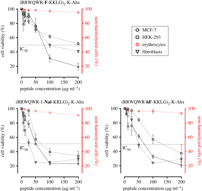
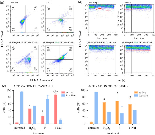
The dimeric peptide 26[F]: (RRWQWRFKKLG)2-K-Ahx has exhibited a potent cytotoxic effect against breast cancer cell lines, with position 26 (F) being the most relevant for anti-cancer activity. In this investigation, six analogues of the 26[F] peptide were synthesized in which the 26th position was replaced by non-natural hydrophobic amino acids, finding that some modifications increased the resistance to proteolytic degradation exerted by trypsin or pepsin. Additionally, these modifications increased the cytotoxic effect against breast cancer cells and generated cell death mediated by apoptosis pathways, activating caspases 8 and 9, and did not compromise the integrity of the cytoplasmic membrane. Finally, it was found that the modified peptides have a broad spectrum of action, since they also have a cytotoxic effect against the HeLa human cervical cancer cell line. Peptide 26[F] was inoculated in mice by ip administration and the lethal dose 50 (LD50) was between 70 and 140 mg kg-1. While for the 26[1-Nal]: (RRWQWR-1-Nal-KKLG)2-K-Ahx peptide, a dose-response test was performed, and the survival rate was 100%. These results suggested that these peptides are safe in this animal model and could be considered as promissory to develop a treatment against breast cancer.

Keywords: LfcinB-derived peptides; MCF-7 cells; cytotoxic activity; non-natural amino acids; proteolytic degradation.


Conflict of interest statement

We declare we have no competing interests.

Figures

Figure 1.
Figure 1.
Cytotoxic effect of dimeric peptides against breast cancer cells. Left: cell viability plots of dimeric peptides with lesser cytotoxic effect than the original peptide 26[F] (black); 26[Dip] (red), 26[4-Abz] (orange), 26[2-Abz] (green). Right: cell viability plots of dimeric peptides with greater cytotoxic effect than the original peptide; 26[Bpa] (blue), 26[1-Nal] (green), 26[hF] (violet). The data represent the mean ± s.e. Three independent experiments with n = 4 each. (ANOVA, Sidak's multiple comparisons test was used, p ≤ 0.05).
Figure 2.
Figure 2.
Pepsin and trypsin digestion of dimeric peptides. Pepsin digestion of: (a) 26[F], (b) 26[1-Nal] and (c) 26[hF]. Trypsin digestion of: (d) 26[F], (e) 26[1-Nal] and (f) 26[hF] The peptides were treated with pepsin for 3 h and trypsin for 5 min, 10 min and 3 h.
Figure 3.
Figure 3.
Cytotoxic effect of dimeric peptides against non-tumorigenic cells HEK-293 and erythrocytes. Breast cancer cells MCF-7 (black solid line ─○─); human kidney non-tumorigenic cell line HEK-293 (black dotted line ─□─); fibroblasts (black dotted line ─∇─) and erythrocytes (red solid line ─○─). The data represent the mean ± s.e. Three independent experiments with n = 4 each. (ANOVA, Sidak's multiple comparisons test was used, p ≤ 0.05).
Figure 4.
Figure 4.
Flow cytometry assays of peptides 26[F] and 26[1-Nal]. (a) Annexin/7AAD assays; negative control: untreated cells. Positive control: ActD 0.5 µM. (b) extracellular cytosolic calcium flux assay. Negative control: vehicle. Positive control: PMA 9 µM. (c) Caspase 8 and 9 activation assays. Negative control: untreated cells. Positive control: H2O2. Statistically significant differences were found between cells with no treatment and the two peptides. The data represents the mean ± s.e. An independent experiment with n = 3 each. (ANOVA, Sidak's multiple comparisons test was used, p ≤ 0.05). For all assays, the peptide concentration used was the IC50 value determined by MTT assay.

Similar articles

Cited by

References

    1. Ritchie H, Roser M. 2015 Cancer. Our World in Data. See https://ourworldindata.org/cancer.
    1. Wilkinson L, Gathani T. 2022. Understanding breast cancer as a global health concern. Br. J. Radiol. 95, 7-9. (10.1259/bjr.20211033) - DOI - PMC - PubMed
    1. Waks AG, Winer EP. 2019. Breast cancer treatment. J. Am. Med. Assoc. 321, 288-300. (10.1001/jama.2018.19323) - DOI - PubMed
    1. Gaspar D, Salomé Veiga A, Castanho MARB. 2013. From antimicrobial to anticancer peptides: a review. Front. Microbiol. 4, 1-16. (10.3389/fmicb.2013.00294) - DOI - PMC - PubMed
    1. de O. Mello É, Taveira GB, de O. Carvalho A, Gomes VM. 2019. Improved smallest peptides based on positive charge increase of the γ-core motif from PνD1 and their mechanism of action against Candida species. Int. J. Nanomedicine 14, 407-420. - PMC - PubMed