. 2023 Jul 7;133(2):158-176.
doi: 10.1161/CIRCRESAHA.123.322875. Epub 2023 Jun 16.

Activation of the Aryl Hydrocarbon Receptor in Muscle Exacerbates Ischemic Pathology in Chronic Kidney Disease

Affiliations

Activation of the Aryl Hydrocarbon Receptor in Muscle Exacerbates Ischemic Pathology in Chronic Kidney Disease

Nicholas Balestrieri et al. Circ Res. .

Abstract

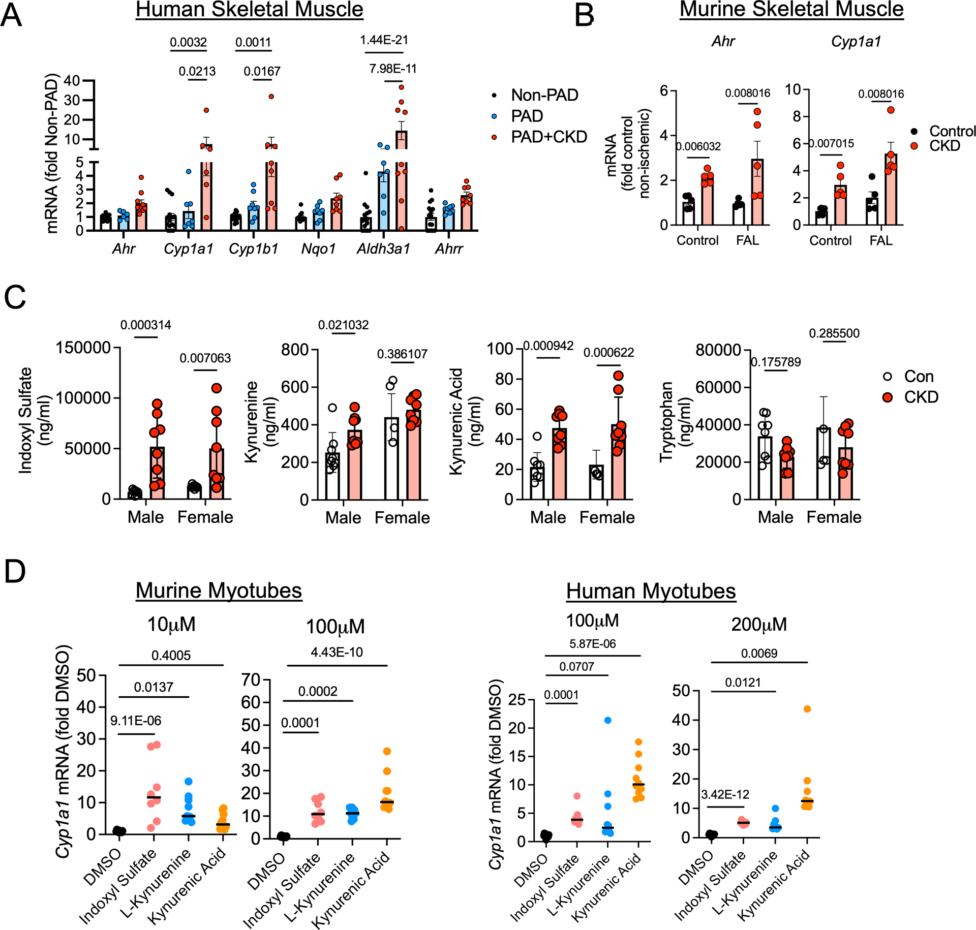
Background: Chronic kidney disease (CKD) accelerates the development of atherosclerosis, decreases muscle function, and increases the risk of amputation or death in patients with peripheral artery disease (PAD). However, the mechanisms underlying this pathobiology are ill-defined. Recent work has indicated that tryptophan-derived uremic solutes, which are ligands for AHR (aryl hydrocarbon receptor), are associated with limb amputation in PAD. Herein, we examined the role of AHR activation in the myopathy of PAD and CKD.

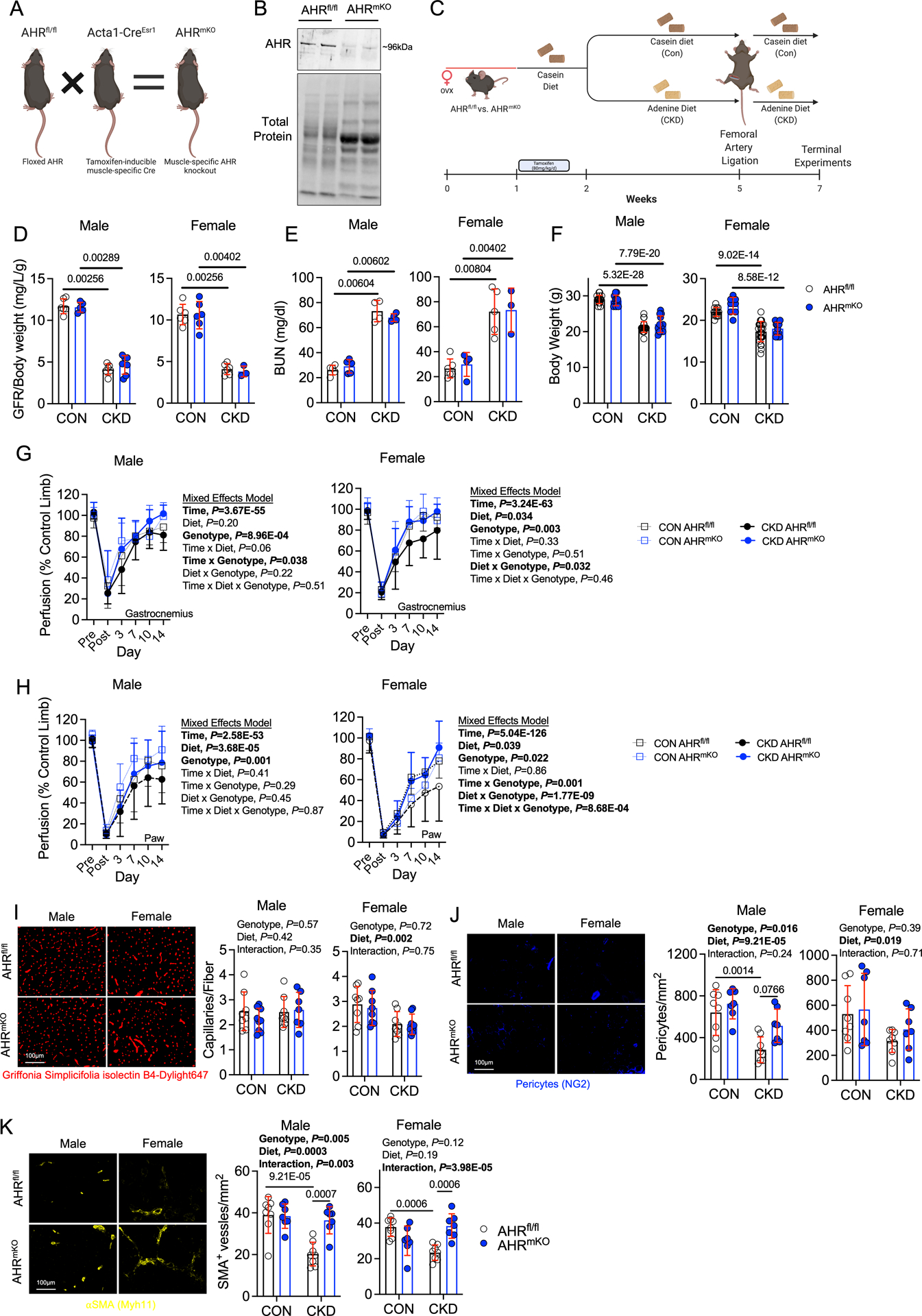
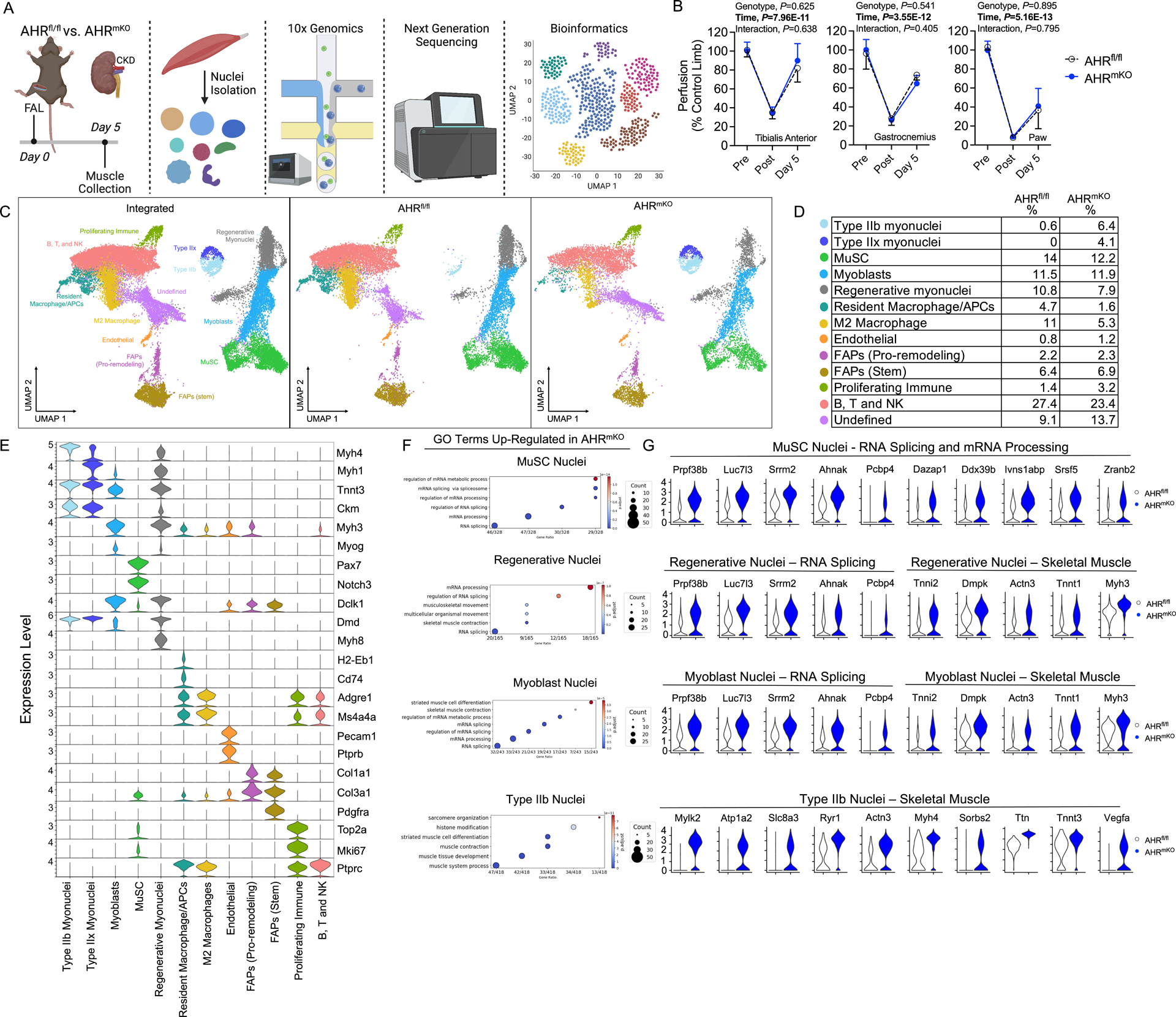
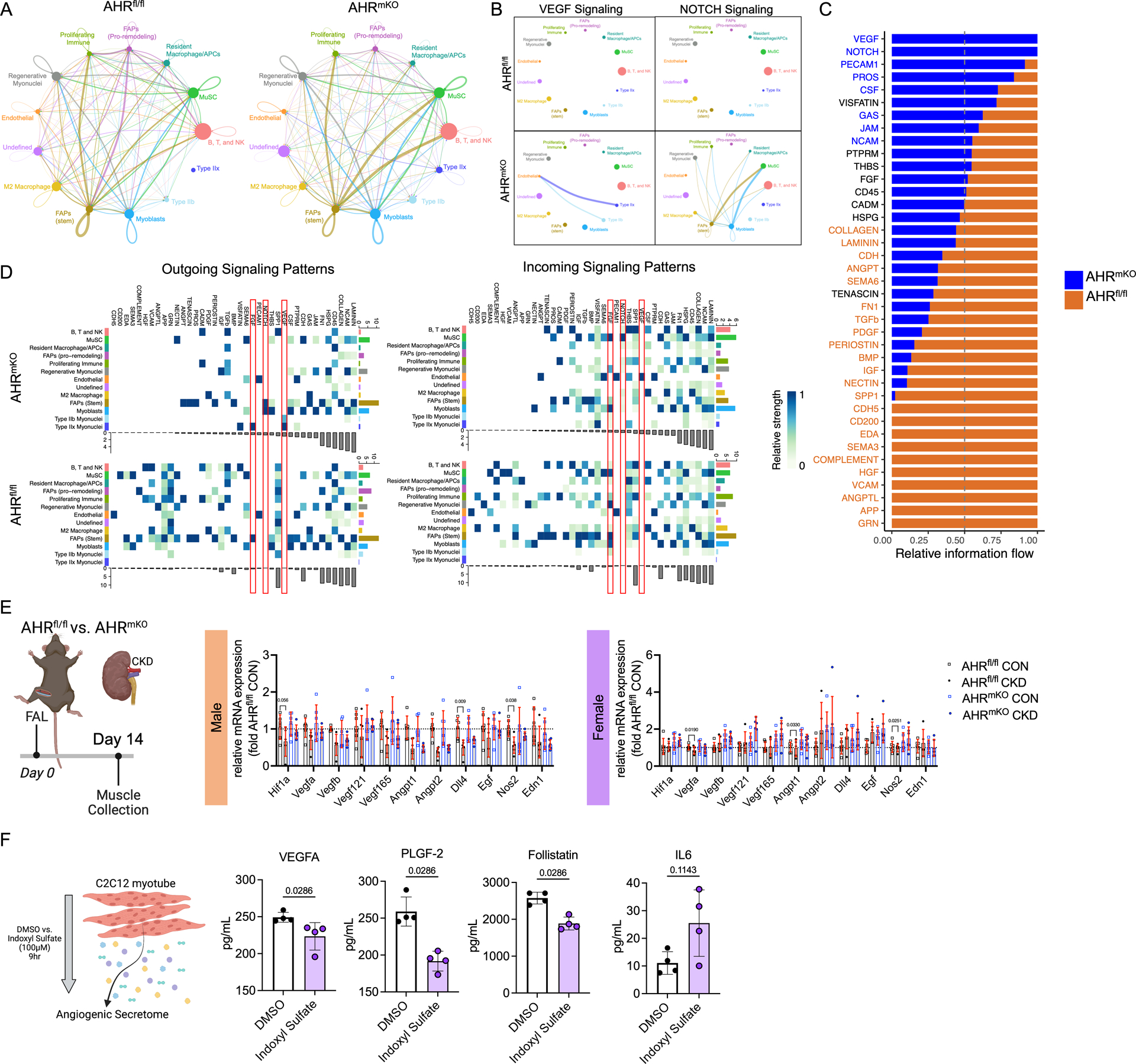
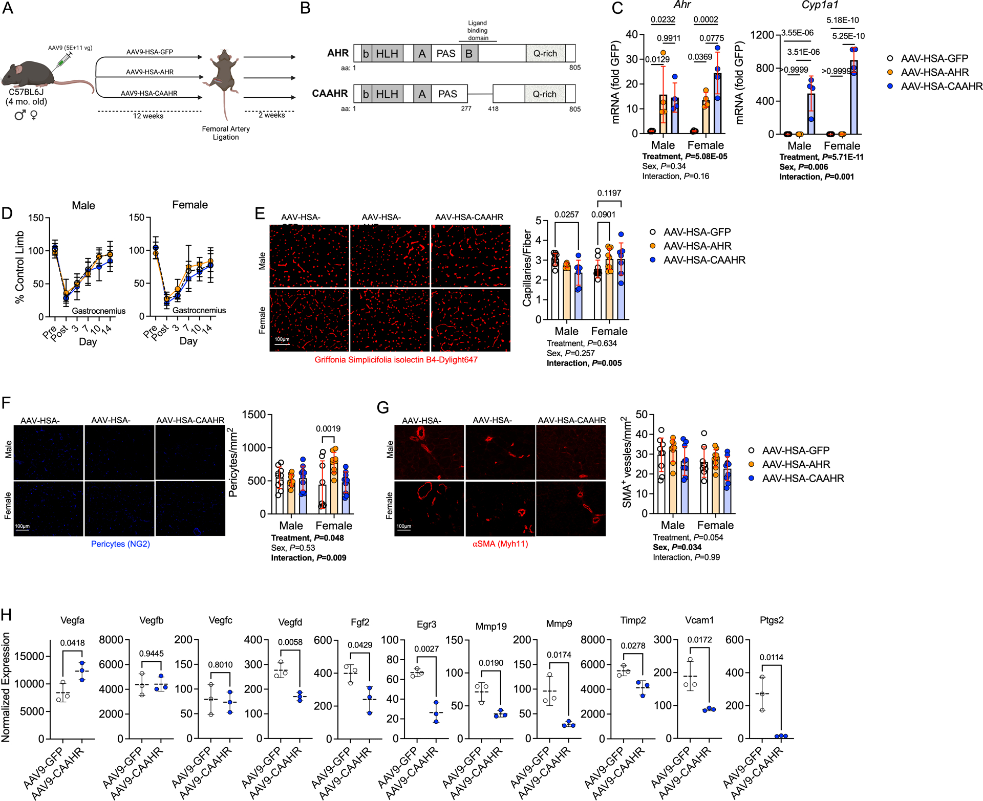
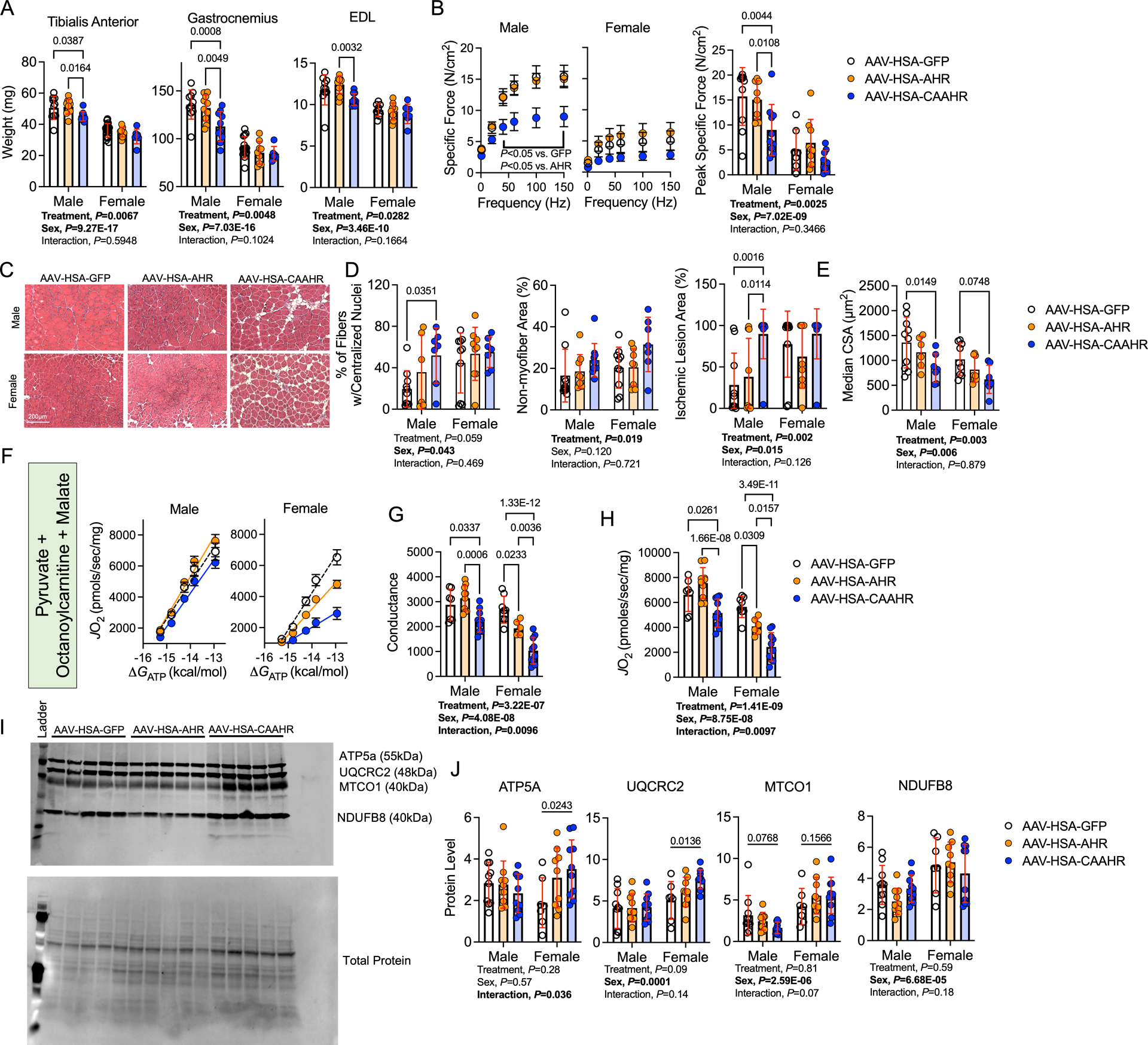
Methods: AHR-related gene expression was evaluated in skeletal muscle obtained from mice and human PAD patients with and without CKD. AHRmKO (skeletal muscle-specific AHR knockout) mice with and without CKD were subjected to femoral artery ligation, and a battery of assessments were performed to evaluate vascular, muscle, and mitochondrial health. Single-nuclei RNA sequencing was performed to explore intercellular communication. Expression of the constitutively active AHR was used to isolate the role of AHR in mice without CKD.

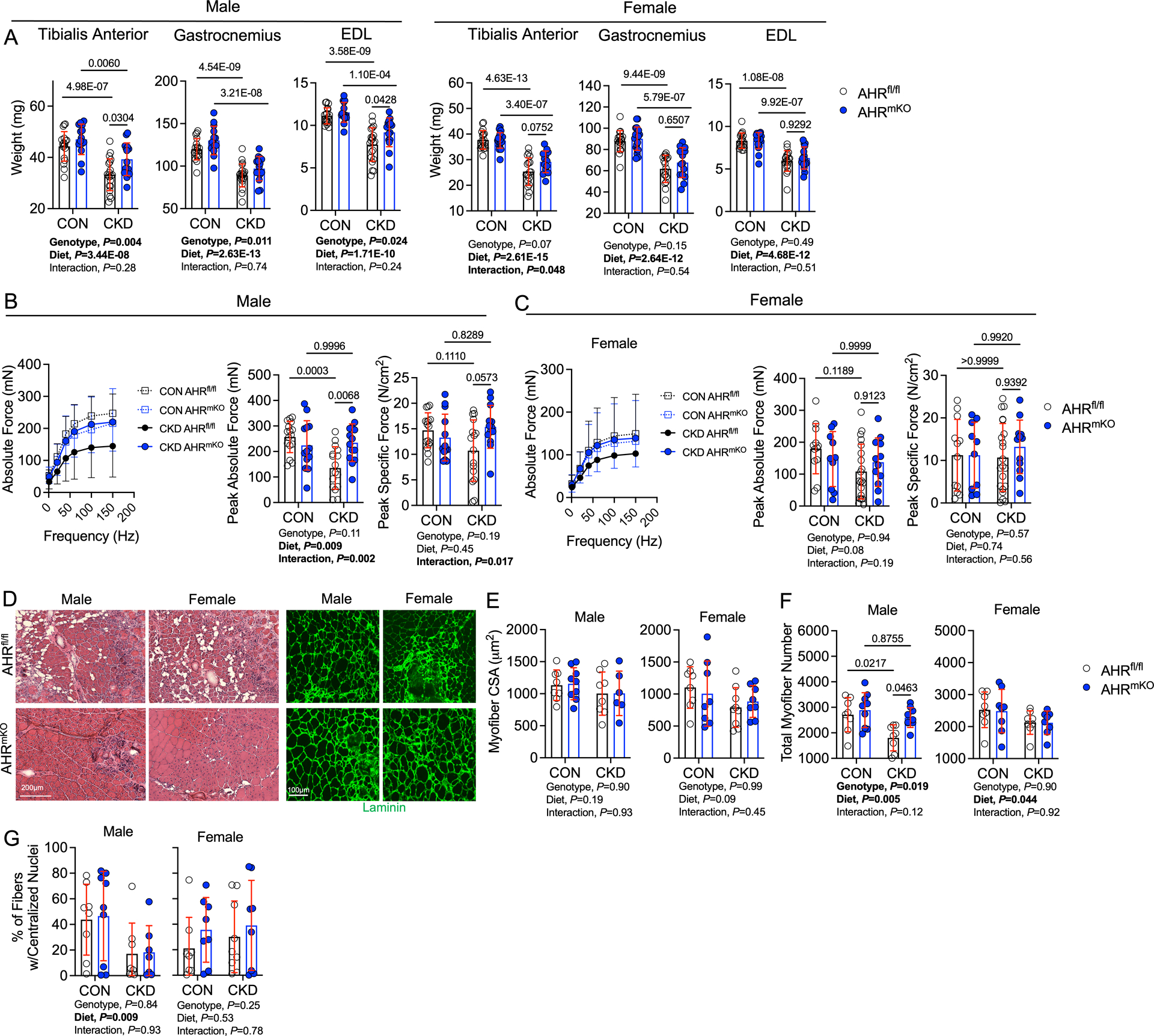
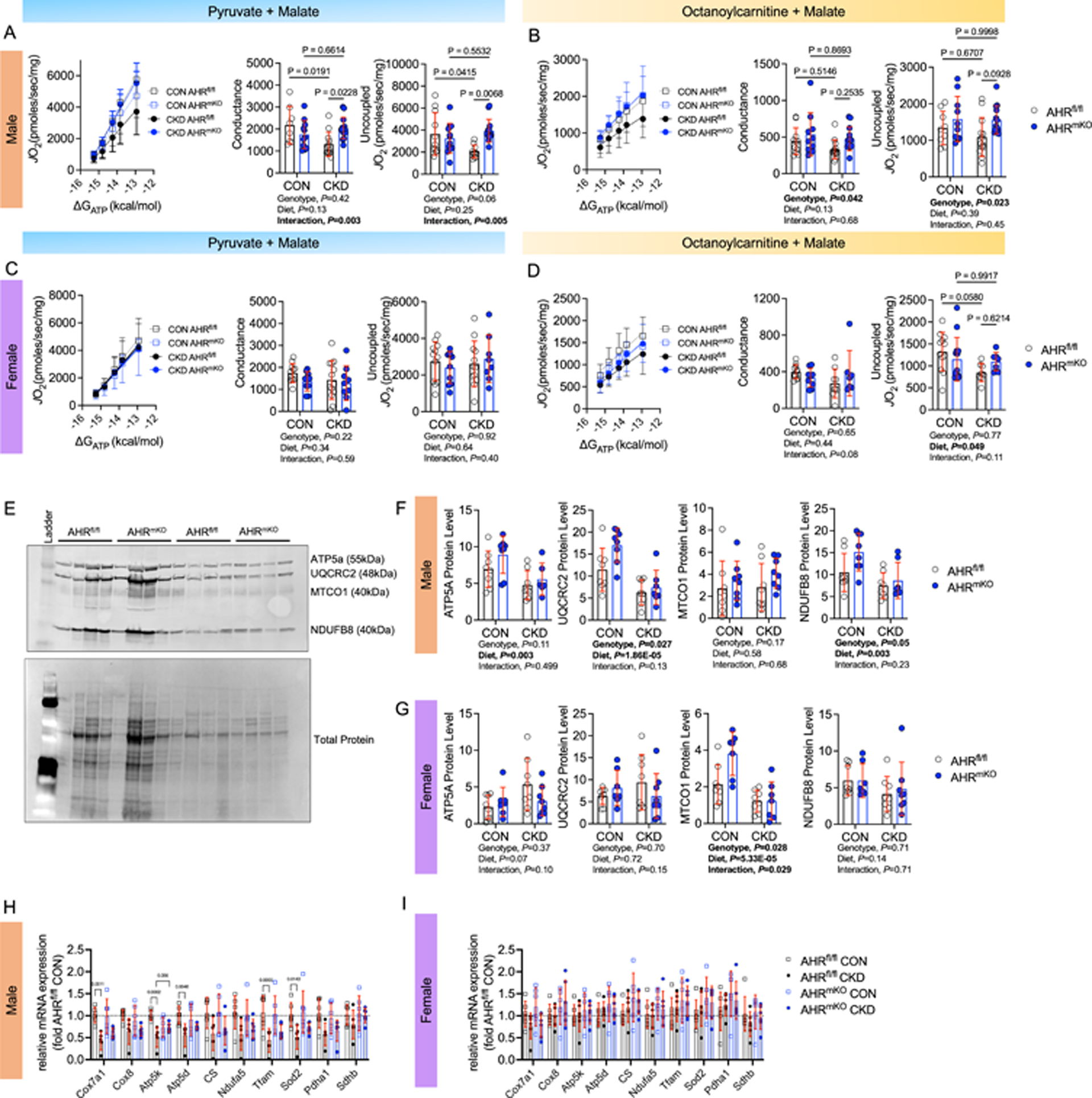
Results: PAD patients and mice with CKD displayed significantly higher mRNA expression of classical AHR-dependent genes (Cyp1a1, Cyp1b1, and Aldh3a1) when compared with either muscle from the PAD condition with normal renal function (P<0.05 for all 3 genes) or nonischemic controls. AHRmKO significantly improved limb perfusion recovery and arteriogenesis, preserved vasculogenic paracrine signaling from myofibers, increased muscle mass and strength, as well as enhanced mitochondrial function in an experimental model of PAD/CKD. Moreover, viral-mediated skeletal muscle-specific expression of a constitutively active AHR in mice with normal kidney function exacerbated the ischemic myopathy evidenced by smaller muscle masses, reduced contractile function, histopathology, altered vasculogenic signaling, and lower mitochondrial respiratory function.

Conclusions: These findings establish AHR activation in muscle as a pivotal regulator of the ischemic limb pathology in CKD. Further, the totality of the results provides support for testing of clinical interventions that diminish AHR signaling in these conditions.

Keywords: humans; kidney; mitochondria; peripheral artery disease; uremia.


Conflict of interest statement

Disclosures None.

Figures

Figure 1.
Figure 1.. Evidence of AHR activation by uremic metabolites in human and mouse skeletal muscle.
(A) Relative mRNA levels of AHR-related genes in gastrocnemius muscle specimens from non-PAD adults (n=15) and PAD patients with (n=10) and without CKD (n=8). (B) Relative expression of Ahr and Cyp1a1 in the gastrocnemius muscle of mice with and without CKD in both control and ischemic limbs (n=5/group). (C) Quantified levels of tryptophan-derived AHR ligands in the serum of mice with and without CKD (n=8 male Con, 4 female Con, 8 male CKD, 8 female CKD). (D) qPCR analysis of relative mRNA levels of Cyp1a1 following acute treatment with AHR ligands in both murine (n= 10 DMSO, 8 indoxyl sulfate, 9 L-kynurenine, and 10 kynurenic acid) and human cultured myotubes (n=10 DMSO, 9 indoxyl sulfate, 10 L-kynurenine, and 10 kynurenic acid). Panels A and D were analyzed using a one-way ANOVA with Sidak’s post hoc testing. Panels B and C were analyzed using Mann-Whitney tests. *P<0.05, Error bars represent the standard deviation.
Figure 2.
Figure 2.. Skeletal Muscle-Specific AHR deletion Promotes Ischemic Muscle Perfusion Recovery and Arteriogenesis in Mice with CKD.
(A) Generation of inducible, muscle-specific AHR knockout mice and (B) immunoblotting confirmation of AHR deletion in muscle. (C) Graphical depiction of the experiment design. (D) Glomerular filtration rate (GFR) in mice with and without CKD (n=6 AHRfl/fl males/group, 5 AHRmKO male CON and 7 CKD, 6 AHRfl/fl female CON and 4 CKD, 6 AHRmKO female CON and 3 CKD). (E) Quantification of blood urea nitrogen (BUN) (n=5 AHRfl/fl male CON and 4 CKD, 5 AHRmKO male CON and 4 CKD, 6 AHRfl/fl female CON and 5 CKD, 4 AHRmKO female CON and 3 CKD). (F) Quantification of body weights (n=17 AHRfl/fl male CON and 21 CKD, 11 AHRmKO male CON and 12 CKD, 13 AHRfl/fl female CON and 25 CKD, 12 AHRmKO female CON and 14 CKD). (G) Quantification of perfusion recovery in the gastrocnemius muscle (n=14 AHRfl/fl male CON and 17 CKD, 13 AHRmKO male CON and 13 CKD, 10 AHRfl/fl female CON and 18 CKD, 9 AHRmKO female CON and 8 CKD). (H) Quantification of perfusion recovery in the paw (n=14 AHRfl/fl male CON and 17 CKD, 13 AHRmKO male CON and 13 CKD, 10 AHRfl/fl female CON and 18 CKD, 9 AHRmKO female CON and 8 CKD). Perfusion recovery was analyzed using mixed model analysis. (I) Quantification of capillary density in the tibialis anterior muscle (n=8 AHRfl/fl males/group, 9 AHRmKO male CON and 7 CKD, and n=8/genotype/diet in females). (J) Quantification of pericyte density in the tibialis anterior muscle (n=8 AHRfl/fl males/group, 7 AHRmKO males/group, and n=8 AHRfl/fl females/group, 7 AHRmKO females/group). (K) Quantification of arteriole density in the tibialis anterior muscle (n=8 AHRfl/fl males/group, 7 AHRmKO males/group, and n=8 AHRfl/fl females/group, 7 AHRmKO females/group). Statistical analyses with normally distributed data were performed using two-way ANOVA with Sidak’s post hoc testing for multiple comparisons when significant interactions were detected. Panels D-E were analyzed using Mann-Whitney tests. Error bars represent the standard deviation.
Figure 3.
Figure 3.. Skeletal Muscle-Specific deletion of the AHR Preserves Ischemic Muscle Mass and Contractile Function in Mice with CKD.
(A) Quantification of muscle weights in wildtype (AHRfl/fl) and AHRmKO mice (n=18 AHRfl/fl male CON and 20 CKD, 14 AHRmKO male CON and 15 CKD, 18 AHRfl/fl females/group, 20 AHRmKO female CON and 13 CKD). (B) Muscle contractile function quantification in male mice (n=16 AHRfl/fl male CON and 16 CKD, 13 AHRmKO male CON and 14 CKD). (C) Muscle contractile function quantification in female mice (n=12 AHRfl/fl female CON and 21 CKD, 10 AHRmKO female CON and 12 CKD). Specific force was calculated as the absolute force normalized to the cross-sectional area of the muscle. (D) Representative hematoxylin & eosin staining and laminin staining immunofluorescence from mice with CKD. (E) Quantification of the mean myofiber cross-sectional area of the tibialis anterior muscle (n=8 AHRfl/fl males/group, 9 AHRmKO male CON and 6 CKD, 8 AHRfl/fl female CON and 9 CKD, 8 AHRmKO females/group). (F) Quantification of the total fiber numbers in the tibialis anterior muscle (n=8 AHRfl/fl males/group, 9 AHRmKO males/group, 8 AHRfl/fl females/group, 8 AHRmKO females/group). (G) Quantification of the percentage of myofibers containing centralized nuclei within the tibialis anterior muscle (n=8 AHRfl/fl males/group, 9 AHRmKO male CON and 7 CKD, 8 AHRfl/fl female CON and 9 CKD, 8 AHRmKO females/group). Statistical analyses performed using two-way ANOVA with Sidak’s post hoc testing for multiple comparisons. Error bars represent the standard deviation.
Figure 4.
Figure 4.. Skeletal Muscle-Specific deletion of the AHR Preserves Ischemic Muscle Mitochondrial Energetics in Mice with CKD.
(A) Relationship between JO2 and ΔGATP when mitochondria were fueled with pyruvate and malate and quantification of the conductance (slope of JO2 and ΔGATP relationship) and uncoupled respiration rates in male mice (n=8 AHRfl/fl male CON and 12 CKD, 11 AHRmKO male CON and 14 CKD). (B) Relationship between JO2 and ΔGATP when mitochondria were fueled with octanoylcarnitine and malate and quantification of the conductance and uncoupled respiration rates in male mice (n=11 AHRfl/fl male CON and 13 CKD, 10 AHRmKO male CON and 13 CKD). (C) Relationship between JO2 and ΔGATP when mitochondria were fueled with pyruvate and malate and quantification of the conductance and uncoupled respiration rates in female mice (n=11 AHRfl/fl female CON and 13 CKD, 10 AHRmKO female CON and 9 CKD). (D) Relationship between JO2 and ΔGATP when mitochondria were fueled with octanoylcarnitine and malate and quantification of the conductance and uncoupled respiration rates in female mice (n=11 AHRfl/fl female CON and 9 CKD, 9 AHRmKO female CON and 7 CKD). (E) Western blotting membrane of mitochondrial electron transport system proteins (n=8/group/sex/genotype). (F) Quantification of mitochondrial protein abundance in male mice n=8/group/sex/genotype). (G) Quantification of mitochondrial protein abundance in female mice n=8/group/sex/genotype). (H) qPCR analysis of mitochondrial transcripts in male mice (n=6/group). (I) qPCR analysis of mitochondrial transcripts in female mice (n=6/group/sex/genotype). Statistical analyses performed using two-way ANOVA with Sidak’s post hoc testing for multiple comparisons. Error bars represent the standard deviation.
Figure 5.
Figure 5.. snRNA sequencing on ischemic muscle from AHRfl/fl and AHRmKO mice with CKD.
(A) Schematic of the experimental design. (B) Quantification of limb perfusion (n=3 males/group). (C) UMAPs visualization of the integrated datasets to identify clusters, as well as UMAPs for AHRfl/fl and AHRmKO muscle nuclei. (D) Percentage of nuclei within each cluster determined by group. (E) Violin plots showing z-score transformed expression of selected marker genes. (F) Dotplots of gene ontology (GO) analysis of significantly upregulated genes in AHRmKO myonuclei populations. (G) Violin plots showing normalized expression levels of select differentially expressed genes.
Figure 6.
Figure 6.. AHRmKO mice with CKD have preserved paracrine vasculogenic signaling between myofibers and vascular cells.
(A) Circle plots showing the overall intercellular communication occurring in AHRfl/fl and AHRmKO muscles. (B) Circle plots showing VEGF and NOTCH signaling communication in AHRfl/fl and AHRmKO muscles. (C) Ranked significant ligand-receptor communications for relative information flow between AHRfl/fl and AHRmKO muscles. (D) Enriched outgoing and incoming signaling patterns according to cell type based on signaling strength. (E) Targeted angiogenic and vascular reactivity qPCR analysis on total RNA from the ischemic hindlimb muscles (n=6/group/sex/genotype). (F) Analysis of the protein abundance of secreted angiogenic growth factors in conditioned media from C2C12 myotubes (n=4/group). Analyses in A-D were performed using CellChat. Panel E analyses involved a two-way ANOVA with Sidak’s post hoc testing for multiple comparisons. Panel F was analyzed with a Mann-Whitney test. Error bars represent the standard deviation.
Figure 7.
Figure 7.. Expression of a Constitutively Active AHR Decreases Capillary Density and alters Angiogenic Signaling in Male Mice with Normal Kidney Function.
(A) Schematic of the experimental design. (B) Generation of a mutant constitutively active AHR (CAAHR). (C) Quantification of mRNA levels of Ahr and Cyp1a1 in mice (n=4/group/sex). (D) Perfusion recovery in the gastrocnemius muscle (n=10/group/sex). (E) Representative images of the tibialis anterior muscle labeled for endothelial cells and quantification of capillary density (n=10 AAV-HSA-GFP/sex, 6 male and 9 female AAV-HSA-AHR, and 7 AAV-HSA-CAAHR/sex). (F) Representative images of the tibialis anterior muscle labeled for pericytes and quantification of pericyte density (n=10 male and 8 female AAV-HSA-GFP, 10 male and 9 female AAV-HSA-AHR, and 10 AAV-HSA-CAAHR/sex). (G) Representative images of the tibialis anterior muscle labeled for arterioles and quantification of arteriole density (n=9 male and 8 female AAV-HSA-GFP, 9male and 10 female AAV-HSA-AHR, and 10 AAV-HSA-CAAHR/sex). (H) Vascular-associated gene expression in AAV-HSA-GFP and AAV-HSA-CAAHR muscle via bulk RNA sequencing analysis (n=3 males/group). Analysis of panels C-G was performed using two-way ANOVA with Sidak’s post hoc testing for multiple comparisons. Analysis in Panel H involved false-discovery rate corrected Wilcoxon test. Error bars represent the standard deviation.
Figure 8.
Figure 8.. Expression of a Constitutively Active AHR in Myofibers Exacerbates Ischemic Myopathy in Mice with Normal Kidney Function.
(A) Quantification of muscle weights from the ischemic limb (n=10 male and 14 female AAV-HSA-GFP, 10 male and 9 female AAV-HSA-AHR, and 10 male and 7 female AAV-HSA-CAAHR). (B) Force frequency curves in male and female mice. Specific force was calculated as the absolute force normalized to the cross-sectional area of the muscle (n=10 male and 7 female AAV-HSA-GFP, 10 male and 9 female AAV-HSA-AHR, and 10 male and 9 female AAV-HSA-CAAHR). (C) Representative hematoxylin & eosin staining of the ischemic tibialis anterior muscles. (D) Quantification of the histopathology of the ischemic tibialis anterior muscles (n=10 AAV-HSA-GFP/sex, 9 AAV-HSA-AHR/sex, and 10 AAV-HSA-CAAHR/sex). (E) Quantification of the mean myofiber CSA in the ischemic tibialis anterior muscles (n=10 male and 9 female AAV-HSA-GFP, 6 male and 8 female AAV-HSA-AHR, and 7 AAV-HSA-CAAHR/sex). (F) Relationship between JO2 and ΔGATP when mitochondria were fueled with pyruvate, octanoylcarnitine, and malate (n=7 male and 8 female AAV-HSA-GFP, 10 male and 6 female AAV-HSA-AHR, and 10 AAV-HSA-CAAHR/sex). (G) Quantification of the conductance (slope of JO2 and ΔGATP relationship) in the ischemic muscle mitochondria (n=7 male and 8 female AAV-HSA-GFP, 10 male and 6 female AAV-HSA-AHR, and 10 AAV-HSA-CAAHR/sex). (H) Maximal uncoupled respiration following the addition of a mitochondrial protonophore/uncoupler (FCCP) (n=7 male and 8 female AAV-HSA-GFP, 10 male and 6 female AAV-HSA-AHR, and 10 AAV-HSA-CAAHR/sex). (I) Western blotting membrane of mitochondrial electron transport system proteins. (J) Quantification of mitochondrial protein abundance in mice (n=10 male and 7 female AAV-HSA-GFP, 10 male and 9 female AAV-HSA-AHR, and 10 AAV-HSA-CAAHR/sex). Analysis performed using two-way ANOVA with Sidak’s post hoc testing for multiple comparisons. Error bars represent the standard deviation.

Update of

Similar articles

Cited by

References

    1. Ostchega Y, Paulose-Ram R, Dillon CF, Gu Q, Hughes JP. Prevalence of peripheral arterial disease and risk factors in persons aged 60 and older: data from the National Health and Nutrition Examination Survey 1999–2004. J Am Geriatr Soc. 2007;55:583–589. doi: 10.1111/j.1532-5415.2007.01123.x - DOI - PubMed
    1. Fowkes FG, Rudan D, Rudan I, Aboyans V, Denenberg JO, McDermott MM, Norman PE, Sampson UK, Williams LJ, Mensah GA, et al. Comparison of global estimates of prevalence and risk factors for peripheral artery disease in 2000 and 2010: a systematic review and analysis. Lancet. 2013;382:1329–1340. doi: 10.1016/S0140-6736(13)61249-0 - DOI - PubMed
    1. Heideman PP, Rajebi MR, McKusick MA, Bjarnason H, Oderich GS, Friese JL, Fleming MD, Stockland AH, Harmsen WS, Mandrekar J, et al. Impact of Chronic Kidney Disease on Clinical Outcomes of Endovascular Treatment for Femoropopliteal Arterial Disease. J Vasc Interv Radiol. 2016;27:1204–1214. doi: 10.1016/j.jvir.2016.04.036 - DOI - PMC - PubMed
    1. Kaminski MR, Raspovic A, McMahon LP, Lambert KA, Erbas B, Mount PF, Kerr PG, Landorf KB. Factors associated with foot ulceration and amputation in adults on dialysis: a cross-sectional observational study. BMC Nephrol. 2017;18:293. doi: 10.1186/s12882-017-0711-6 - DOI - PMC - PubMed
    1. O’Hare AM, Sidawy AN, Feinglass J, Merine KM, Daley J, Khuri S, Henderson WG, Johansen KL. Influence of renal insufficiency on limb loss and mortality after initial lower extremity surgical revascularization. J Vasc Surg. 2004;39:709–716. doi: 10.1016/j.jvs.2003.11.038 - DOI - PubMed

Publication types

MeSH terms

Substances