. 2023 Aug 31;14(4):e0017423.
doi: 10.1128/mbio.00174-23. Epub 2023 Jun 16.

A cathepsin C-like protease mediates the post-translation modification of Toxoplasma gondii secretory proteins for optimal invasion and egress

Affiliations

A cathepsin C-like protease mediates the post-translation modification of Toxoplasma gondii secretory proteins for optimal invasion and egress

L Brock Thornton et al. mBio. .

Abstract

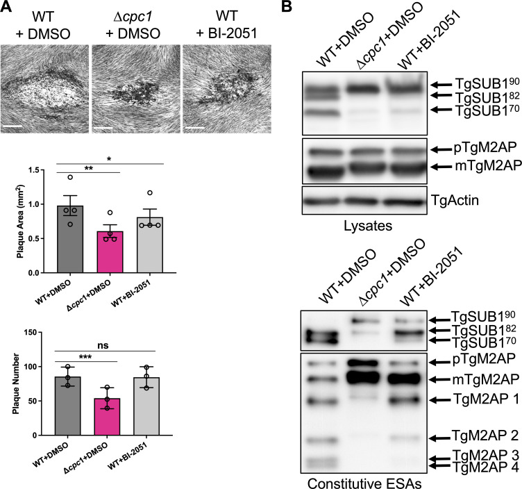
Microbial pathogens use proteases for their infections, such as digestion of proteins for nutrients and activation of their virulence factors. As an obligate intracellular parasite, Toxoplasma gondii must invade host cells to establish its intracellular propagation. To facilitate invasion, the parasites secrete invasion effectors from microneme and rhoptry, two unique organelles in apicomplexans. Previous work has shown that some micronemal invasion effectors experience a series of proteolytic cleavages within the parasite's secretion pathway for maturation, such as the aspartyl protease (TgASP3) and the cathepsin L-like protease (TgCPL), localized within the post-Golgi compartment and the endolysosomal system, respectively. Furthermore, it has been shown that the precise maturation of micronemal effectors is critical for Toxoplasma invasion and egress. Here, we show that an endosome-like compartment (ELC)-residing cathepsin C-like protease (TgCPC1) mediates the final trimming of some micronemal effectors, and its loss further results in defects in the steps of invasion, egress, and migration throughout the parasite's lytic cycle. Notably, the deletion of TgCPC1 completely blocks the activation of subtilisin-like protease 1 (TgSUB1) in the parasites, which globally impairs the surface-trimming of many key micronemal invasion and egress effectors. Additionally, we found that Toxoplasma is not efficiently inhibited by the chemical inhibitor targeting the malarial CPC ortholog, suggesting that these cathepsin C-like orthologs are structurally different within the apicomplexan phylum. Collectively, our findings identify a novel function of TgCPC1 in processing micronemal proteins within the Toxoplasma parasite's secretory pathway and expand the understanding of the roles of cathepsin C protease. IMPORTANCE Toxoplasma gondii is a microbial pathogen that is well adapted for disseminating infections. It can infect virtually all warm-blooded animals. Approximately one-third of the human population carries toxoplasmosis. During infection, the parasites sequentially secrete protein effectors from the microneme, rhoptry, and dense granule, three organelles exclusively found in apicomplexan parasites, to help establish their lytic cycle. Proteolytic cleavage of these secretory proteins is required for the parasite's optimal function. Previous work has revealed that two proteases residing within the parasite's secretory pathway cleave micronemal and rhoptry proteins, which mediate parasite invasion and egress. Here, we demonstrate that a cathepsin C-like protease (TgCPC1) is involved in processing several invasion and egress effectors. The genetic deletion of TgCPC1 prevented the complete maturation of some effectors in the parasites. Strikingly, the deletion led to a full inactivation of one surface-anchored protease, which globally impaired the trimming of some key micronemal proteins before secretion. Therefore, this finding represents a novel post-translational mechanism for the processing of virulence factors within microbial pathogens.

Keywords: Toxoplasma gondii; aminopeptidase; apicomplexan; cathepsin C; digestive vacuole; egress; invasion; lysosome; protease; protein trafficking.


Conflict of interest statement

The authors declare no conflict of interest.

Figures

FIG 1
FIG 1
Toxoplasma cathepsin C-like protease 1 (TgCPC1) is an endolysosomal protease. (A) Both TgCPC1-3xmycc and TgCPC1-3xmyci strains were co-stained with antibodies recognizing the myc epitope and either the PLVAC marker (TgCPL) or ELC markers (TgNHE3 and proTgM2AP). Immunofluorescence microscopy (IFA) of pulse-invaded parasites revealed that ~70–75% of TgCPC1 is localized in the ELC, while ~25–30% of TgCPC1 resides within the PLVAC. Co-localization analysis was quantified in ~80 parasites per biological replicate for four independent trials. Bar = 2 µm. A one-way analysis of variance (ANOVA) test was used to determine statistical significance; *, P < 0.05; ***, P < 0.001. (B) Expression patterns of epitope-tagged TgCPC1 in Toxoplasma parasites. A 3xmyc tag was inserted at the C-terminus or within the light chain of TgCPC1, which created TgCPC1-3xmycc and TgCPC1-3xmyci strains, respectively. Immunoblotting analysis showed that TgCPC1 was cleaved into a few species via multiple proteolytic cleavages. Based on the cleavage patterns of TgCPC1 seen in the immunoblots, TgCPC1 can be labeled into five domains. The domain division was deduced from the domain annotation of human cathepsin C protease via homologous alignment between TgCPC1 and human cathepsin C protease. The apparent molecular weights of TgCPC1 intermediates and final cleavage products were calculated based on their migration distances within SDS-PAGE. The intermediates and final products corresponding to individual molecular were annotated in the schematic. The polypeptides derived from TgCPC1-3xmycc and TgCPC1-3xmyci were marked in blue and red, respectively. The bands denoted by asterisks represented putative intermediates during TgCPC1 maturation or degradation products. The band labeled by the number sign was a degradation product from the putative heavy chain. TgActin was probed as the loading control. (C) TgCPC1 was mainly located in the ELC within replicated parasites. Only a minute amount of TgCPC1 was observed to overlap with TgCPL. The co-localization between TgCPC1 with TgCPL (the PLVAC marker) or proTgM2AP/TgNHE3 (the ELC markers) was denoted by white arrowheads. Bar = 5 µm.
FIG 2
FIG 2
TgCPC1 plays an important role in the lytic cycle of Toxoplasma parasites and their acute virulence. (A) The TgCPC1-deletion mutant displayed fewer and smaller plaques than WT and ∆cpc1CPC1 parasites. A noteworthy characteristic of ∆cpc1 plaques is the lack of a clear central region, suggesting that the mutant cannot migrate efficiently. Three independent assays were completed. Statistical analysis was completed using one-way analysis of variance (ANOVA), and WT was used as the control for comparison. Bar = 500 µm and 50 µm in the 25× and 200× amplification images, respectively. (B) Parasite motility was chemically induced by adding 100 mM zaprinast and recorded by time-lapse videos using an inverted fluorescence microscope with a CCD camera. The circular motility and the total distance traveled revealed that the motility of the ∆cpc1 parasites was significantly reduced compared to WT and ∆cpc1CPC1. Data shown here were derived from at least four independent trials. One-way ANOVA was used for statistical analysis. (C) Parasite invasion was reduced by ~50% in ∆cpc1 compared to WT and ∆cpc1CPC1. Six fields of view were counted for each strain per biological replicate in a total of six individual trials. (D) Lactate dehydrogenase release-based egress assay revealed that egress in ∆cpc1 was reduced by ~50% compared to WT and ∆cpc1CPC1. Data from five trials were combined for statistical calculation. (E) Replication assays were performed by quantifying the number of parasites per PV in WT, ∆cpc1, and ∆cpc1CPC1 at 28 h post-infection. One hundred PVs were enumerated per replicate in a total of three replicates and plotted. The average numbers of parasites for individual strains were compared for statistical significance calculation. All strains displayed comparable replication rates. Statistical significance for assays listed in panels C through E was determined using unpaired Student’s t-test. (F) Acute virulence was evaluated in a murine model via subcutaneous infection. One hundred parasites from each strain were used to infect outbred CD-1 mice (n = 5 per strain). Mice infected with ∆cpc1 had a modest yet significant increase in survival time. Data were recorded and presented using the Kaplan–Meier plot. Statistical analysis was performed using the Log-rank (Mantel–Cox) test. *, P < 0.05; **, P < 0.01; ***, P < 0.001; n.s., not significant.
FIG 3
FIG 3
The protein secretion patterns are altered in ∆cpc1. (A) Several microneme proteins were not properly trimmed and released in excretory secretory antigens (ESA). ESA fractions were prepared by standard constitutive and 1% ethanol-induced protein secretion. Purified ESAs were probed against a few representative microneme proteins, such as TgMIC2, TgM2AP, TgAMA1, TgPLP1, and TgMIC5. (B) Evaluation of dense granule secretion in TgCPC1-deficient parasites via immunoblotting. TgActin was probed against the lysates as loading controls. At least three independent preparations of constitutive and induced ESA samples were generated for this assay.
FIG 4
FIG 4
Intracellular trimming of some micronemal proteins is altered in ∆cpc1, while their intracellular trafficking is not changed. (A) The micronemal proteins probed in Fig. 3A were also probed in the lysates to assess the roles of TgCPC1 in micronemal protein trimming. (B) A few rhoptry proteins were also probed in the lysates to assess if TgCPC1 is involved in rhoptry protein maturation. (C) Some representative micronemal proteins were stained in pulse-invaded and replicated WT, ∆cpc1, and ∆cpc1CPC1 parasites to test if defective intracellular trimming impairs their delivery to the micronemes. (D) Abnormal intracellular trimming of TgM2AP did not cause its higher accumulation in the ELC and PLVAC. Bar = 2 µm or 5 µm in pulse-invaded and replicated parasites, respectively, in (C) and (D). All assays were repeated at least in triplicate.
FIG 5
FIG 5
Altered microneme protein secretion in ∆cpc1 is due to blocked maturation of TgSUB1. (A) Constitutive and induced ESAs as well as lysates from WT, ∆cpc1, and ∆cpc1CPC1 were probed with a TgSUB1-recognizing antibody. The majority of TgSUB1 cannot be maturated into its mature form in ∆cpc1 parasites. Accordingly, the TgSUB1 on the parasite surface is not fully active within the TgCPC1-deletion mutant. (B) To evaluate the abundance of surface-localized TgSUB1, extracellular, non-permeabilized parasites were immunostained and imaged. TgSAG1 was included as a positive control. Immunofluorescence microscopy revealed that TgSUB1 still trafficked normally to the surface of ∆cpc1 parasites, albeit in an inactive form. (C) TgSUB1 staining in fully permeabilized, pulse-invaded, and replicated ∆cpc1 mutant parasites showed that the immature TgSUB1 still traffics to the micronemes properly. Bar = 2 µm. (D) Some TgSUB1 accumulated in the ELC prior to trafficking to micronemes. The loss of TgCPC1 highly blocked the maturation of TgSUB1 but did not result in its accumulation in the ELC to a greater extent than that in WT and ∆cpc1CPC1. Bar = 2 µm.
FIG 6
FIG 6
Chemical inhibition of TgCPC1 recapitulates the phenotypes seen within ∆cpc1. WT parasites were treated with 10 µM BI-2051, a potent inhibitor against PfDPAP1, for 48 h before (A) plaque assay and (B) the preparation of lysates and ESAs. The plaque assay and immunoblotting showed that the proteolytic activity of TgCPC1 is important for the parasite’s lytic cycle, TgSUB1 maturation, and the final trimming of TgM2AP. Bar = 500 µm. Statistical significance in panel A was calculated by unpaired Student’s t-test. *, P < 0.05; **, P < 0.01; ***, P < 0.001; n.s., not significant.
FIG 7
FIG 7
Working model of the post-translational modification of micronemal effectors by TgCPC1 in Toxoplasma. Post ER biosynthesis, microneme proteins traffic through the Golgi apparatus and are cleaved within a post-Golgi compartment by TgASP3 (3), a major maturase for micronemal invasion effectors. Additionally, a minute amount of TgCPL makes an additional contribution to the maturation of some micronemal proteins in the ELC (8). Our findings suggest that TgCPC1, a dipeptidyl aminopeptidase, mediates post-translational modification on some micronemal proteins before they reach their final forms, such as TgM2AP, or gets involved in initial trimming of some micronemal proteins before subsequent cleavages, such as TgSUB1. Properly processed micronemal effectors are further delivered to microneme before subsequent processing on the parasite’s surface, followed by secretion. In the absence of TgCPC1, some incorrectly processed micronemal proteins are delivered to the surface and secreted from the parasites. Most importantly, the majority of TgSUB1 is kept as a zymogen on the parasite’s surface and cannot cleave multiple key micronemal effectors required for parasite invasion and egress. ELC, endosome-like compartment; ER, endoplasmic reticulum; M, microneme; N, nucleus; PLVAC, plant-like vacuolar compartment. This figure was created with BioRender.com.

Update of

Similar articles

References

    1. Blader IJ, Coleman BI, Chen C-T, Gubbels M-J. 2015. Lytic cycle of Toxoplasma gondii: 15 years later. Annu Rev Microbiol 69:463–485. doi:10.1146/annurev-micro-091014-104100 - DOI - PMC - PubMed
    1. Tenter AM, Heckeroth AR, Weiss LM. 2000. Toxoplasma gondii: from animals to humans. Int J Parasitol 30:1217–1258. doi:10.1016/s0020-7519(00)00124-7 - DOI - PMC - PubMed
    1. Dogga SK, Mukherjee B, Jacot D, Kockmann T, Molino L, Hammoudi P-M, Hartkoorn RC, Hehl AB, Soldati-Favre D. 2017. A druggable secretory protein maturase of Toxoplasma essential for invasion and egress. Elife 6:e27480. doi:10.7554/eLife.27480 - DOI - PMC - PubMed
    1. Lagal V, Binder EM, Huynh M-H, Kafsack BFC, Harris PK, Diez R, Chen D, Cole RN, Carruthers VB, Kim K. 2010. Toxoplasma gondii protease TgSUB1 is required for cell surface processing of micronemal adhesive complexes and efficient adhesion of tachyzoites. Cell Microbiol 12:1792–1808. doi:10.1111/j.1462-5822.2010.01509.x - DOI - PMC - PubMed
    1. Miller SA, Binder EM, Blackman MJ, Carruthers VB, Kim K. 2001. A conserved subtilisin-like protein TgSUB1 in microneme organelles of Toxoplasma gondii. J Biol Chem 276:45341–45348. doi:10.1074/jbc.M106665200 - DOI - PubMed

Publication types