. 2023 Dec;12(31):e2301683.
doi: 10.1002/adhm.202301683. Epub 2023 Jun 25.

Panthenol Citrate Biomaterials Accelerate Wound Healing and Restore Tissue Integrity

Affiliations

Panthenol Citrate Biomaterials Accelerate Wound Healing and Restore Tissue Integrity

Huifeng Wang et al. Adv Healthc Mater. 2023 Dec.

Abstract

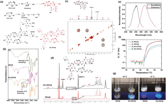
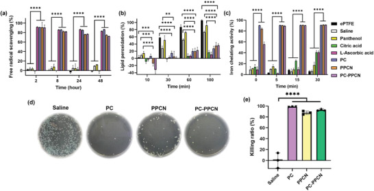
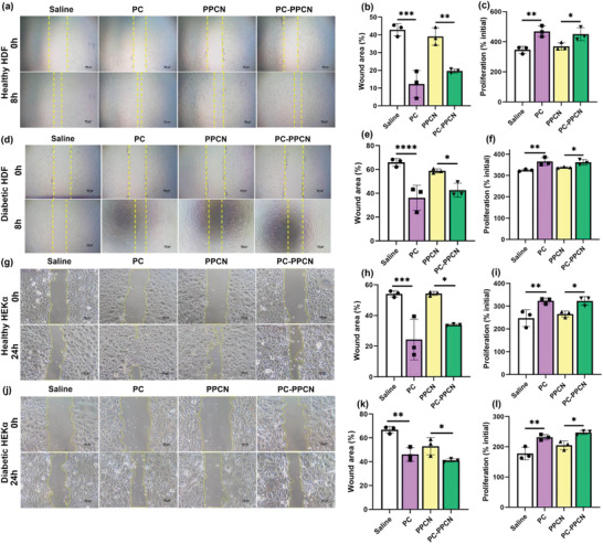
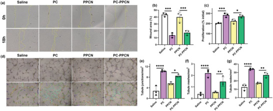
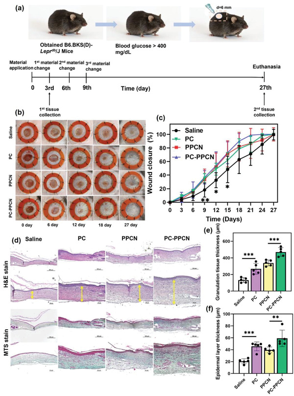
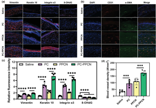
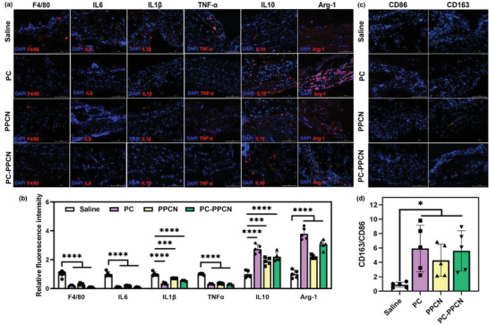
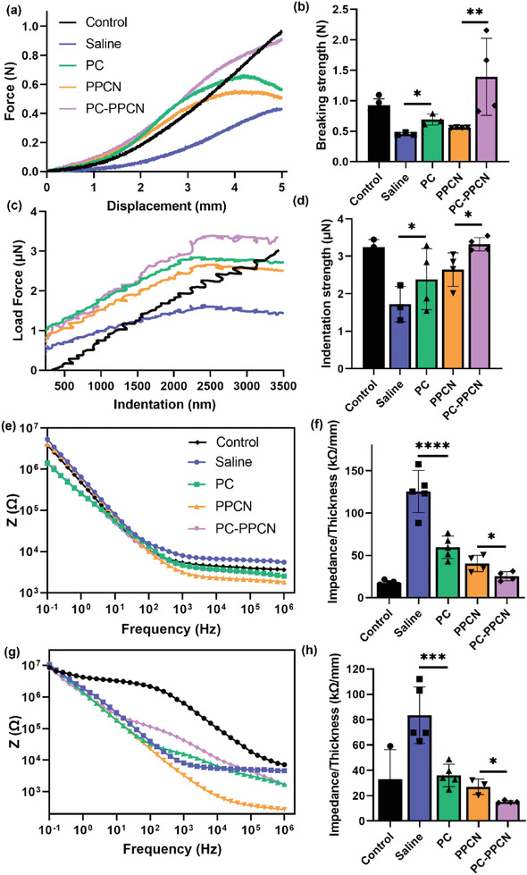
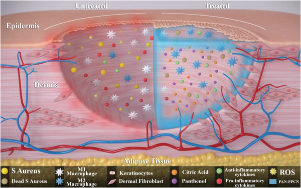
Impaired wound healing is a common complication for diabetic patients and effective diabetic wound management remains a clinical challenge. Furthermore, a significant problem that contributes to patient morbidity is the suboptimal quality of healed skin, which often leads to reoccurring chronic skin wounds. Herein, a novel compound and biomaterial building block, panthenol citrate (PC), is developed. It has interesting fluorescence and absorbance properties, and it is shown that PC can be used in soluble form as a wash solution and as a hydrogel dressing to address impaired wound healing in diabetes. PC exhibits antioxidant, antibacterial, anti-inflammatory, and pro-angiogenic properties, and promotes keratinocyte and dermal fibroblast migration and proliferation. When applied in a splinted excisional wound diabetic rodent model, PC improves re-epithelialization, granulation tissue formation, and neovascularization. It also reduces inflammation and oxidative stress in the wound environment. Most importantly, it improves the regenerated tissue quality with enhanced mechanical strength and electrical properties. Therefore, PC could potentially improve wound care management for diabetic patients and play a beneficial role in other tissue regeneration applications.

Keywords: diabetic wound healing; panthenol citrate; provitamin B5; regenerative engineering and medicine.


Conflict of interest statement

G.A.A. is an inventor on patents that disclose PC and PC‐PPCN. The remaining authors declare no conflict of interest.

Figures

Figure 1
Figure 1
Synthesis and characterization of PC and PC‐PPCN. a) Scheme of the chemical reactions to synthesize PC and PC‐PPCN. b) FT‐IR spectra of PC, PPCN, and PC‐PPCN. c) 1H NMR and 2D NMR spectra of PC. d) 1H NMR spectra of PPCN and PC‐PPCN. e) Photoluminescent properties of PC. f) The thermally‐induced phase change of PPCN and PC‐PPCN. The crossover temperature of storage modulus (G′) and loss modulus (G″) defined their LCST as 28 °C for PPCN and 30 °C for PC‐PPCN. g) Digital image of PPCN and PC‐PPCN at 25 °C and 37 °C, respectively. PPCN and PC‐PPCN are liquids at 25 °C and become gels at 37 °C. PC‐PPCN emits blue fluorescence under UV exposure at 365 nm.
Figure 2
Figure 2
Antioxidant and antibacterial properties of PC and PC‐PPCN. a–c) Antioxidant properties of PC, PC‐PPCN, and PPCN were confirmed via ABTS free radical scavenging, β‐carotene lipid peroxidation, and Fe2+ ion chelating assays. All three materials are capable of scavenging free radicals, inhibiting lipid peroxidation, and chelating Fe2+ ions. d) Digital images of S. aureus colonies on culture plates after incubation with saline, PC, PPCN, and PC‐PPCN. e) Quantification of the images in (d) describing the resulting bacteria killing ratio against S. aureus. All data are presented as mean ± SD (n = 3 for antioxidant assays; ns: not significant; *p < 0.05; **p < 0.01; ***p < 0.001, ****p < 0.0001).
Figure 3
Figure 3
PC and PC‐PPCN promote the migration and proliferation of human dermal fibroblasts (HDF) and human epidermal keratinocytes (HEKα) from healthy and diabetic individuals. a) Images of healthy HDF migration after exposure to saline, PC, PPCN, and PC‐PPCN for 8 h. b) Quantification of percentage wound area remaining for healthy HDF. c) Healthy HDF proliferation at 72 h. d) Images of diabetic HDF migration after exposure to saline, PC, PPCN, and PC‐PPCN for 8 h. e) Quantification of percentage of wound area remaining for diabetic HDF. f) Diabetic HDF proliferation at 72 h. g) Images of healthy HEKα migration after exposure to saline, PC, PPCN, and PC‐PPCN for 24 h. h) Quantification of percentage of wound area remaining for healthy HEKα. i) Healthy HEKα proliferation at 72 h. j) Images of diabetic HEKα migration after exposure to saline, PC, PPCN, and PC‐PPCN for 24 h. k) Quantification of percentage of wound area remaining for diabetic HEKα. l) Diabetic HEKα proliferation at 72 h (n = 3; ns, not significant; *p < 0.05; **p < 0.01; ***p < 0.001, ****p < 0.0001).
Figure 4
Figure 4
PC and PC‐PPCN promote HMVEC migration, proliferation, and tubule formation in vitro. a) Images of HMVEC migration after treatment with saline, PC, PPCN, and PC‐PPCN. b) Quantification of percentage wound area remaining on HMVEC. c) HMVEC proliferation at 72 h. d) Digital images of endothelial cell tubulogenesis after treatment with saline, PC, PPCN, and PC‐PPCN. Quantification of e) tubule junctions, f) tubule meshes, and g) tubule nodes. All data are presented as mean ± SD (n = 3; ns, not significant; *p < 0.05; **p < 0.01; ***p < 0.001, ****p < 0.0001).
Figure 5
Figure 5
PC and PC‐PPCN accelerate wound closure and stimulate granulation tissue and keratinocyte layer formation in a diabetic mouse model. a) Experimental design and timeline of interventions. b) Digital images of the wounds after treatment with saline, PC, PPCN, and PC‐PPCN at different time points. c) Quantification of wound areas at different time points. d) Representative tissue sections from the wound sites collected on day 27 and stained with H&E and MTS. The yellow arrows indicate the thickness of granulation tissue. e) Quantification of granulation tissue thickness. f) Quantification of epidermal layer thickness. All data are presented as mean ± SD (n = 5; ns, not significant; *p < 0.05; **p < 0.01; ***p < 0.001, ****p < 0.0001).
Figure 6
Figure 6
PC and PC‐PPCN promote keratinocyte and fibroblasts migration and proliferation and enhance blood vessel formation in vivo. Representative immunofluorescence images of tissues obtained at day 27 post‐wounding that were probed for: a) vimentin, keratin 10, integrin α3, and 8‐OHdG and b) CD31 and α‐smooth muscle actin (α‐SMA). Blue fluorescence corresponds to cell nuclei stained with 4′,6‐diamidino‐2‐phenylindole (DAPI); green fluorescence corresponds to the expression of α‐SMA, red fluorescence corresponds to the expression of vimentin, keratin 10, integrin α3, 8‐OHdG, and CD31 as indicated. c) Relative fluorescence intensity from the immunofluorescence images for vimentin, keratin 10, integrin α3, and 8‐OHdG at day 27 after treatment with saline, PC, PPCN, and PC‐PPCN. d) Quantification of blood vessel density for CD31 and α‐SMA staining. All data are presented as mean ± SD (n = 5; ns, not significant; *p < 0.05; **p < 0.01; ***p < 0.001, ****p < 0.0001).
Figure 7
Figure 7
PC and PC‐PPCN modulate inflammatory responses and facilitate the transition from M1 to M2 macrophage phenotype in vivo. a) Representative immunofluorescence images of tissues sampled at 3 days post‐wounding and probed for F4/80, IL6, IL1β, TNF‐α, IL10, and Arg‐1. Blue fluorescence corresponds to cell nuclei stained with 4′,6‐diamidino‐2‐phenylindole (DAPI); red fluorescence corresponds to the expression of F4/80, IL6, IL1β, TNF‐α, IL10, and Arg‐1 as indicated. b) Relative fluorescence intensity from the immunofluorescence images in panel (a). c) Representative immunofluorescence images of tissues sampled at 3 days post‐wounding and probed for CD86 and CD163. d) Quantification of the ratio of CD163 and CD86. All data are presented as mean ± SD (n = 5; ns, not significant; *p < 0.05; **p < 0.01; ***p < 0.001, ****p < 0.0001).
Figure 8
Figure 8
PC and PC‐PPCN improve mechanical and electrical properties of regenerated tissue. a) Tensile test of regenerated skin treated with saline, PC, PPCN, and PC‐PPCN. Uninjured db/db mouse skin was used as control. b) Breaking strength reached with 5 mm displacement. c) Indentation measurement of regenerated skin. d) Indentation strength achieved with 10 µm indentation. e) Electrochemical impedance spectra of regenerated skin in the direct configuration. f) The direct impedance of the regenerated skin at 0.1 Hz. g) Bode plots of the regenerated skin in the capacitive configuration. h) The capacitive impedance of the regenerated skin at 0.1 Hz. All data are presented as mean ± SD (n = 5; ns, not significant; *p < 0.05; **p < 0.01; ***p < 0.001, ****p < 0.0001).
Figure 9
Figure 9
Illustration of the potential mechanisms for PC and PC‐PPCN to accelerate diabetic wound closure, including facilitating macrophage polarization from M1 to M2, inhibiting bacterial infection, promoting skin cell migration, and stimulating angiogenesis in the db/db mouse model.

Similar articles

Cited by

References

    1. Lavery L. A., Higgins K. R., Lanctot D. R., Constantinides G. P., Zamorano R. G., Athanasiou K. A., Armstrong D. G., Agrawal C. M., Diabetes Care 2007, 30, 14. - PubMed
    1. Ndosi M., Wright‐Hughes A., Brown S., Backhouse M., Lipsky B. A., Bhogal M., Reynolds C., Vowden P., Jude E. B., Nixon J., Nelson E. A., Diabetic Med. 2018, 35, 78. - PMC - PubMed
    1. Armstrong D. G., Boulton A. J. M., Bus S. A., N. Engl. J. Med. 2017, 376, 2367. - PubMed
    1. Bekele F., Chelkeba L., J. Foot Ankle Res. 2020, 13, 65. - PMC - PubMed
    1. Deng L., Du C., Song P., Chen T., Rui S., Armstrong D. G., Deng W., Oxid. Med. Cell. Longevity 2021, 2021, 8852759. - PMC - PubMed

Publication types

LinkOut - more resources