. 2023 Jun 16;9(24):eadf2161.
doi: 10.1126/sciadv.adf2161. Epub 2023 Jun 16.

A Plasmodium membrane receptor platform integrates cues for egress and invasion in blood forms and activation of transmission stages

Affiliations

A Plasmodium membrane receptor platform integrates cues for egress and invasion in blood forms and activation of transmission stages

Ronja Marie Kuehnel et al. Sci Adv. .

Abstract

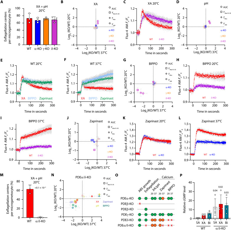
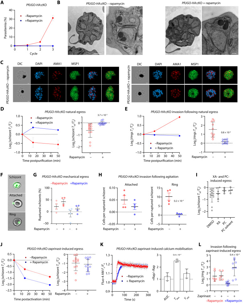
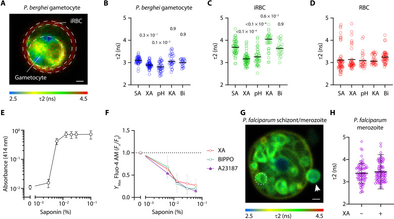
Critical events in the life cycle of malaria-causing parasites depend on cyclic guanosine monophosphate homeostasis by guanylyl cyclases (GCs) and phosphodiesterases, including merozoite egress or invasion of erythrocytes and gametocyte activation. These processes rely on a single GCα, but in the absence of known signaling receptors, how this pathway integrates distinct triggers is unknown. We show that temperature-dependent epistatic interactions between phosphodiesterases counterbalance GCα basal activity preventing gametocyte activation before mosquito blood feed. GCα interacts with two multipass membrane cofactors in schizonts and gametocytes: UGO (unique GC organizer) and SLF (signaling linking factor). While SLF regulates GCα basal activity, UGO is essential for GCα up-regulation in response to natural signals inducing merozoite egress and gametocyte activation. This work identifies a GC membrane receptor platform that senses signals triggering processes specific to an intracellular parasitic lifestyle, including host cell egress and invasion to ensure intraerythrocytic amplification and transmission to mosquitoes.


Figures

Fig. 1.
Fig. 1.. XA and a rise in extracellular pH activate calcium mobilization at 37° and 20°C in P. berghei gametocytes, while a drop in temperature is essential later for DNA replication.
(A) Fluorescence response kinetics of gametocytes loaded with Fluo-4 AM upon stimulation with 100 μM XA and illustration of the different parameters used for the quantification of calcium mobilization (SD; n = 3 technical replicates). (B and C) Fluorescence response upon stimulation with 100 μM XA or a rise from pH 7.2 to 7.8 at 20°C (B) and 37°C (C) (SD; n = 3 technical replicates). (D) Relative changes in calcium response at 37°C compared to 20°C upon stimulation with 100 μM XA or rise from pH 7.2 to 7.8 indicate that the time to reach the maximum calcium response is faster at 37°C (SD; n = 2 biological replicates, unpaired two-tailed t test). (E) Gametocyte activation with 100 μM XA or rise from pH 7.2 to 7.8 does not allow microgamete formation at 37°C, whereas the same stimuli at 20°C leads to formation of microgametes [SD; n = 3 technical replicates, one-way analysis of variance (ANOVA)]. (F) Proportion of haploid or multiploid gametocytes in nonactivated and 8-min activated gametocytes at 20° or 37°C shows that DNA replication is blocked at 37°C (SD; n = 3 biological replicates, one-way ANOVA). (G) Dilution of gametocytes maintained in SA medium (SA) at 37°C into a cold SA medium leading to a rapid decrease to 20.5°C is not sufficient to induce a rapid calcium response. Dilution into the same SA containing either 100 μM XA or at a pH of 7.8 leads to normal calcium mobilization.
Fig. 2.
Fig. 2.. PDEα and PDEδ are active at 37°C in gametocytes and epistatic interactions between both enzymes that prevent premature and abortive activation.
(A) KOs of nonessential PDEs show normal exflagellation rates (SD; n = 3 biological replicates). (B) Comparison of the Ca2+ response of each PDE-KO to the wild type (WT). A shift toward the left reflects a decrease at 37°C in the KO compared to the WT. A shift below the horizontal line reflects a decrease at 20°C in the KO compared to the WT. The parameters used correspond to the parameters described in Fig. 1A (SD; n = 3 or n = 2 biological replicates at 20° and 37°C, respectively). (C) Fluorescence kinetics of WT and PDEδ-KO gametocytes upon stimulation with 100 μM XA. (D) A rise to pH 7.8 leads to similar response profiles observed with XA for each PDE-KO line. (E and F) Fluorescence kinetics of WT gametocytes upon stimulation at 20°C (E) or 37°C (F) (SD; n = 3 biological replicates). (G) Upon stimulation with 5 μM 5-benzyl-3-isopropyl-1H-pyrazolo[4,3-d]pyrimidin-7(6H)-one (BIPPO), a strong decrease of PDEδ-KO response is observed. (H and I) Fluorescence kinetics of WT and PDEδ-KO gametocytes upon stimulation with 5 μM BIPPO at 20°C (H) and 37°C (I) (SD; n = 3 technical replicates). (J) Upon stimulation with 500 μM zaprinast, a decrease of PDEα-KO response is observed at 37°C. (K and L) Fluorescence kinetics of WT and PDEα-KO gametocytes upon stimulation with 500 μM zaprinast at 20°C (K) or 37°C (L). (M) The simultaneous deletions of PDEα and PDEδ prevent microgametogenesis (SD; n = 3 biological replicates, unpaired two-tailed t test). (N) Comparison of the calcium response of PDEα/δ-KO gametocytes with WT at 37° and 20°C. (O) Summary of assessed PDE phenotypes. ABS, asexual blood stages; red crosses, affected; orange circles, mildly affected; green circles, not affected. (P) Relative basal and induced cGMP levels in WT and PDEα/δ-KO gametocytes (SD; n = 2 biological replicates with technical duplicates each, one-way ANOVA).
Fig. 3.
Fig. 3.. In P. berghei, GCα is part of a signaling platform whose basal and induced activities are differentially regulated by SLF and UGO, respectively.
(A) GCα-AID/HA is not detectable anymore after 1 hour of degradation with auxin. Tir1 is tagged by c-Myc and serves as a loading control. (B) A strong reduction of exflagellation in response to XA is observed upon GCα-AID/HA degradation (SD; n = 6 biological replicates, unpaired two-tailed t test). (C) Fluorescence kinetics of gametocytes upon GCα-AID/HA degradation in response to 100 μM XA. (D) The relative AUC of Fluo-4 AM fluorescence upon GCα degradation compared to untreated gametocytes (SD; n = 3 biological replicates, unpaired two-tailed t test). (E) Mass spectrometry identification of proteins immunoprecipitated (IP) from lysates of UGO-HA and GCα-HA gametocytes. ND, not detected (n = 2 biological replicates). (F) Depletion of SLF-AID/HA upon auxin treatment (expected size is 144 kDa). (G) A twofold reduction of exflagellation in response to XA is observed upon SLF-AID/HA degradation (SD; n = 3 biological replicates, unpaired two-tailed t test). (H) AUC of the fluorescence response upon SLF-AID/HA degradation compared to untreated gametocytes (SD; n = 3 biological replicates, unpaired two-tailed t test). (I) Relative basal and induced cGMP levels upon treatment with XA and BIPPO (Bi) in SLF-AID/HA gametocytes in presence or absence of IAA (SD; n = 3 biological replicates, one-way ANOVA). (J) A strong reduction of exflagellation in response to XA is observed upon UGO-AID/HA degradation (SD; n = 6 biological replicates, unpaired two-tailed t test). (K) AUC of the fluorescence response upon UGO degradation compared to untreated gametocytes (SD; n = 3 biological replicates, unpaired two-tailed t test). (L) Relative basal and induced cGMP levels upon treatment with XA and BIPPO in UGO-AID/HA gametocytes in presence or absence of auxin (SD; n = 2 biological replicates with technical duplicates, one-way ANOVA).
Fig. 4.
Fig. 4.. UGO is essential for XA-dependent activation of gametogenesis in P. falciparum.
Conditional deletion of PfUGO in sexual ring stages does not affect the sex ratio (A) but significantly affects female gametogenesis (B) and male gametogenesis (C) [SD; n = 3 (A) and n = 5 (B) and (C) biological replicates, unpaired two-tailed t test]. In (B), representative images from the fluorescence microscopy–based female gamete activation assay are shown. DMSO (top)– or rapamycin (bottom)–treated gametocytes were stained with SYBR Green and α-Pfs25 antibodies. Activated females form clusters of cells and are Pfs25-positives. Scale bars, 30 μm, insets 2 μm.
Fig. 5.
Fig. 5.. UGO interacts with GCα in P. berghei schizonts and shows differential requirement for zaprinast-induced egress.
(A) Mass spectrometric identification of proteins immunoprecipitated from lysates of UGO-HA and GCα-HA P. berghei schizonts (n = 2 biological replicates). (B) Fluorescence response kinetics of schizonts of the non–gametocyte-producing ANKA 2.33 (NGP) line loaded with Fluo-4 AM. Cells are stimulated with zaprinast, BIPPO, or A23187. (C) The relative AUC of Fluo-4 AM fluorescence upon stimulation. Error bars show SD from the mean from three independent infections. (D) Change in the numbers of schizonts, as assessed by Giemsa staining, following 50 min of treatment of cells with DMSO or zaprinast (SD; n = 5 independent inductions from two biological replicates, unpaired two-tailed t test). (E) Change in the numbers of schizonts, as assessed by Giemsa staining, following 50 min of treatment of cells with zaprinast upon UGO-AID/HA degradation at the onset of the culture (16 hours) or 1 hour before treatment (1 hour) [SD; n = 5 independent inductions from four biological replicates (16 hours) and two biological replicates (1 hour), unpaired two-tailed t test).
Fig. 6.
Fig. 6.. UGO is essential for P. falciparum merozoite egress and invasion but can be bypassed by PDE inhibition.
(A) Replication of DMSO- and rapamycin-treated PfUGO-HA:cKO parasites (SD; n = 3 independent infections). (B and C) PfUGO-deleted parasites reach late schizogony as observed by electron microscopy (B). Scale bars, 1 μm (left) and 200 nm (right). AMA1 and MSP1 localization by immunofluorescence (C). Scale bars, 2 μm. (D) Conditional deletion of PfUGO leads to the accumulation of schizonts (left: data from two sister cultures; right: replicates from six independent cultures; unpaired two-tailed t test). (E) Conditional deletion of PfUGO leads to a diminution of ring stage formation (left: duplicates from two sister cultures; right: replicates from five independent cultures; two-tailed t test). (F) Representative images used to quantify schizonts, attached merozoites, and ring stages. Bright-field and SYBR Green (green) channels are shown. Scale bar, 7 μm. (G) Agitation leads to the rupture of 19% of UGO-blocked schizonts (n = 4 biological replicates, unpaired two-tailed t test). (H) PfUGO conditional deletion reduces merozoite attachment and ring formation following mechanical disruption of schizonts (n = 4 biological replicates, unpaired two-tailed t test). (I) Treatment of P. falciparum–segmented schizonts with 100 μM XA or 50 μM PC treatments does not induce egress. (J) Treatment of segmented schizonts with the PDE inhibitor zaprinast bypasses the requirement of PfUGO for egress (left: data from two sister cultures; right: replicates from six independent cultures; unpaired two-tailed t test). (K) PfUGO-deleted schizonts are responsive to zaprinast (SD; left: n = 3 technical replicates; right: n = 3 biological replicates; unpaired two-tailed t test). (L) Invasion following zaprinast-induced egress is not affected (SD; n = 3 biological replicates with technical duplicates; one-way ANOVA).
Fig. 7.
Fig. 7.. XA and a rise in extracellular pH similarly lower membrane tension in gametocytes but not in schizonts.
(A) Lifetime fluorescence in nanoseconds of the Flipper-TR probe in membranes of a P. berghei gametocyte in an erythrocyte. Dashed lines illustrate the regions of interest analyzed. Scale bar, 1 μm. (B to D) Lifetime fluorescence quantification of Pama1ICM1 gametocytes (B), infected erythrocytes (C), and noninfected erythrocytes (D) in presence of SA (pH 7.2), XA (pH 7.2), SA (pH 7.8), KA (pH 7.2), and BIPPO [(Bi), pH 7.2]. Circles represent values from single cells from at least three independent infections; one-way ANOVA. (E) Hemoglobin release upon exposure of a P. berghei–infected RBC to increasing concentration of saponin as measured by absorbance at 414 nm (SD; n = 3 biological replicates). (F) No difference in maximum calcium mobilization (YMax) is observed upon treatment with XA, BIPPO, and A23187 at a given saponin concentration (SD; n = 3 biological replicates). (G) Lifetime fluorescence in nanoseconds of the Flipper-TR probe in membranes of a P. falciparum schizont in an erythrocyte and an extracellular merozoite (white arrow). Dashed lines illustrate the regions of interest analyzed. Scale bar, 1 μm. (H) Lifetime fluorescence quantification of P. falciparum schizonts in the absence or presence of XA. Circles represent values from singles cells from at least three independent infections.
Fig. 8.
Fig. 8.. Current model showing how cGMP homeostasis is regulated in P. berghei gametocytes and P. falciparum schizonts.
In gametocytes (left), cGMP synthesis depends on GCα whose steady-state activity requires GEP1 and SLF. UGO induces GCα activity in response to XA or a rise in extracellular pH (pHex) possibly via lowered membrane tension. In nonactivated gametocytes, cGMP hydrolysis depends at least on PDEα and PDEδ that are both active at 37°C. PDEδ is also active at 20°C and dampens the induced signal. PDEα is targeted by zaprinast, and PDEδ is the main target of BIPPO in gametocytes. In schizonts (right), the same architecture is conserved, where UGO also induces GCα activity in response to unknown signals. Unidentified zaprinast- and BIPPO-sensitive PDEs prevent premature egress.

References

    1. C. R. Collins, F. Hackett, M. Strath, M. Penzo, C. Withers-Martinez, D. A. Baker, M. J. Blackman, Malaria parasite cGMP-dependent protein kinase regulates blood stage merozoite secretory organelle discharge and egress. PLOS Pathog. 9, e1003344 (2013). - PMC - PubMed
    1. R. E. Sinden, The cell biology of sexual development in Plasmodium. Parasitology 86, 7–28 (1983). - PubMed
    1. O. Billker, V. Lindo, M. Panico, A. E. Etienne, T. Paxton, A. Dell, M. Rogers, R. E. Sinden, H. R. Morris, Identification of xanthurenic acid as the putative inducer of malaria development in the mosquito. Nature 392, 289–292 (1998). - PubMed
    1. D. S. Yamamoto, M. Sumitani, M. Hatakeyama, H. Matsuoka, Malaria infectivity of xanthurenic acid-deficient anopheline mosquitoes produced by TALEN-mediated targeted mutagenesis. Transgenic Res. 27, 51–60 (2018). - PubMed
    1. M. Arai, O. Billker, H. R. Morris, M. Panico, M. Delcroix, D. Dixon, S. V. Ley, R. E. Sinden, Both mosquito-derived xanthurenic acid and a host blood-derived factor regulate gametogenesis of Plasmodium in the midgut of the mosquito. Mol. Biochem. Parasitol. 116, 17–24 (2001). - PubMed