. 2023 Jun 26;164(8):bqad096.
doi: 10.1210/endocr/bqad096.

The Clock-modulatory Activity of Nobiletin Suppresses Adipogenesis Via Wnt Signaling

Affiliations

The Clock-modulatory Activity of Nobiletin Suppresses Adipogenesis Via Wnt Signaling

Xuekai Xiong et al. Endocrinology. .

Abstract

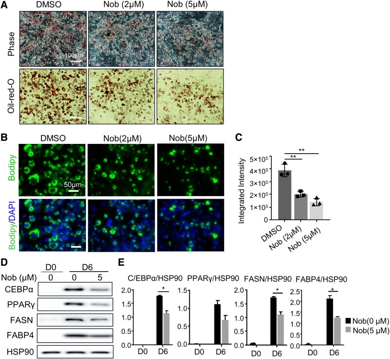
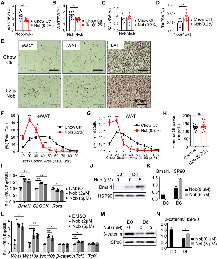
The circadian clock machinery exerts transcriptional control to modulate adipogenesis and its disruption leads to the development of obesity. Here, we report that Nobiletin, a circadian clock amplitude-enhancing molecule, displays antiadipogenic properties via activation of Wnt signaling pathway that is dependent on its clock modulation. Nobiletin augmented clock oscillatory amplitude with period lengthening in the adipogenic mesenchymal precursor cells and preadipocytes, accompanied by an induction of Bmal1 and clock components within the negative feedback arm. Consistent with its clock-modulatory activity, Nobiletin strongly inhibited the lineage commitment and terminal differentiation of adipogenic progenitors. Mechanistically, we show that Nobiletin induced the reactivation of Wnt signaling during adipogenesis via transcriptional up-regulation of key components within this pathway. Furthermore, Nobiletin administration in mice markedly reduced adipocyte hypertrophy, leading to a significant loss of fat mass and reduction of body weight. Last, Nobiletin inhibited the differentiation of primary preadipocytes, and this effect was dependent on a functional clock regulation. Collectively, our findings uncover a novel activity of Nobiletin in suppressing adipocyte development in a clock-dependent manner, implicating its potential application in countering obesity and associated metabolic consequences.

Keywords: Wnt signaling; adipocyte development; adipogenesis; circadian clock; differentiation.


Figures

Figure 1.
Figure 1.
Nobiletin promotes clock oscillation in adipocytes. (A-C) Baseline-adjusted tracing plots of average bioluminescence activity of 3T3-L1 cells with stable expression of a Per2::dLuc reporter for 5 days (A), with quantitative analysis of clock cycling amplitude (B) and period length (C), with Nobiletin (Nob) treatment at indicated concentrations. (D–F) Baseline-adjusted tracing plots of average bioluminescence activity of C3H10T1/2 cells with stable expression of a Per2::dLuc reporter for 5 days (D), with quantitative analysis of clock cycling amplitude (E) and period length (F), with Nob treatment at indicated concentrations. Data are presented as mean ± SD of n = 4 replicates for each concentration tested, with 3 independent repeat experiments. (G, H) Real-time quantitative PCR analysis of clock gene expression at indicated concentrations of Nobiletin in 3T3-L1 cells (G) and C3H10T1/2 cells (H). Data are presented as mean ± SD of n = 3 replicates. *P < .05 and **P < 0.01 Nob vs DMSO by Student t test.
Figure 2.
Figure 2.
Effect of Nobiletin on terminal differentiation of 3T3-L1 preadipocyte. (A–C) Representative images of Oil-Red-O (A) and Bodipy staining of lipids (B) with quantification (C), at day 6 of 3T3-L1 preadipocyte differentiation with Nobiletin treatment at indicated concentrations. Scale bars: 100 μm (A) and 50 μm (B). (D, E) Immunoblot analysis of adipogenic factors and mature adipocyte markers at days 0 and 6 of 3T3-L1 differentiation with Nobiletin treatment at indicated concentration, with quantification shown (E). Each lane represents a pooled sample of 3 replicates.
Figure 3.
Figure 3.
Effect of Nobiletin on adipogenic differentiation of C3H10T1/2 mesenchymal precursor cells. (A, B) Representative images of phase contrast and Oil-Red-O staining of lipid accumulation at day 5 (A) and day 8 (B) of C3H10T1/2 differentiation with Nobiletin treatment at indicated concentrations. Scale bars: 100 μm. (C, D) Representative images of Bodipy staining of lipids (C) with quantification (D) at day 8 of C3H10T1/2 differentiation with Nobiletin treatment at indicated concentrations. (E-H) Immunoblot analysis of adipogenic factors (E) with quantification (F), and mature adipocyte markers (G) with quantification (H), at days 0 and 8 of C3H10T1/2 differentiation in response to Nobiletin treatment at indicated concentration. Each lane represents a pooled sample of 3 replicates.
Figure 4.
Figure 4.
Nobiletin inhibits adipogenic differentiation via Wnt signaling pathway. (A, B) Real-time quantitative PCR analysis of expression of genes involved in the Wnt signaling pathway with Nobiletin treatment at indicated concentrations in 3T3-L1 preadipocytes (A) and C3H10T1/2 mesenchymal precursors (B). Data are presented as mean ± SD of n = 3 replicates. *P < .05 and **P < 0.01 Nob vs DMSO by Student t test. (C, D) Immunoblot analysis of β-catenin at days 0 and 8 of C3H10T1/2 adipogenic differentiation with Nobiletin treatment (C) with quantitative analysis shown (D). Each lane represents a pooled sample of 3 replicates. (E) Analysis of Nobiletin effect on TOPFlash luciferase reporter activity in C3H10T1/2 the presence or absence of 10% Wnt3a media at indicated concentrations. Data are presented as mean ± SD of n = 4 replicates. **P < .05 and P < .01 Nob vs DMSO by Student t test.
Figure 5.
Figure 5.
Nobiletin displays antiobesity effect in vivo. (A–D) Analysis of the effect of 0.1% Nob-supplemented diet feeding for 4 weeks on body weight (A), change in body weight (B), epidydimal white adipose tissue (eWAT, C), and gastrocnemius (GN) muscle weight to body weight ratio (D) in 48-week-old male mice (n = 7/group). (E–H) Effect of 0.2% Nob-supplemented diet feeding for 4 weeks on body weight (E), nuclear magnetic resonance analysis of total fat mass (F), total fat mass to body weight ratio (G), and lean mass to body weight ratio (H) in 10-week-old male mice (n = 5-10/group). (I–N) Analysis of the effect of 0.2% Nob-supplemented diet feeding on whole-body energy homeostasis. Average plots with quantitative analysis of food intake (I, J), oxygen consumption (K, L), and total activity monitoring (M, N) were shown for control and Nobiletin-treated mice. Day and night times are indicated by open and closed bars, respectively. n = 5 mice/group. Data are presented as mean ± SE. *P < .05 and **0P < .01 Nob vs control diet by Student t test.
Figure 6.
Figure 6.
Nobiletin administration in mice reduces adipose tissue expansion. (A–D) Analysis of effect of 0.2% Nob-supplemented diet feeding on tissue weight after 4 weeks of treatment in epidydimal white adipose tissue (eWAT, A), inguinal subcutaneous white adipose tissue (iWAT, B), interscapular brown adipose tissue (BAT, C), and tibialis anterior (TA, D) (n = 10/group). (E) Representative hematoxylin and eosin histology images of eWAT, iWAT, and BAT in chow and 0.2% Nob-supplemented diet mice after 4 weeks. Scale bar: 100 μm. (F, G) Analysis of adipocyte cell size distribution of eWAT (F) and iWAT (G) in normal chow control and 0.2% Nob-supplemented diet mice. (H) Analysis of serum glucose levels at 9 Am in control and 0.2% Nob diet-treated mice (n = 10/group). (I) Real-time quantitative PCR analysis of circadian clock genes induction by Nob in isolate primary preadipocytes at indicated concentrations. Data are presented as mean ± SD of n = 3 replicates. *P < .05 and **P < .01 Nob vs DMSO by Student t test. (J, K) Immunoblot analysis of Bmal1 protein expression by 5 μM Nob treatment at days 0 and 6 of adipogenic induction in primary preadipocytes (J) with quantification (K). (L) Real-time quantitative PCR analysis of Wnt pathway genes by Nob in isolate primary preadipocytes at indicated concentrations. Data are presented as mean ± SD of 3 replicates. *P < .05 and **P < .01 Nob vs DMSO by Student t test. (M, N) Immunoblot analysis of β-catenin protein expression by 5 μM Nob treatment at days 0 and 6 of adipogenic induction of primary preadipocytes (M) with quantification (N). Each lane represents a pooled sample of 3 replicates.
Figure 7.
Figure 7.
Effect of Nobiletin on primary preadipocyte differentiation. (A, B) Representative images of Oil-Red-O staining of adipogenic differentiation of isolated primary preadipocyte from the stromal vascular fraction of subcutaneous inguinal adipose tissue treated at indicated concentration of Nobiletin at day 4 (A) and day 6 (B). Scale bar: 100 μm. (C–F) Immunoblot analysis of adipogenic factors (C) with quantification (D), and mature adipocyte markers (E) with quantification (F), at days 0 and 6 of primary preadipocyte differentiation in response to Nobiletin treatment. Each lane represents a pooled sample of 3 replicates. (G) Representative images of Bodipy staining at day 6 of differentiation of primary preadipocytes isolated from floxed control (BMCtr) and Bmal1-deficient (BMKO) mice treated at indicated concentration of Nobiletin. Scale bar: 50 μm. (H, I) Quantitative analysis of Bodipy staining of day 4 (H) and day 6 (I) of BMCtr and BMKO primary adipocyte differentiation (n = 3).

Update of

References

    1. Takahashi JS. Transcriptional architecture of the mammalian circadian clock. Nature Rev Genet. 2017;18(3):164‐179. - PMC - PubMed
    1. Bass J, Takahashi JS. Circadian integration of metabolism and energetics. Science. 2010;330(6009):1349‐1354. - PMC - PubMed
    1. Panda S, Antoch MP, Miller BH, et al. . Coordinated transcription of key pathways in the mouse by the circadian clock. Cell. 2002;109(3):307‐320. - PubMed
    1. Eckel-Mahan KL, Patel VR, Mohney RP, Vignola KS, Baldi P, Sassone-Corsi P. Coordination of the transcriptome and metabolome by the circadian clock. Proc Natl Acad Sci USA. 2012;109(14):5541‐5546. - PMC - PubMed
    1. Stenvers DJ, Scheer F, Schrauwen P, La Fleur SE, Kalsbeek A. Circadian clocks and insulin resistance. Nat Rev Endocrinol. 2019;15(2):75‐89. - PubMed

Publication types