. 2023 Jun 22;186(13):2880-2896.e17.
doi: 10.1016/j.cell.2023.05.026. Epub 2023 Jun 15.

Structural specializations of the sperm tail

Affiliations

Structural specializations of the sperm tail

Miguel Ricardo Leung et al. Cell. .

Abstract

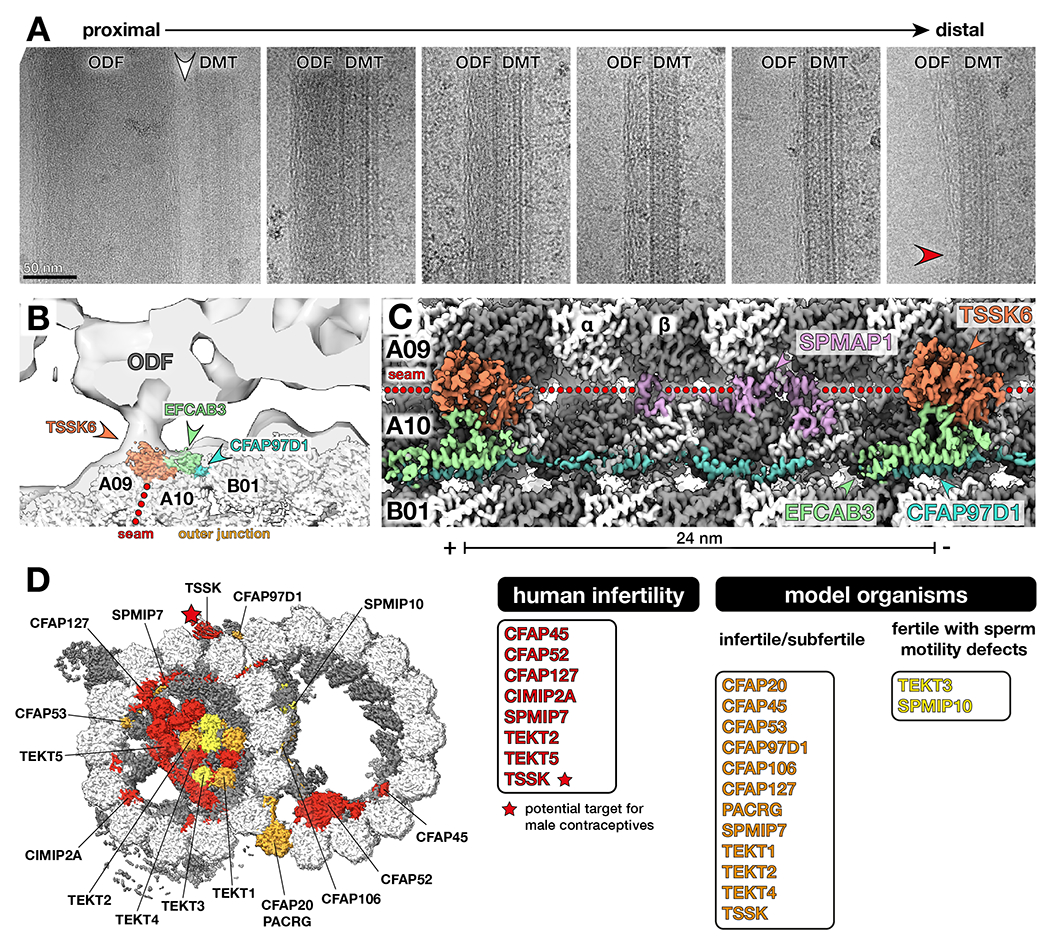
Sperm motility is crucial to reproductive success in sexually reproducing organisms. Impaired sperm movement causes male infertility, which is increasing globally. Sperm are powered by a microtubule-based molecular machine-the axoneme-but it is unclear how axonemal microtubules are ornamented to support motility in diverse fertilization environments. Here, we present high-resolution structures of native axonemal doublet microtubules (DMTs) from sea urchin and bovine sperm, representing external and internal fertilizers. We identify >60 proteins decorating sperm DMTs; at least 15 are sperm associated and 16 are linked to infertility. By comparing DMTs across species and cell types, we define core microtubule inner proteins (MIPs) and analyze evolution of the tektin bundle. We identify conserved axonemal microtubule-associated proteins (MAPs) with unique tubulin-binding modes. Additionally, we identify a testis-specific serine/threonine kinase that links DMTs to outer dense fibers in mammalian sperm. Our study provides structural foundations for understanding sperm evolution, motility, and dysfunction at a molecular level.

Keywords: cryoelectron microscopy; microtubule associated proteins; microtubule inner proteins; motile cilia; sperm.


Conflict of interest statement

Declaration of interests The authors declare no competing interests.

Figures

Figure 1.
Figure 1.. Cryo-electron microscopy of native sperm axonemal doublet microtubules (DMTs).
(A-B) Schematic diagrams of (A) sea urchin and (B) bovine sperm. (C-D) Electron micrographs of (C) sea urchin and (D) bovine sperm. Scale bars: left panels – 10 μm, right panels – 50 nm. (E-F) Cross sections (viewed from the plus end) of cryo-EM maps of the 48-nm DMT repeat from (E) sea urchin sperm, with a total of 55 microtubule inner proteins (MIPs) and microtubule-associated proteins (MAPs) colored individually; and from (F) bovine sperm, with 65 MIPs/MAPs colored individually. Unassigned proteins are colored in grey.
Figure 2.
Figure 2.. Structural comparisons of DMTs from different species and cell types.
(A) Venn diagram of MIPs and MAPs identified in cryo-EM structures of DMTs from sea urchin and bovine sperm (this study), bovine respiratory cilia (PDB 7RRO) , and Chlamydomonas flagella (PDB 6U42) . Full lists of proteins corresponding to all regions of the Venn diagram are provided in Table S4. (B) Lists of core-MIPs, sperm-MIPs, and MAPs identified in this study. Notes: (1) SAXO proteins are present in all DMT structures to date, although individual proteins may be species- or cell type-specific. (2) CCDC81 and SPATA45 are also present in our bovine sperm proteome, but definitive density is currently only resolved in the cryo-EM map of sea urchin sperm. (3) CIMAP3 (Pitchfork) density is weaker in cryo-EM structures of bovine sperm, but it is present in our proteomics data. (C) Comparison of mammalian DMTs from different cell types, with MIPs specific to trachea colored in green and MIPs specific to sperm colored in pink. (D) Comparison of sperm DMTs from different species, with MIPs resolved only in sea urchin colored in blue and MIPs resolved only in bovine colored in pink.
Figure 3.
Figure 3.. Evolution and structural expansion of the tektin bundle.
A simplified phylogenetic tree with nodes indicating the number of tektin genes found in each species. (A-D) Top panels show cryo-EM maps of DMTs from (A) bovine sperm (this study), (B) bovine respiratory cilia (EMD-24664) , (C) sea urchin sperm (this study), and (D) Chlamydomonas (this study). Tektins and TEKTL1, a tektin-like protein, are colored. Middle panels show ribbon representations of the tektin bundle and TEKTL1 filament. Bottom panels show schematic diagrams of the tektin bundle. (E) Longitudinal slice showing cryo-EM density colored by protein for the expanded tektin bundle in bovine sperm DMTs. TEKT5-F runs diagonally perpendicular to other tektins. (F) Zoomed-in ribbon diagrams showing the intermolecular β-sheet formed by the N-termini of TEKT5-F, TEKT3-1, and CIMIP2A.
Figure 4.
Figure 4.. TEKTL1 is a conserved tektin-like sperm-MIP.
(A-B) Longitudinal slices from cryo-EM density maps of (A) sea urchin and (B) bovine sperm DMTs showing the molecular environment of the TEKTL1 filament. TEKTL1 interacts with both the outer junction core-MIP CFAP77 and the sperm-MIP SPMIP10 (TEX43). (C) Comparison of the tertiary (left) and quaternary (right) structures of TEKTL1 (upper) and tektin filaments (lower, tektin 5-A as an example) from bovine sperm DMTs. In the left panels, proteins are colored in a rainbow palette from N- (blue) to C-terminus (red). TEKTL1 secondary structures are annotated by analogy to tektin. In the right panel, one copy of each protein is colored in a rainbow palette from N to C. The arrowheads indicate differences in the L12 loop at the inter-protomer interface. For the TEKTL1 filament, SPMIP10 molecules that bind at the inter-protomer interfaces are shown as transparent surfaces.
Figure 5.
Figure 5.. The Mn-motif is a universal microtubule-binding motif.
(A-C) Location and identities of SAXO proteins in (A) sea urchin sperm DMTs, (B) bovine sperm DMTs, and (C) bovine sperm endpiece singlet microtubules (SMTs). SAXO proteins are classified based on how they bind the microtubule lattice: through a single Mn-motif (blue circles), through multiple Mn-motifs along a single protofilament (violet circles), or through multiple Mn-motifs across neighboring protofilaments (orange circles). Asterisks indicate that SAXO1 is tentatively assigned based on in-cell cross-links to SPACA9 (see also Fig. S5 and Table S6). (D-F) Representative SAXO proteins from each of the aforementioned classes. Density and models are shown for selected sperm-MIPs from sea urchin sperm DMTs. Other examples are listed below and colored based on whether they are conserved (black), sperm-specific (pink), or Chlamydomonas-specific (green). Notes: (1) SAXO3 and SAXO4 (PPP1R32) are present in sea urchin sperm, bovine sperm, and bovine respiratory cilia, but not in Chlamydomonas. (2) CFAP68 and CFAP107 have only one Mn motif in Chlamydomonas. (G) The sperm-MIP SAXO5 (TEX45) has 11 Mn-motifs in sea urchin (top) and Bos taurus (bottom) and 96-nm periodicity (as shown in Fig. S6).
Figure 6.
Figure 6.. Axonemal microtubule-associated proteins (MAPs) interact with DMTs through unique tubulin binding modes.
(A-B) Axonemal MAPs identified in cryo-EM structures of (A) sea urchin and (B) bovine sperm DMTs. MAPs are shown in licorice cartoon representation. (C) Cross section views comparing microtubule-binding modes of neuronal MAPs Tau (PDB 6CVN) and MAP7 (PDB 7SGS) to the axonemal wedge-MAPs (CIMAP1, CIMAP2, CIMAP3, CFAP96) and arc-MAP (SPMAP2) identified in sperm DMTs. (D) The axonemal MAP CFAP97D1 specifically recognizes the outer junction, a site of atypical inter-tubulin contacts. (E) CFAP97D1 has an overall 24-nm repeat and consists of four helices joined by linkers that zig-zag between the surface helices of protofilament A10 and the neighboring protofilament B01. (F) Binding of CFAP97D1 to protofilament B01 appears to be mediated by positively charged patches on CFAP97D1 interacting with negatively charged pockets at the minus end of β-tubulin molecules that are exposed by the unique inter-tubulin geometry at the outer junction.
Figure 7.
Figure 7.. Testis-specific serine kinase 6 (TSSK6) is a seam-binding axonemal MAP that links DMTs to outer dense fibers (ODFs) in mammalian sperm.
(A) Electron micrographs of DMT-ODF pairs from bovine sperm showing that ODFs possess a peripheral filamentous substructure that persists as the ODFs taper (red arrowhead). Note that the largest (and therefore most proximal) regions of the ODFs are not directly connected to DMTs (white arrowhead). Scale bar: 50 nm. (B) Cryo-EM map of the bovine sperm DMT fit into an in-situ subtomogram average of porcine sperm DMTs (EMD-12071) . TSSK6 localizes to the base of the DMT-ODF linkage. (C) Longitudinal view of cryo-EM density of bovine sperm DMTs showing MAPs at the seam and outer junction (TSSK6, SPMAP1, EFCAB3, and CFAP97D1). (D) TSSK (starred) is linked to male infertility and a target for novel male contraceptives. Other proteins linked to human infertility are shown in red. Proteins that impair fertility when perturbed in model organisms are colored in orange. Proteins that do not reduce overall fertility but affect sperm motility are colored in yellow. See Table S5 for a full list of phenotypes.

Comment in

References

    1. Suarez SS, and Pacey AA (2006). Sperm transport in the female reproductive tract. Hum. Reprod. Update 12, 23–37. 10.1093/humupd/dmi047. - DOI - PubMed
    1. De Jonge C, and Barratt CLR (2019). The present crisis in male reproductive health: an urgent need for a political, social, and research roadmap. Andrology 7, 762–768. 10.1111/andr.12673. - DOI - PubMed
    1. Ravitsky V, and Kimmins S (2019). The forgotten men: rising rates of male infertility urgently require new approaches for its prevention, diagnosis and treatment. Biol. Reprod 101, 872–874. 10.1093/biolre/ioz161. - DOI - PMC - PubMed
    1. Hamada A, Esteves SC, and Agarwal A (2011). Unexplained male infertility. Hum. Androl 1, 2–16. 10.1097/01.xha.0000397686.82729.09. - DOI
    1. Hemmerling A, Christopher E, and Young Holt B (2020). Towards a roadmap to advance non-hormonal contraceptive multipurpose prevention technologies: strategic insights from key stakeholders†. Biol. Reprod 103, 289–298. 10.1093/biolre/ioaa092. - DOI - PMC - PubMed

Publication types

LinkOut - more resources