. 2023 Jul;299(7):104925.
doi: 10.1016/j.jbc.2023.104925. Epub 2023 Jun 14.

SAMHD1 impairs type I interferon induction through the MAVS, IKKε, and IRF7 signaling axis during viral infection

Affiliations

SAMHD1 impairs type I interferon induction through the MAVS, IKKε, and IRF7 signaling axis during viral infection

Constanza E Espada et al. J Biol Chem. 2023 Jul.

Abstract

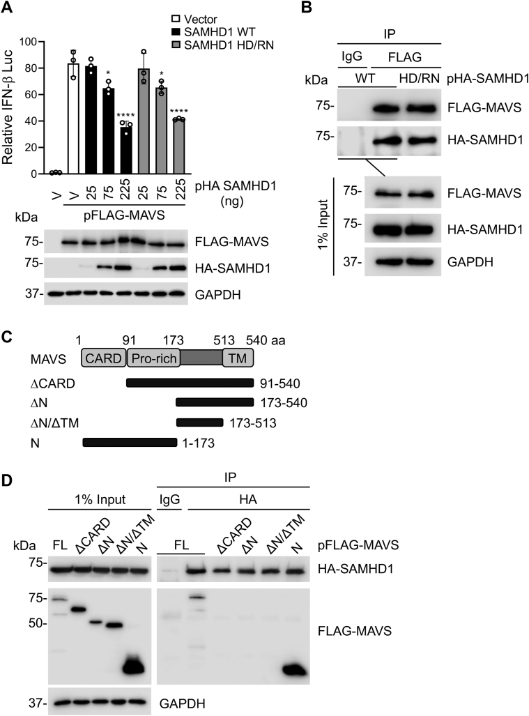
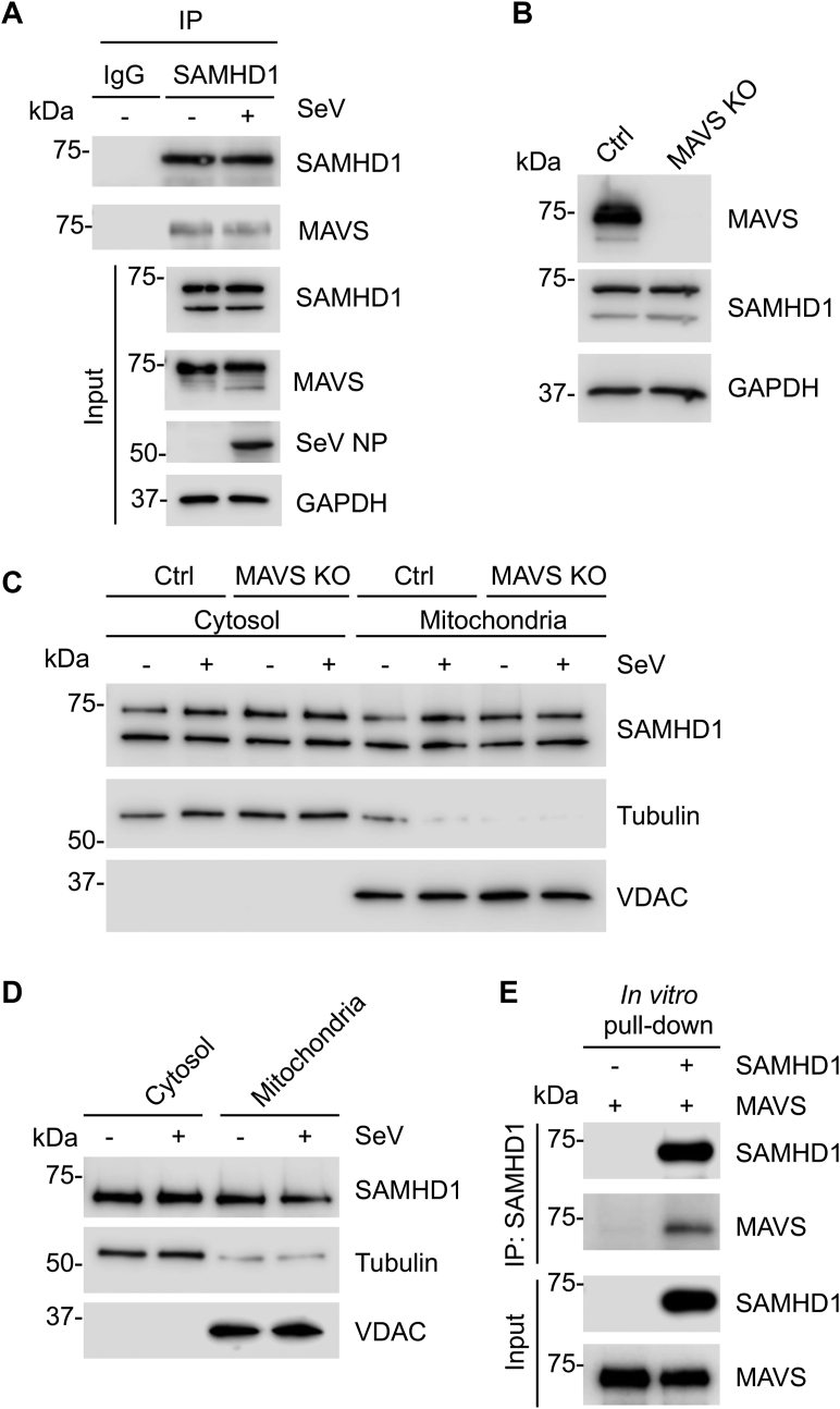
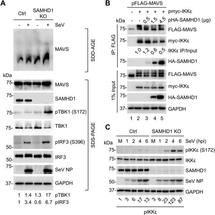
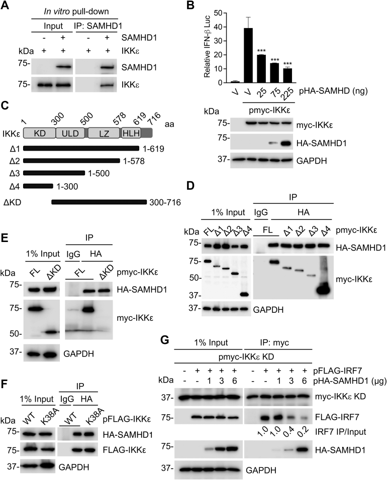
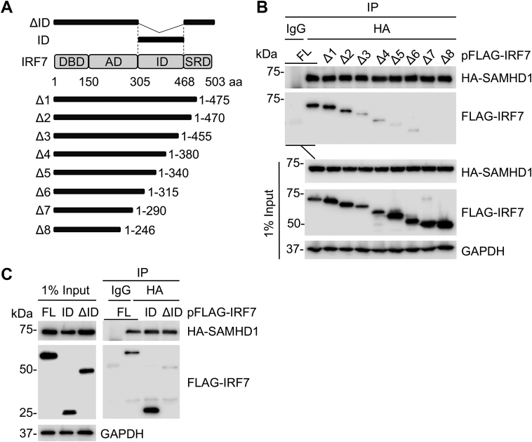
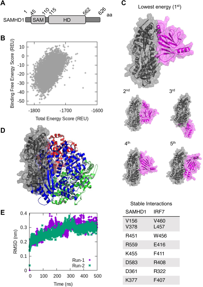
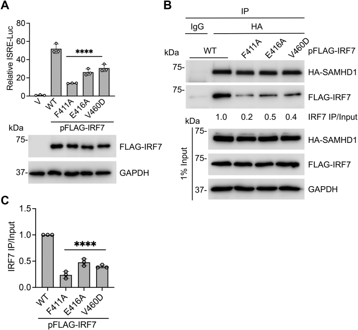
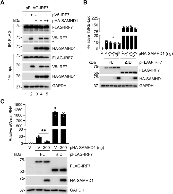
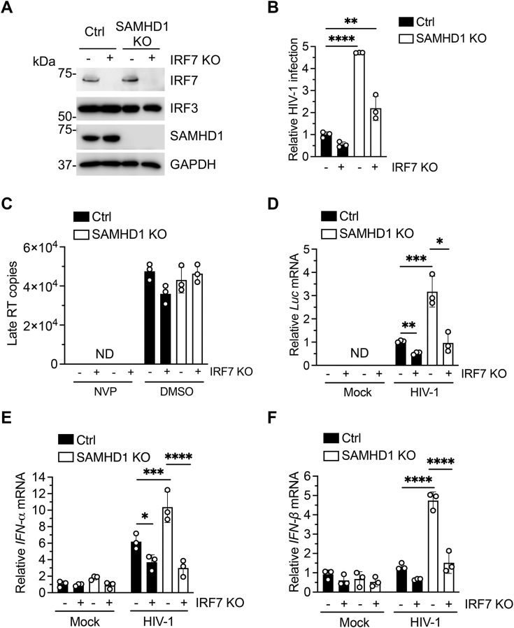
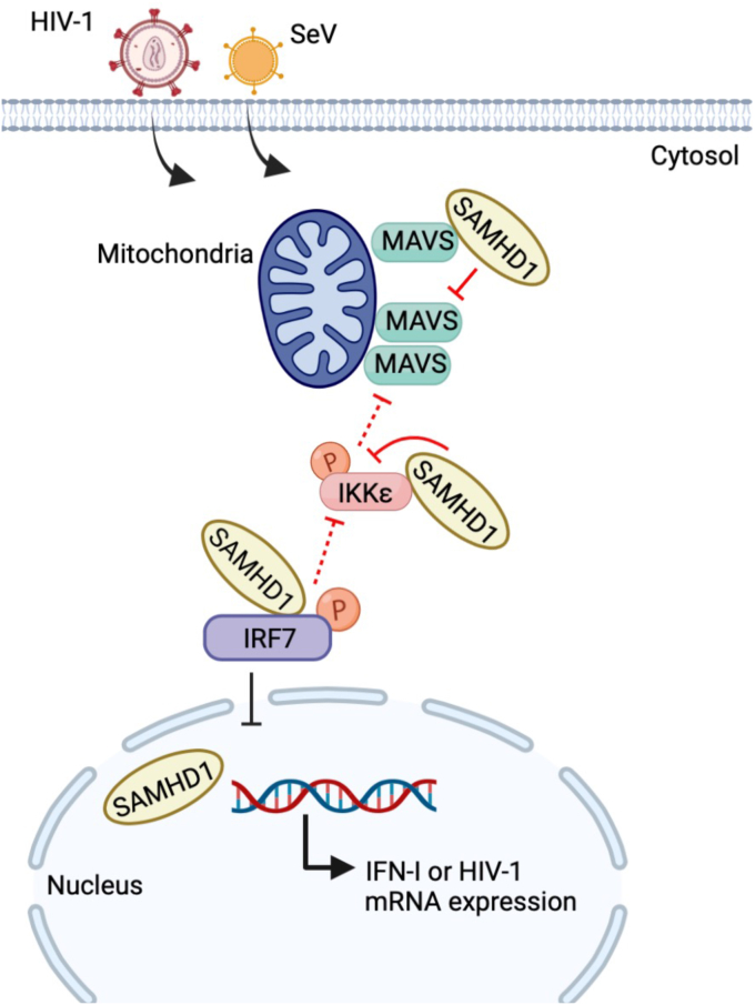
Sterile alpha motif and HD domain-containing protein 1 (SAMHD1) restricts human immunodeficiency virus type 1 (HIV-1) infection by reducing the intracellular dNTP pool. We have shown that SAMHD1 suppresses nuclear factor kappa-B activation and type I interferon (IFN-I) induction by viral infection and inflammatory stimuli. However, the mechanism by which SAMHD1 inhibits IFN-I remains unclear. Here, we show that SAMHD1 inhibits IFN-I activation induced by the mitochondrial antiviral-signaling protein (MAVS). SAMHD1 interacted with MAVS and suppressed MAVS aggregation in response to Sendai virus infection in human monocytic THP-1 cells. This resulted in increased phosphorylation of TANK binding kinase 1 (TBK1), inhibitor of nuclear factor kappa-B kinase epsilon (IKKε), and IFN regulatory factor 3 (IRF3). SAMHD1 suppressed IFN-I activation induced by IKKε and prevented IRF7 binding to the kinase domain of IKKε. We found that SAMHD1 interaction with the inhibitory domain (ID) of IRF7 (IRF7-ID) was necessary and sufficient for SAMHD1 suppression of IRF7-mediated IFN-I activation in HEK293T cells. Computational docking and molecular dynamics simulations revealed possible binding sites between IRF7-ID and full-length SAMHD1. Individual substitution of F411, E416, or V460 in IRF7-ID significantly reduced IRF7 transactivation activity and SAMHD1 binding. Furthermore, we investigated the role of SAMHD1 inhibition of IRF7-mediated IFN-I induction during HIV-1 infection. We found that THP-1 cells lacking IRF7 expression had reduced HIV-1 infection and viral transcription compared to control cells, indicating a positive role of IRF7 in HIV-1 infection. Our findings suggest that SAMHD1 suppresses IFN-I induction through the MAVS, IKKε, and IRF7 signaling axis.

Keywords: HIV-1; IKKε; IRF; MAVS; SAMHD1; innate immunity; interferon; molecular docking.


Conflict of interest statement

Conflict of interest The authors declare that they have no conflicts of interest with the contents of this article.

Figures

Figure 1
Figure 1
SAMHD1 inhibits MAVS-mediated IFN-I signaling and interacts with the CARD of MAVS.A, HEK293T cells were cotransfected with increasing amounts of plasmid encoding HA-SAMHD1 WT or HD/RN, IFN-β-luciferase reporter, renilla-TK, and FLAG-tagged MAVS. Dual luciferase assay was performed at 24 h posttransfection, and cell lysates were harvested for IB. Error bars represent mean ± SD. Statistical significance was determined using one-way ANOVA; ∗∗p < 0.01; ∗∗∗∗p < 0.0001 compared with the vector control in the same group. B, HEK293T cells were cotransfected with plasmids encoding HA-SAMHD1 WT or HD/RN and FLAG-MAVS. Cells were lysed and IP was performed using anti-FLAG antibody at 36 h posttransfection. Nonspecific IgG was used as a negative control in IP, and the indicated proteins were detected by IB. C, schematic representation of full-length (FL) MAVS and MAVS mutants. The amino acid (aa) numbers of MAVS are shown. The CARD, Pro-rich, and TM domains are indicated. D, HEK293T cells were cotransfected with plasmids encoding HA-SAMHD1 and FLAG-MAVS or MAVS mutants. Cells were lysed and IP was performed using anti-HA antibody at 36 h posttransfection. Nonspecific IgG was used as a negative control in IP, and the indicated proteins were detected by IB. A, B and D, representative data from three independent experiments are shown. CARD, caspase-recruitment domain; HD, histidine-aspartate; IB, immunoblot; IFN, interferon; MAVS, mitochondrial antiviral-signaling protein; Pro-rich, proline-rich; SAMHD, sterile alpha motif and HD domain-containing protein; TM, transmembrane; V, vector controls.
Figure 2
Figure 2
SAMHD1 localizes to the mitochondria and directly interacts with MAVS.A, THP-1 control cells were infected with SeV (MOI of 10) and harvested at 6 hpi for IP with SAMHD1 antibody or IgG control. IB was performed to detect the indicated proteins. Two bands of SAMHD1 in input samples represent the splicing variants. B, IB analysis of MAVS and SAMHD1 expression in THP-1 Ctrl and MAVS KO cells. GAPDH was used as a loading control. C, IB analysis of endogenous SAMHD1 from the cytosol and mitochondrial fraction of THP-1 Ctrl and MAVS KO mock-infected or infected with SeV for 6 h (MOI of 10). D, IB analysis of endogenous SAMHD1 from the cytosol and mitochondrial fraction of HEK293T cells mock-infected or infected with SeV for 6 h (MOI of 1). C and D, tubulin and VDAC were used as cytosolic and mitochondrial markers, respectively. Equal amounts of protein for both fractions were loaded. E, recombinant FL SAMHD1 and MAVS without TM purified from E. coli were pulled down with an anti-SAMHD1 antibody and analyzed by IB. A and CE, representative data from three independent experiments are shown. FL, full-length; hpi, hours postinfection; IB, immunoblot; MAVS, mitochondrial antiviral-signaling protein; MOI, multiplicity of infection; SAMHD, sterile alpha motif and HD domain-containing protein; SeV, Sendai virus; SeV NP, nucleoprotein of SeV; TM, transmembrane; VDAC, voltage-dependent anion channel.
Figure 3
Figure 3
SAMHD1 suppresses MAVS aggregation and disrupts MAVS interaction with IKKε.A, THP-1 Ctrl and SAMHD1 KO cells were mock-infected or infected with SeV (MOI of 10) for 8 h. Crude mitochondria were isolated and subjected to SDD-AGE (top). Whole cell lysates were analyzed by SDS-PAGE (bottom). Quantification of pTBK1 (S172) and pIRF3 (S396) levels was performed by densitometry and normalized relative to total TBK1 and IRF3, respectively. B, HEK293T cells were transfected with expression plasmids encoding the indicated proteins (FLAG-MAVS, myc-IKKε, and increasing amounts of HA-SAMHD1). Cells were lysed 36 h posttransfection, and IP was performed using an anti-FLAG antibody. Indicated proteins were detected by IB. C, THP-1 Ctrl and SAMHD1 cells were mock-infected (M) or infected with SeV (MOI of 10) and harvested at the indicated time points. Cell lysates were analyzed by IB with the indicated antibodies. Quantification of pIKKε (S172) levels was performed by densitometry and expressed relative to total IKKε. AC, representative results from three independent experiments are shown. IB, immunoblot; IKKε, inhibitor of nuclear factor kappa-B kinase epsilon; IRF, IFN regulatory factor; M, mock infection; MAVS, mitochondrial antiviral-signaling protein; MOI, multiplicity of infection; pIKKε, phospho-IKKε; SAMHD, sterile alpha motif and HD domain-containing protein; SDD-AGE, semidenaturing detergent agarose gel electrophoresis; SeV, Sendai virus; SeV NP, nucleoprotein of SeV; TBK1, TANK binding kinase 1.
Figure 4
Figure 4
SAMHD1 inhibits IKKε-mediated IFN-I signaling and disrupts IRF7 binding to the IKKε kinase domain.A, recombinant SAMHD1 and IKKε purified from Escherichia coli and HEK293T cells, respectively, were pulled down with an anti-SAMHD1 antibody and analyzed by IB. Representative data from two independent experiments are shown. B, HEK293T cells were cotransfected with increasing amounts of plasmid encoding HA-SAMHD1, IFN-β-luciferase reporter, renilla-TK, and myc-tagged IKKε. Dual luciferase assay was performed at 24 h posttransfection, and cell lysates were harvested for IB. Error bars represent mean ± SD. Statistical significance was determined using one-way ANOVA; ∗∗∗p < 0.001 compared with the vector control in the same group. C, schematic representation of full-length (FL) human IKKε (top). Five myc-tagged C-terminal and N-terminal deletion mutants are represented by the solid bars below the FL IKKε. The aa numbers of IKKε are shown. DF, HEK293T cells were cotransfected with plasmids encoding HA-SAMHD1 and myc-tagged IKKε FL or C-terminal deletion mutants (D), IKKε N-terminal deletion lacking the kinase domain (ΔKD) (E), or IKKε kinase-inactive mutant K38A (F). Cells were lysed, and IP was performed using anti-HA antibody at 36 h posttransfection. Nonspecific IgG was used as a negative control in IP, and the indicated proteins were detected by IB. G, HEK293T cells were cotransfected with myc-IKKε KD, FLAG-IRF7, and HA-SAMHD1. Cells were lysed, and IP was performed using anti-myc antibody at 36 h posttransfection. The indicated proteins were detected by IB. A and B and DG, representative data from three independent experiments are shown. HLH, helix-loop-helix domain; IB, immunoblot; IFN, interferon; IKKε, inhibitor of nuclear factor kappa-B kinase epsilon; IRF, IFN regulatory factor; KD, kinase domain; LZ, leucine zipper domain; SAMHD, sterile alpha motif and HD domain-containing protein; ULD, ubiquitin-like domain; V, vector controls; WT, wild-type.
Figure 5
Figure 5
IRF7 inhibitory domain is necessary and sufficient for SAMHD1 binding.A, Human IRF7 is illustrated schematically. The aa numbers of IRF7 are shown. A series of FLAG-tagged C-terminal truncated mutants, ID alone, and ID-deleted mutant (ΔID) are represented by the solid bars. B, HEK293T cells were cotransfected with plasmids encoding HA-SAMHD1 and FLAG-tagged IRF7 full length or C-terminal deletion mutant. Cells were lysed and IP was performed using anti-HA antibody at 36 h posttransfection. Nonspecific IgG was used as a negative control in IP, and the indicated proteins were detected by IB. C, HEK293T cells were cotransfected with the indicated plasmids. IP was performed as described in (B). B and C, representative results from three independent experiments are shown. AD, activating domain; DBD, DNA-binding domain; IB, immunoblot; ID, inhibitory domain; IRF, IFN regulatory factor; SAMHD, sterile alpha motif and HD domain-containing protein; SRD, serine-rich domain.
Figure 6
Figure 6
Computational prediction of IRF7-ID–binding interactions with SAMHD1.A, schematic representation of full-length human SAMHD1. The aa numbers are shown. B, global docking, showing total energy score versus binding free energy score in Rosetta Energy Unit (REU) of 50,000 sampling poses. C, the five poses with minimum binding free energy scores. Gray, SAMHD1 monomer; Purple, IRF7-ID. Note that the fourth best pose is almost identical to the best pose (the first pose), with an RMSD of 1.7 Å. The second and fifth poses have very similar SAMHD1 binding sites with different orientations of IRF7-ID. Only the third best pose has a different SAMHD1 binding within the oligomeric interface. D, tetrameric SAMHD1 complex (4bzc in the PDB database) with the same orientation of the gray monomer as in (B), showing that all best poses in (B) bind IRF7 in the oligomeric interface. Green, blue, and red structures are the three other monomers of SAMHD1 in the tetramer. E, molecular dynamics simulations of the top pose, indicating the stability of the complex at 300 K (left panel), along with the observed stabilizing interactions between SAMHD1 and IRF7-ID (Table; note that the first interaction consists of two amino acids from each protein). Amino acid numbers correspond to full-length SAMHD1 and IRF7. ID, inhibitory domain; IRF, IFN regulatory factor; SAMHD, sterile alpha motif and HD domain-containing protein.
Figure 7
Figure 7
Three key residues of IRF7 important for its transactivation activity and SAMHD1 binding.A, HEK293T cells were cotransfected with ISRE-luciferase reporter, renilla-TK, and FLAG-tagged IRF7 WT or individual mutants as indicated. Dual luciferase assay was performed at 24 h posttransfection, and cell lysates were harvested for IB. Error bars represent mean ± SD. Statistical significance was determined using one-way ANOVA. ∗∗∗∗p < 0.0001 compared with IRF7 WT. B, HEK293T cells were transfected with expression plasmids encoding HA-SAMHD1 and FLAG-IRF7 WT or individual mutants as indicated. Cells were lysed at 36 h posttransfection, and IP was performed using anti-HA antibody. Indicated proteins were detected by IB. Quantification of IRF7 IP/Input levels was performed by densitometry and expressed relative to IRF7 WT. C, average results of relative IRF7 IP/Input levels based on three independent experiments. Error bars represent mean ± SD. Statistical significance was determined using one-way ANOVA. ∗∗∗∗p < 0.0001 compared with IRF7 WT. IB, immunoblot; IRF, IFN regulatory factor; ISRE, IFN-sensitive response element; SAMHD, sterile alpha motif and HD domain-containing protein; V, vector control.
Figure 8
Figure 8
SAMHD1 binding to IRF7-ID is required for suppression of IRF7-mediated IFN-I induction.A, HEK293T cells were transfected with expression plasmids encoding FLAG-IRF7, V5-IRF7, and HA-SAMHD1. IP and IB was performed as described above. (∗) IgG Heavy chain. B, HEK293T cells were cotransfected with increasing amounts of plasmid encoding HA-SAMHD1, ISRE-luciferase reporter, renilla-TK, and FLAG-tagged IRF7 FL or ΔID. Dual luciferase assay was performed at 24 h posttransfection, and cell lysates were harvested for IB. Error bars represent mean ± SD. Statistical significance was determined using one-way ANOVA. ∗p < 0.05 compared with the vector control in the same group. C, HEK293T cells were cotransfected with plasmids encoding FLAG-IRF7 FL or ΔID and HA-SAMHD1. IFN-α mRNA levels were measured by RT-qPCR at 24 h posttransfection. HPRT mRNA was used as housekeeping control for normalization. Error bars represent mean ± SD. t test was used for statistical significance. ∗∗p < 0.01. AC, representative data from three independent experiments are shown. FL, full-length; HPRT, hypoxanthine phosphoribosyl transferase; IB, immunoblot; ID, inhibitory domain; IFN, interferon; IRF, IFN regulatory factor; ISRE, IFN-sensitive response element; qPCR, quantitative PCR; SAMHD, sterile alpha motif and HD domain-containing protein; V, vector controls.
Figure 9
Figure 9
Endogenous IRF7 promotes HIV-1 infection and viral transcription in THP-1 cells.A, immunoblot analysis of IRF7 expression in THP-1 Ctrl and SAMHD1 KO in combination with IRF7 KO. GAPDH was used as a loading control. BF, THP-1 Ctrl and SAMHD1 KO in combination with IRF7 KO cells were infected with HIV-1-Luc/VSV-G (MOI of 2). B, HIV-1 infection was determined by luciferase assay 24 hpi. Luciferase values were expressed relative to THP-1 Ctrl and normalized to 10 μg of total protein. C, genomic DNA was isolated at 14 hpi, and late RT products were quantified by qPCR. Serial dilutions 108 to 102 of an HIV-1 proviral plasmid (pNL4-3) were used to calculate late RT copy numbers. Unspliced GAPDH was used as an endogenous control. DF, levels of Luciferase (Luc) and IFN-α/β (E and F) mRNA levels were evaluated by RT-qPCR at 18 hpi and 2 hpi, respectively. HPRT mRNA was used as a housekeeping control. Values were expressed relative to HIV-1–infected THP-1 Ctrl cells (D) or mock-infected THP-1 Ctrl cells (E and F). AF, representative data from three independent experiments are shown. Error bars represent mean ± SD. Statistical significance was determined using two-way ANOVA with multiple comparisons. ∗p < 0.05; ∗∗p < 0.01; ∗∗∗p < 0.001, ∗∗∗∗p < 0.0001. HIV, human immunodeficiency virus; hpi, hours postinfection; HPRT, hypoxanthine phosphoribosyl transferase; IFN, interferon; IRF, IFN regulatory factor; MOI, multiplicity of infection; ND, not detectable; NVP, nevirapine; qPCR, quantitative PCR; RT, reverse transcription; SAMHD, sterile alpha motif and HD domain-containing protein; VSV-G, vesicular stomatitis virus G protein.
Figure 10
Figure 10
SAMHD1 impairs IFN-I induction through the MAVS, IKKε, and IRF7 signaling axis during viral infection. SAMHD1 inhibits MAVS- and IKKε-mediated IFN-I induction in monocytic cells. Mechanistically, SAMHD1 interacts with the CARD of MAVS and suppresses MAVS aggregation, resulting in reduced IKKε recruitment to MAVS and IKKε phosphorylation (indicated with a letter P). SAMHD1 also binds to the kinase domain of IKKε and disrupts binding of IRF7 to IKKε KD. SAMHD1 suppression of the IRF7-mediated antiviral response depends on its interaction with the inhibitory domain of IRF7. Our results also indicated that endogenous IRF7 in THP-1 cells is required for efficient HIV-1 infection and viral gene expression. The red T-shaped symbols indicate inhibitory functions confirmed in experiments, while the black T-shaped symbols indicate proposed inhibitory mechanisms. This figure was created with BioRender.com. CARD, caspase-recruitment domain; HIV, human immunodeficiency virus; IFN, interferon; IKKε, inhibitor of nuclear factor kappa-B kinase epsilon; IRF, IFN regulatory factor; KD, kinase domain; MAVS, mitochondrial antiviral-signaling protein; SAMHD, sterile alpha motif and HD domain-containing protein.

Similar articles

Cited by

References

    1. Hollenbaugh J.A., Gee P., Baker J., Daly M.B., Amie S.M., Tate J., et al. Host factor SAMHD1 restricts DNA viruses in non-dividing myeloid cells. PLoS Pathog. 2013;9 - PMC - PubMed
    1. Sommer A.F., Riviere L., Qu B., Schott K., Riess M., Ni Y., et al. Restrictive influence of SAMHD1 on Hepatitis B Virus life cycle. Sci. Rep. 2016;6 - PMC - PubMed
    1. Zhang K., Lv D.W., Li R. Conserved herpesvirus protein kinases target SAMHD1 to facilitate virus replication. Cell Rep. 2019;28:449–459.e445. - PMC - PubMed
    1. Goldstone D.C., Ennis-Adeniran V., Hedden J.J., Groom H.C., Rice G.I., Christodoulou E., et al. HIV-1 restriction factor SAMHD1 is a deoxynucleoside triphosphate triphosphohydrolase. Nature. 2011;480:379–382. - PubMed
    1. Powell R.D., Holland P.J., Hollis T., Perrino F.W. Aicardi-Goutieres syndrome gene and HIV-1 restriction factor SAMHD1 is a dGTP-regulated deoxynucleotide triphosphohydrolase. J. Biol. Chem. 2011;286:43596–43600. - PMC - PubMed

Publication types

MeSH terms