Review
. 2023 Sep 5;29(5):521-544.
doi: 10.1093/humupd/dmad012.

Uterus transplantation: from research, through human trials and into the future

Affiliations
Review

Uterus transplantation: from research, through human trials and into the future

Mats Brännström et al. Hum Reprod Update. .

Abstract

Women suffering from absolute uterine factor infertility (AUFI) had no hope of childbearing until clinical feasibility of uterus transplantation (UTx) was documented in 2014 with the birth of a healthy baby. This landmark accomplishment followed extensive foundational work with a wide range of animal species including higher primates. In the present review, we provide a summary of the animal research and describe the results of cases and clinical trials on UTx. Surgical advances for graft removal from live donors and transplantation to recipients are improving, with a recent trend away from laparotomy to robotic approaches, although challenges persist regarding optimum immunosuppressive therapies and tests for graft rejection. Because UTx does not involve transplantation of the Fallopian tubes, IVF is required as part of the UTx process. We provide a unique focus on the intersection between these two processes, with consideration of when oocyte retrieval should be performed, whether, and for whom, preimplantation genetic testing for aneuploidy should be used, whether oocytes or embryos should be frozen and when the first embryo transfer should be performed post-UTx. We also address the utility of an international society UTx (ISUTx) registry for assessing overall UTx success rates, complications, and live births. The long-term health outcomes of all parties involved-the uterus donor (if live donor), the recipient, her partner and any children born from the transplanted graft-are also reviewed. Unlike traditional solid organ transplantation procedures, UTx is not lifesaving, but is life-giving, although as with traditional types of transplantation, costs, and ethical considerations are inevitable. We discuss the likelihood that costs will decrease as efficiency and efficacy improve, and that ethical complexities for and against acceptability of the procedure sharpen the distinctions between genetic, gestational, and social parenthood. As more programs wish to offer the procedure, we suggest a scheme for setting up a UTx program as well as future directions of this rapidly evolving field. In our 2010 review, we described the future of clinical UTx based on development of the procedure in animal models. This Grand Theme Review offers a closing loop to this previous review of more than a decade ago. The clinical feasibility of UTx has now been proved. Advancements include widening the criteria for acceptance of donors and recipients, improving surgery, shortening time to pregnancy, and improving post-UTx management. Together, these improvements catalyze the transition of UTx from experimental into mainstream clinical practice. The procedure will then represent a realistic and accessible alternative to gestational surrogacy for the treatment of AUFI and should become part of the armamentarium of reproductive specialists worldwide.

Keywords: IVF; animal models; assisted reproduction; ethics; human; infertility; surgery; transplantation; uterine factor infertility; uterus.


Conflict of interest statement

No author reports any conflict of interest regarding the contents of this manuscript.

Figures

None
Live birth is achievable after uterus transplantation: key features in the development and application of uterus transplantation to human. ET: embryo transfer.
Figure 1.
Figure 1.
Time-line of animal-based research and major accomplishments in human uterus transplantation. Animal research in uterus transplantation followed a natural sequence, from rodents to large domestic species, and then to non-human primates. The specific references connected to the start of research in each animal species are: mouse (Racho El-Akouri et al., 2002), rat (Wranning et al., 2008a), sheep (Wranning et al., 2008b), pig (Avison et al., 2009), non-human primate (Enskog et al., 2010). UTx, uterus transplantation; LD, live donor; DD, deceased donor.
Figure 2.
Figure 2.
Flowchart of uterus transplantation in human. The arrows on the left indicate that pregnancy and delivery (by Caesarean section) can be repeated several times. LD, live donor; DD, deceased donor; ET, embryo transfer; UTx, uterus transplantation.
Figure 3.
Figure 3.
Schematic drawing of the operating field of the donor’s right pelvic side at hysterectomy. The left panel is a close up of the surgical field at the distal aspect of the ureter. The middle panel is an overview of the entire surgical field. The right panel is a close up of the surgical field of the ureteric tunnel. In all panels, arteries are red, veins are blue, ureter, and bladder are yellow, and uterus is grey. The squared grid illustrates those areas covered in connective tissue.
Figure 4.
Figure 4.
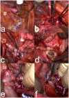
Photographs showing specific steps in live donor hysterectomy by laparotomy. (a) A large peritoneal flap (#) is dissected from the bladder dome to be included in the graft after the round ligaments have been divided and tagged. (b) The right ureter (*) is dissected free. (c) Image showing completed dissection on the right pelvic sidewall of the ureter (*), a deep uterine vein (v), and the uterine artery (a) extending from the anterior internal iliac artery. (d) The vagina of the donor is transected as the last step before vascular clamping with transection of vessels and extraction of the organ. (e) The right anterior division of the internal iliac artery (ia) is clamped, leaving the major posterior branch, before transection of the major vessels and removal of the uterine graft from the donor. The bifurcation (bf) of the right common iliac artery into the external and internal iliac arteries is seen to the left. (f) A vascular clamp is placed over the left internal iliac vein, to include a segment of this vein together with the deep uterine vein (v) of the graft.
Figure 5.
Figure 5.
Photographs showing specific steps in live donor hysterectomy by robotics. (a) The peritoneal flap (#), to be included in the graft, is dissected off from the bladder dome. (b) Image showing dissection of the right ureteric tunnel with the proximal ureter (*) in a blue rubber band. (c) Dissection of the distal portion of the right ureter (*) before the inlet to the bladder. (d) The vaginal vault (vv) is opened. (e) A bulldog clamp (bc) after being positioned on a major branch of the right anterior branch of the internal iliac artery (ia) before transection of major graft vessels. The upper rubber band is around the arterial trunk (a) leading to the uterine artery. The lower rubber band is placed around the ureter. (f) The uterus is placed in a laparoscopic bag and is extracted through the vagina.
Figure 6.
Figure 6.
Schematic drawing of the vascular and vaginal anastomoses in the recipient. The tissues of the recipient are covered by a lined grid. The anterior portions of the internal iliac arteries are anastomosed end-to-side to the external iliac arteries on both sides. On the recipient’s left side, one deep uterine vein and the uterine branch of the utero-ovarian vein are anastomosed end-to-side to the external iliac vein. On the recipient’s right side, the utero-ovarian vein is anastomosed end-to-side to the external iliac vein.
Figure 7.
Figure 7.
Photographs showing specific steps in recipient surgery at uterus transplantation. (a) The midline uterine rudiment (ur) in a patient with Mayer–Rokitansky–Küster–Hauser syndrome is lifted by forceps with simultaneous dissection to separate the bladder (bl) from the rudiment and the vaginal vault. (b) Image showing the uterine rudiment after cleavage in the midline down to the vaginal vault, with fixation sutures placed through the sacrouterine ligaments (su) and through both sides of the cleaved uterine rudiment (ur). (c) Image showing end-to-end vascular anastomose under completion between a deep uterine vein (v) of the graft and the external iliac vein of the recipient. (d) An end-to-end vascular anastomose is completed between the artery (a) of the graft and the external iliac artery of the recipient. A bulldog clamp is placed proximally to the completed venous anastomosis. (e) The vaginal vault (vv) is opened longitudinally by monopolar diathermy. (f) The grafted uterus (ut) is seen after completed transplantation, with a peritoneal flap (pf) overlaying the recipient’s bladder.

References

    1. Akouri R, Maalouf G, Abboud J, Nakad T, Bedran F, Hajj P, Beaini C, Cricu L, Aftimos G, Hajj C. et al. First live birth after uterus transplantation in the Middle East. MEFS J 2020;25:30–35.
    1. Alghrani A. Uterus transplantation in and beyond cisgender women: revisiting procreative liberty in light of emerging reproductive technologies. J Law Biosci 2018;5:301–328. - PMC - PubMed
    1. Andraus W, Ejzenberg D, Santos RM, Mendes LR, Arantes RM, Baracat EC, D'Albuquerque LA.. Sheep model for uterine transplantation: the best option before starting a human program. Clinics (Sao Paulo) 2017;72:178–182. - PMC - PubMed
    1. Api M, Boza A, Ceyhan M.. Could the female-to-male transgender population be donor candidates for uterus transplantation? Turk J Obstet Gynecol 2017;14:233–237. - PMC - PubMed
    1. Arantes RM, Nacif LS, Pinheiro RS, Rocha-Santos V, de Martino RB, Waisberg DR, Pantanali CAR, Fortunato A, Lima MR, Ducatti L. et al. Novel technique in a sheep model of uterine transplantation. Transplant Proc 2020;52:1399–1401. - PubMed