. 2023 Jun 16;9(1):92.
doi: 10.1038/s41531-023-00520-1.

Synaptic mechanisms underlying onset and progression of memory deficits caused by hippocampal and midbrain synucleinopathy

Affiliations

Synaptic mechanisms underlying onset and progression of memory deficits caused by hippocampal and midbrain synucleinopathy

Attilio Iemolo et al. NPJ Parkinsons Dis. .

Abstract

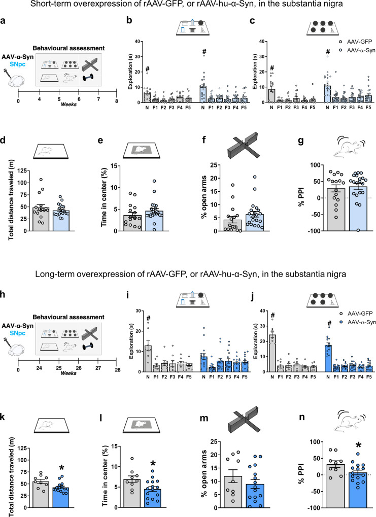
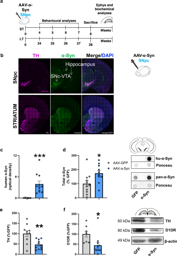
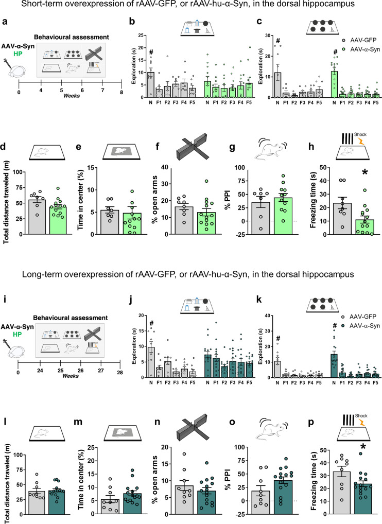
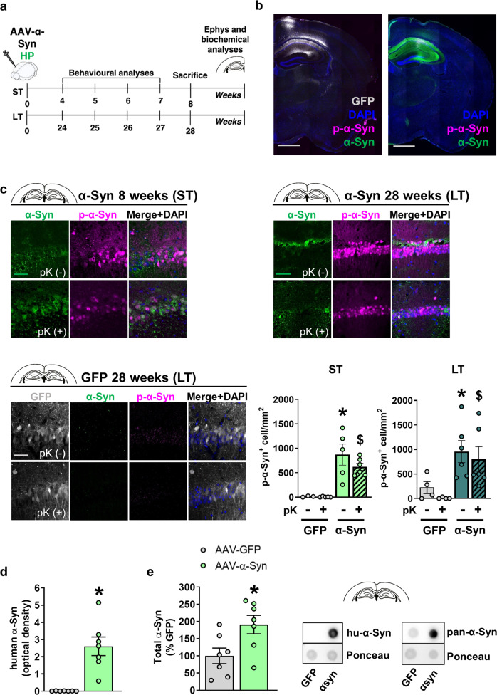
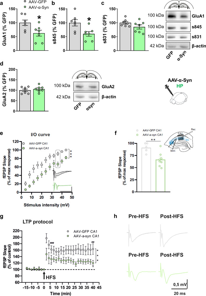
Cognitive deficits, including working memory, and visuospatial deficits are common and debilitating in Parkinson's disease. α-synucleinopathy in the hippocampus and cortex is considered as the major risk factor. However, little is known about the progression and specific synaptic mechanisms underlying the memory deficits induced by α-synucleinopathy. Here, we tested the hypothesis that pathologic α-Synuclein (α-Syn), initiated in different brain regions, leads to distinct onset and progression of the pathology. We report that overexpression of human α-Syn in the murine mesencephalon leads to late onset memory impairment and sensorimotor deficits accompanied by reduced dopamine D1 expression in the hippocampus. In contrast, human α-Syn overexpression in the hippocampus leads to early memory impairment, altered synaptic transmission and plasticity, and decreased expression of GluA1 AMPA-type glutamate receptors. These findings identify the synaptic mechanisms leading to memory impairment induced by hippocampal α-synucleinopathy and provide functional evidence of the major neuronal networks involved in disease progression.


Conflict of interest statement

The authors declare no competing interests.

Figures

Fig. 1
Fig. 1. Short and long-term rAAV-mediated overexpression of GFP and hu-α-Syn in the midbrain.
a, h Schematic representations of behavioral assessment at 4- and 24-weeks after injections with rAAV-hu-α-Syn or rAAV-GFP vectors. b, c SNpc-hu-α-mice compared to SNpc-GFP mice do not show differences in the exploration of the novel object (N) compared to all the familiar ones (F1-F5) in both the DOT and IOT tasks (ST rAAV-GFP 6-DOT repeated measures ANOVA: F(5,70) = 9.75; P < 0.001; ST rAAV-hu-α-Syn 6-DOT repeated measures ANOVA: F(5,90) = 14.99; P < 0.001; ST rAAV-GFP 6-IOT repeated measures ANOVA: F(5,70) = 12.82; P < 0.001; ST rAAV-hu-α-Syn 6-IOT repeated measures ANOVA: F(5,95) = 17.45; p < 0.001), (d, e) exploratory behavior in the open field or (f) in the elevated plus maze, (g) as well as in pre-pulse inhibition in the startling reflex-box. in In LT-experiment, rAAV-GFP explored significantly more the novel object (N) compared to all the familiar ones (F1-F5) in both the DOT and IOT tasks. However, rAAV-hu-α-Syn showed a specific impairment in recognition of the novel object in the DOT but not in the IOT task. Indeed, the novel object was not significantly more explored then all the familiars in the 6-DOT (LT rAAV-GFP 6-DOT repeated measures ANOVA: F(5,35) = 9.79; P < 0.001; LT rAAV-hu-α-Syn 6-DOT repeated measures ANOVA: F(5,70) = 2.94; P = 0.018; LT rAAV-GFP 6-IOT repeated measures ANOVA: F(5,35) = 102.99; P < 0.001; LT rAAV-hu-α-Syn 6-IOT repeated measures ANOVA: F(5,70) = 74.36; p < 0.001). k, l SNpc-hu-α-Syn mice, as compared to SNpc-GFP, also showed reduction in distance traveled and time spent in center of the open field (unpaired t test, t = 2.65, P < 0.05 and t = 2.39, P < 0.05, respectively, SNpc -GFP n = 9; SNpc -hu-α-Syn, n = 15), (m), but not in the percentage time spent in the open arm, suggesting hypokinesia and increased avoidance behavior. Finally, (n) SNpc-hu-α-Syn, compared to SNpc-GFP mice, showed impaired pre-pulse inhibition, an endophenotype of psychosis (unpaired t test, t = 2.08, P < 0.05, SNpc-GFP, n = 9; SNpc-hu-α-Syn, n = 15). Data are presented as mean ± SEM. *P < 0.05 different from SNpc-GFP control. #P < 0.05 N different from F1–F5 (Dunnett’s test).
Fig. 2
Fig. 2. rAAV-mediated overexpression of hu-α-Syn in the midbrain leads to reduced TH and D1 receptor expression in the hippocampus.
a Schematic representation of the experimental design for the short-term and long-term experiments, including behavioral testing starting 4- or 24-weeks after intra-SNpc injections as described in. After behavioral testing animals were sacrificed for immunohistochemical, biochemical and ex-vivo electrophysiological analysis (for the short-term time point). b Representative coronal section of hemibrain of SNpc-hu-α-Syn mice showing wide expression of hu-α-Syn in the target regions of the VTA/SNpc, including the hippocampus (dorsal and ventral), the septum and the striatum (Scale bar = 1 mm). Detailed histological analysis of this group of animals has been previously reported in ref. . c, d Dot-blot analysis of hu-α-Syn (unpaired t test, t = −4.55, P = 0.0002, SNpc-GFP, n = 11; SNpc -hu-α-Syn, n = 11) and of pan-α-Syn expression (unpaired t test, t = −2.53, P = 0.019, SNpc-GFP, n = 11; SNpc -hu-α-Syn, n = 11) in mesencephalic tissue of SNpc-hu-α-Syn mice and SNpc-GFP mice. e, f The overexpression of hu-α-Syn in the SNpc/VTA significantly decreased the expression of TH (unpaired t test, t = 3.451, P = 0.0027, SNpc-GFP, n = 10; SNpc-hu-α-Syn, n = 11) and D1 dopamine receptor (unpaired t test, t = 2.647, P = 0.0169, SNpc-GFP, n = 8; SNpc-hu-α-Syn, n = 11) in the hippocampus. Representative bands for each condition are reported. Data are presented as mean ± SEM. *P < 0.05, **P < 0.01, ***P < 0.001 different from SNpc-GFP.
Fig. 3
Fig. 3. Short and long-term rAAV mediated overexpression of GFP and hu-α-Syn in the dorsal hippocampus.
a, i Schematic representations of behavioral assessment at 4- and 24-weeks after injections with rAAV-hu-α-Syn or rAAV-GFP vectors. b, c Short-term rAAV-GFP injected animals explored significantly more the novel object (N) compared to all the familiar ones (F1–F5) in both the DOT and IOT tasks, while hu-α-Syn injected animals showed impairment in the 6-DOT but not in 6-IOT (ST rAAV-GFP 6-DOT repeated measures ANOVA: F(5,35) = 5.23; P = 0.0011; ST rAAV-hu-α-Syn 6-DOT repeated measures ANOVA: F(5,50) = 0.974; P = 0.44; ST rAAV-GFP 6-IOT repeated measures ANOVA: F(5,35) = 6.74; P < 0.001; ST rAAV-hu-α-Syn 6-IOT repeated measures ANOVA: F(5,60) = 47.59; P < 0.001). d No significant changes in measures of exploratory behavior, such as the distance traveled in the open field or (e) percentage of time spent in the center of the arena, (f) percentage of time in the open arm in the plus maze task, and (g) in the pre-pulse inhibition were observed in HP-hu-α-Syn mice when compared to GFP. In contrast, (h) HP-hu-α-Syn mice show a significant reduction in freezing time, during the testing day, in the contextual fear-conditioning task, suggesting the presence of highly specific memory impairment (Group, F(1,19) = 6.67, P < 0.05, Time, F(5, 95) = 8.44, P < 0.001, Group × Time, F(5,95) = 5.19, P < 0.001). j Long-term overexpression of hu-α-Syn in the dorsal hippocampus leads to impairing effects identical to the short-term expression in the novel object exploration in conditions of high load (LT rAAV-GFP 6-DOT repeated measures ANOVA: F(5,40) = 9.87; P < 0.001; LT rAAV-hu-α-Syn 6-DOT repeated measures ANOVA: F(5,70) = 2.86; P = 0.02; LT rAAV-GFP 6-IOT repeated measures ANOVA: F(5,40) = 16.92; P < 0.001; LT rAAV-hu-α-Syn 6-IOT repeated measures ANOVA: F(5,70) = 48.0; P < 0.001), and (p) in the freezing time during the fear-conditioning test (Group, F(1,22) = 5.05, P < 0.05: Time, F(5, 110) = 6.42, P < 0.001), (ko) without affecting performance in any of the other behavioral tests. Data are presented as mean ± SEM. *P < 0.05 different from HP-GFP control. #P < 0.05 N different from F1-F5 (Dunnett’s test).
Fig. 4
Fig. 4. rAAV vector-mediated expression of hu-α-Syn in the dorsal hippocampus leads to sustained overexpression of pK-resistant α-Synuclein.
a Schematic representation of the experimental design for the short-term and long-term experiments, including behavioral testing starting 4- or 24-weeks after intra-brain injection and lasting for about 4 weeks. After behavioral testing, animals were sacrificed for immunohistochemical, biochemical and ex-vivo electrophysiological analysis (for the short-term time point). b Representative sagittal brain section (mosaic reconstruction) of the injection placement of rAAV-GFP (left panel) and rAAV-hu-α-Syn (right panel) vectors, showing a wide distribution of GFP and hu-α-Syn in the whole hippocampus (Scale bar: 1 mm). c Representative confocal images showing α-Syn (green) and p-α-Syn (magenta) expression in the CA1 of the HP injected with rAAV-hu-α-Syn or r-AAV-GFP, in basal immunostaining conditions (pK-) and after the treatment with proteinase K (pK+), for mice sacrificed at 8 weeks (ST) or 28 weeks (LT) after intra-brain injection (Scale bar: 50 μm). Confocal images at the bottom show undetectable α-Syn (green) and p-α-syn (magenta) expression in the CA1 of the HP of an rAAV-GFP mouse sacrificed at 28 weeks after injection, under basal conditions and after treatment with pK (Scale bar: 50 μm). Quantitation of the number of p-α-Syn positive cells (p-α-Syn+ cell/mm2) showed a significant effect for vector injection in the ST experiment (Injection, F(1,14) = 29.244, P < 0.0001, pK treatment, F(1,14) = 0.890, P = 0.3615, Injection × pK treatment, F(1,14) = 0.811, P = 0.3830), and a significant difference between rAAV-GFP and rAAV hu-α-Syn pK- (unpaired t test, t = 2.978, P = 0.0247, rAAV-GFP, n = 3; rAAV-hu-α-Syn, n = 5) and pK+ (unpaired t test, t = 6.789, P = 0.0001, rAAV-GFP, n = 5; rAAV-hu-α-Syn, n = 6). A similar effect was found in the LT experiment (Injection, F(1,16) = 12.433, P = 0.0028, pK treatment, F(1,16) = 0.728, P = 0.4062, Injection × pK treatment, F(1,16) = 0.022, P = 0.8846), again with a significant difference between rAAV-GFP and rAAV hu-α-Syn pK- (unpaired t test, t = 2.448, P = 0.0401, rAAV-GFP, n = 4; rAAV-hu-α-Syn, n = 6) and pK+ (Mann–Whitney test, P = 0.0095, rAAV-GFP, n = 4; rAAV-hu-α-Syn, n = 6). *P < 0.05 vs. HP-GFP pK-. $P < 0.05 vs. HP-GFP pK+. d, e Dot-blot analysis of hu-α-Syn (unpaired t test, t = −4.81, P = 0.0004, rAAV-GFP, n = 7; rAAV-hu-α-Syn, n = 7) and pan-α-Syn expression (unpaired t test, t = −2.58, P = 0.023, rAAV-GFP, n = 7; rAAV-hu-α-Syn, n = 7) in hippocampal tissue of HP-hu-α-Syn and HP-GFP mice. Data are presented as mean ± SEM. *P < 0.01 different from HP-GFP.
Fig. 5
Fig. 5. Short-term overexpression of hu-α-Syn in the hippocampus leads to reduced GluA1 AMPA receptors expression and phosphorylation associated with impaired long-term potentiation.
a Western blot analysis of the hippocampal tissue showing that administration of rAAV-hu-α-Syn vector leads to reduced expression of GluA1 (unpaired t test, t = 2.582, P = 0.02, HP-GFP, n = 7; HP-hu-α-Syn, n = 7) and (b) serine 845 phosphorylation (unpaired t test, t = 2.807, P = 0.01, HP-GFP, n = 7; HP-hu-α-Syn, n = 7) (c) but not serine 831 phosphorylation. d Intra-hippocampal overexpression of rAAV-hu-α-Syn did not affect the expression of the GluA2 subunit. Representative bands for each condition are reported. β-actin reported in 5d belong to the same blot reported in Supplementary Fig. 2a. e, f Field EPSPs input–output curves revealed a significant decrease in excitability in the HP-hu-α-Syn mice with respect to HP-GFP mice (two-way ANOVA: HP-hu-α-Syn n = 11, HP-GFP n = 11, F (11,220) = 5.090, Bonferroni’s post hoc test ***P < 0.001). g High-frequency stimulation (HFS)-induced LTP is reduced in hippocampal slices of HP-hu- α-Syn mice (green circles), compared to the HP-GFP group (gray circles) (two-way ANOVA: HP-hu-α-Syn n = 8, HP-GFP n = 11, F(23,391) = 2.690, Bonferroni’s post hoc test ***P < 0.001). Paired Student t test 2 min pre vs. 6 min post HFS: rAAV-GFP n = 11, t = 5.674, df = 10, ###P < 0.001; rAAV-hu-α-Syn n = 8, t = 2.749, df = 7, +P < 0.05; 2 min pre vs. 38 min post HFS: rAAV-GFP n = 11, t = 3.995, df = 10, ##P < 0.01; rAAV-hu-α-Syn n = 8, t = 2.450, df = 7, +P < 0.05. h Representative traces of evoked fEPSPs recorded before and 40 min after a high-frequency stimulation (HFS) protocol in HP-GFP CA1 (gray traces) and HP-α-Syn CA1 (green traces) animals. Data are presented as mean ± SEM. *P < 0.05 different from HP-GFP.

Similar articles

Cited by

References

    1. Menšíková K, et al. Lewy body disease or diseases with Lewy bodies? npj Parkinsons Dis. 2022;8:1–11. - PMC - PubMed
    1. De Risi M, et al. Mechanisms by which autophagy regulates memory capacity in ageing. Aging Cell. 2020;19:e13189. - PMC - PubMed
    1. Decressac M, Mattsson B, Lundblad M, Weikop P, Björklund A. Progressive neurodegenerative and behavioural changes induced by AAV-mediated overexpression of α-synuclein in midbrain dopamine neurons. Neurobiol. Dis. 2012;45:939–953. doi: 10.1016/j.nbd.2011.12.013. - DOI - PubMed
    1. Spillantini MG, et al. α-Synuclein in Lewy bodies. Nature. 1997;388:839–840. doi: 10.1038/42166. - DOI - PubMed
    1. Mattila PM, et al. Alpha-synuclein-immunoreactive cortical Lewy bodies are associated with cognitive impairment in Parkinson’s disease. Acta Neuropathol. 2000;100:285–290. doi: 10.1007/s004019900168. - DOI - PubMed